The role of lateral tension in calcium-induced DPPS vesicle rupture
- PMID: 22799521
- PMCID: PMC3422639
- DOI: 10.1021/la301976s
The role of lateral tension in calcium-induced DPPS vesicle rupture
Abstract
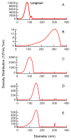
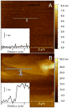
We assess the role of lateral tension in rupturing anionic dipalmitoylphosphatidyserine (DPPS), neutral dipalmitoylphosphatidylcholine (DPPC), and mixed DPPS-DPPC vesicles. Binding of Ca(2+) is known to have a significant impact on the effective size of DPPS lipids and little effect on the size of DPPC lipids in bilayer structures. In the present work we utilized laser transmission spectroscopy (LTS) to assess the effect of Ca(2+)-induced stress on the stability of the DPPS and DPPC vesicles. The high sensitivity and resolution of LTS has permitted the determination of the size and shape of liposomes in solution. The results indicate a critical size after which DPPS single shell vesicles are no longer stable. Our measurements indicate Ca(2+) promotes bilayer fusion up to a maximum diameter of ca. 320 nm. These observations are consistent with a straightforward free-energy-based model of vesicle rupture involving lateral tension between lipids regulated by the binding of Ca(2+). Our results support a critical role of lateral interactions within lipid bilayers for controlling such processes as the formation of supported bilayer membranes and pore formation in vesicle fusion. Using this free energy model we are able to infer a lower bound for the area dilation modulus for DPPS (252 pN/nm) and demonstrate a substantial free energy increase associated with vesicle rupture.
Figures


Similar articles
-
Magnesium-induced lipid bilayer microdomain reorganizations: implications for membrane fusion.J Phys Chem B. 2009 Jul 23;113(29):9932-41. doi: 10.1021/jp9011944. J Phys Chem B. 2009. PMID: 19603842 Free PMC article.
-
Model of an asymmetric DPPC/DPPS membrane: effect of asymmetry on the lipid properties. A molecular dynamics simulation study.J Phys Chem B. 2006 Feb 9;110(5):2358-63. doi: 10.1021/jp0562680. J Phys Chem B. 2006. PMID: 16471825
-
Interfacial charges drive the organization of supported lipid membranes and their interaction with nanoparticles.Colloids Surf B Biointerfaces. 2018 Dec 1;172:254-261. doi: 10.1016/j.colsurfb.2018.08.018. Epub 2018 Aug 16. Colloids Surf B Biointerfaces. 2018. PMID: 30173092
-
Lipid flip-flop in binary membranes composed of phosphatidylserine and phosphatidylcholine.J Phys Chem B. 2013 Dec 5;117(48):15041-50. doi: 10.1021/jp409672q. Epub 2013 Nov 22. J Phys Chem B. 2013. PMID: 24200035
-
Elementary Processes and Mechanisms of Interactions of Antimicrobial Peptides with Membranes-Single Giant Unilamellar Vesicle Studies.Adv Exp Med Biol. 2019;1117:17-32. doi: 10.1007/978-981-13-3588-4_3. Adv Exp Med Biol. 2019. PMID: 30980351 Review.
Cited by
-
Annexin A5 stabilizes matrix vesicle-biomimetic lipid membranes: unravelling a new role of annexins in calcification.Eur Biophys J. 2023 Nov;52(8):721-733. doi: 10.1007/s00249-023-01687-4. Epub 2023 Nov 8. Eur Biophys J. 2023. PMID: 37938350 Free PMC article.
-
Lamellarity-Driven Differences in Surface Structural Features of DPPS Lipids: Spectroscopic, Calorimetric and Computational Study.Membranes (Basel). 2023 Jan 9;13(1):83. doi: 10.3390/membranes13010083. Membranes (Basel). 2023. PMID: 36676890 Free PMC article.
-
Surface enhanced Raman correlation spectroscopy of particles in solution.Anal Chem. 2014 Mar 4;86(5):2625-32. doi: 10.1021/ac403882h. Epub 2014 Feb 17. Anal Chem. 2014. PMID: 24502388 Free PMC article.
-
Physiological Calcium Concentrations Slow Dynamics at the Lipid-Water Interface.Biophys J. 2018 Oct 16;115(8):1541-1551. doi: 10.1016/j.bpj.2018.08.044. Epub 2018 Sep 6. Biophys J. 2018. PMID: 30269885 Free PMC article.
References
-
- Blumenthal R, Clague MJ, Durell SR, Epand RM. Membrane Fusion. Chem Rev. 2003;103(1):53–70. - PubMed
-
- Jahn R, Lang T, Südhof TC. Membrane Fusion. Cell. 2003;112(4):519–533. - PubMed
-
- Cohen FS, Melikyan GB. The Energetics of Membrane Fusion from Binding through Hemifusion Pore Formation, and Pore Enlargement. J Membr Biol. 2004;199(1):1–14. - PubMed
Publication types
MeSH terms
Substances
Grants and funding
LinkOut - more resources
Full Text Sources
Miscellaneous

