. 2012 Oct;33(29):6943-51.
doi: 10.1016/j.biomaterials.2012.06.057. Epub 2012 Jul 15.

The alignment and fusion assembly of adipose-derived stem cells on mechanically patterned matrices

Affiliations

The alignment and fusion assembly of adipose-derived stem cells on mechanically patterned matrices

Yu Suk Choi et al. Biomaterials. 2012 Oct.

Abstract

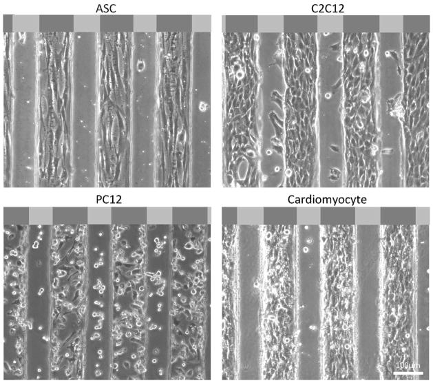
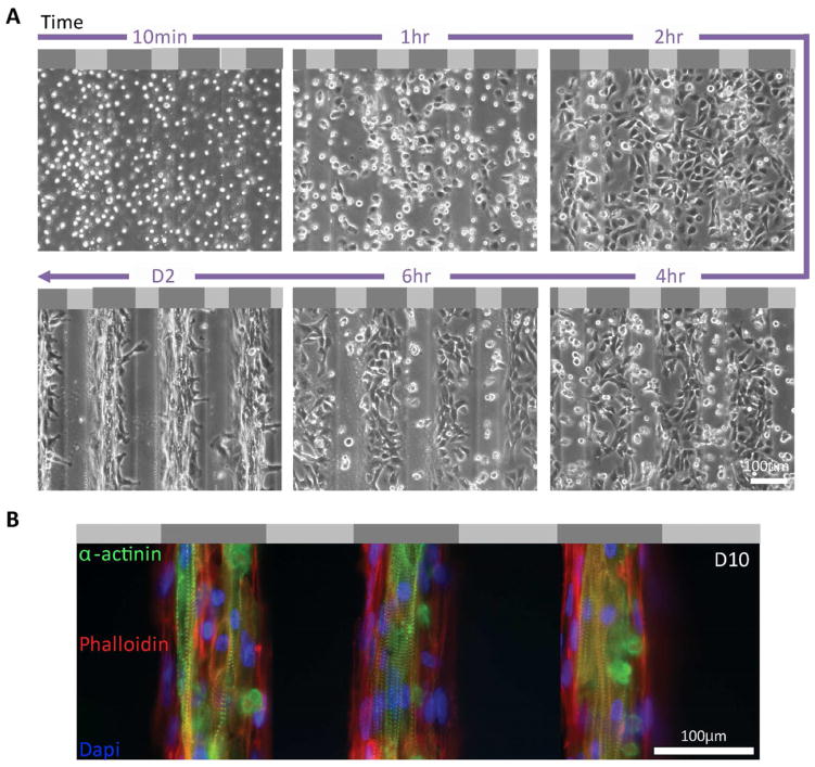
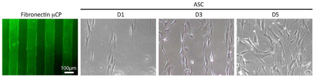
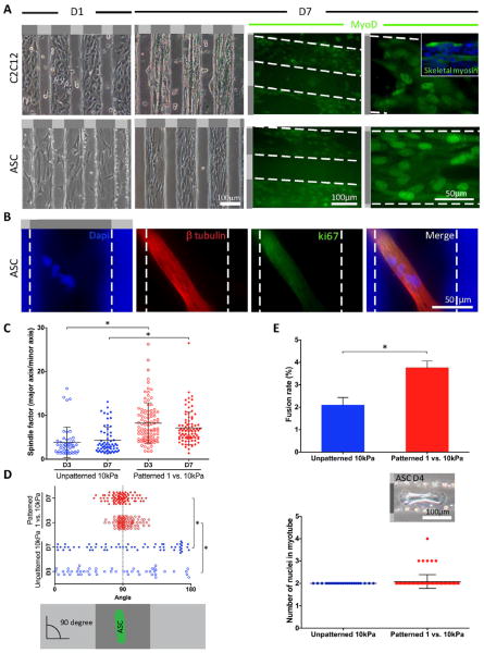
Cell patterning is typically accomplished by selectively depositing proteins for cell adhesion only on patterned regions; however in tissues, cells are also influenced by mechanical stimuli, which can also result in patterned arrangements of cells. We developed a mechanically-patterned hydrogel to observe and compare it to extracellular matrix (ECM) ligand patterns to determine how to best regulate and improve cell type-specific behaviors. Ligand-based patterning on hydrogels was not robust over prolonged culture, but cells on mechanically-patterned hydrogels differentially sorted based on stiffness preference: myocytes and adipose-derived stem cells (ASCs) underwent stiffness-mediated migration, i.e. durotaxis, and remained on myogenic hydrogel regions. Myocytes developed aligned striations and fused on myogenic stripes of the mechanically-patterned hydrogel. ASCs aligned and underwent myogenesis, but their fusion rate increased, as did the number of cells fusing into a myotube as a result of their alignment. Conversely, neuronal cells did not exhibit durotaxis and could be seen on soft regions of the hydrogel for prolonged culture time. These results suggest that mechanically-patterned hydrogels could provide a platform to create tissue engineered, innervated micro-muscles of neural and muscle phenotypes juxtaposed next to each other in order better recreate a muscle niche.


Figures

Figure 1
Figure 1. Mechanically-patterned hydrogel fabrication and characterization
(A) Schematic of photolithography and 2-step polyacrylamide gel fabrication. (B) Matrix stiffness (bottom) was measured for neurogenic-myogenic (blue) and myogenic-osteogenic (red) mechanically-patterned hydrogels (n≥6) fabricated using the indicated polyacrylamide concentrations for top and bottom hydrogels (top). Light and dark gray regions in the bottom schematic represent soft and stiff regions, respectively. (C) Matrix stiffness, height, and surface roughness of mechanically-patterned hydrogels were visualized with three adjacent 90 μm × 90 μm force maps. Matrix stiffness of adjacent soft and stiff strips was overlaid with surface height in a 3D reconstruction of the surface features (top). 2D height topography is shown individually (middle). Values for stiffness and roughness are shown for stiff and soft stripes as well as the interface. Overall height difference was also computed and shown. Light and dark gray regions in the schematics represent soft (1 kPa) and stiff (10 kPa) regions, respectively.
Figure 2
Figure 2. Cell adhesion preference
Different cell types preferentially localized to the stiffer regions of the neurogenic-myogenic mechanically-patterned hydrogels after 48 hours in culture. Phase contrast images show representative images with very confluent cells on the stiffer myogenic lanes for ASC, C2C12, and chicken cardiomyocytes. A portion of the PC12s remained less well-spread and adherent on the softer neurogenic stripes. Light and dark gray regions in the schematic at the top of each image represent soft (1 kPa) and stiff (10 kPa) regions, respectively.
Figure 3
Figure 3. Attachment and alignment of cardiomyocytes on neurogenic-myogenic mechanically-patterned hydrogel
(A) Time lapse images of chicken cardiomyocyte attachment to neurogenic-myogenic mechanically-patterned hydrogel showed adhesion and preferential migration and spreading on stiffer myogenic stripes. Light and dark gray regions in the schematic at the top of each image represent soft (1 kPa) and stiff (10 kPa) regions, respectively. (B) Three consecutive myogenic stripes were fluorescently stained by rhodamin-phalloidin (red), α–actinin (green), and DAPI (blue), revealing an aligned network of cardiomyocytes on myogenic stripes of mechanically-patterned hydrogel at day 10 with adjacent myofibroblasts that lack α–actinin.
Figure 4
Figure 4. ASC on fibronectin-microcontact printing
μCP was confirmed by anti-fibronectin staining (right). ASCs recognized pattern initially at day 1 and proliferated and aligned on pattern at day 3, however, pattern was failed from day 5 and cells attached on both printed and non-printed area.
Figure 5
Figure 5. Mechanically-patterned hydrogel enhances myotube fusion of ASC
(A) Representative cell alignment on neurogenic-myogenic mechanically-patterned hydrogels at day 1 and 7 for both C2C12 and ASCs shown in phase contrast (top) and with MyoD staining at day 7 (bottom). Inset image shows C2C12 myoblasts, which further differentiated into skeletal fast myosin-expressing myotubes. White dashed lines indicate the edges of each region of mechanically-patterned hydrogel. (B) Representative multi-nucleated myotube stained with Dapi that was identified with β-tubulin (red) and negative for ki-67 staining (green), which is indicative of quiescent cells. (C) Spindle factor from ASCs on mechanically-patterned hydrogel were significantly greater than those on unpatterned 10 kPa PA hydrogels at both day 3 and 7. (D) To show the alignment of ASCs on gels, cell angle was measured by phalloidin staining (90° = perfectly aligned on mechanically-patterned hydrogel). ASCs on mechanically-patterned hydrogel showed higher level of alignment that those on unpatterned 10 kPa PA hydrogels at day 7. (E) A higher fusion rate was observed on the mechanically-patterned hydrogel compared to unpatterned 10 kPa PA gels (top) [7]. The number of nuclei per mechanically-patterned hydrogel or unpatterned hydrogel myotube was also quantified (bottom). The inset image shows characteristic multi-nucleated myotube in phase contrast. Light and dark gray regions in the schematic next to or on top of each image represent soft (1 kPa) and stiff (10 kPa) regions, respectively. *p <0.05 for all indicated comparisons.

References

    1. Pittenger MF, Mackay AM, Beck SC, Jaiswal RK, Douglas R, Mosca JD, et al. Multilineage potential of adult human mesenchymal stem cells. Science. 1999;284:143–7. - PubMed
    1. Zuk PA, Zhu M, Mizuno H, Huang J, Futrell JW, Katz AJ, et al. Multilineage cells from human adipose tissue: implications for cell-based therapies. Tissue Eng. 2001;7:211–28. - PubMed
    1. Atala A. Advances in tissue and organ replacement. Curr Stem Cell Res Ther. 2008;3:21–31. - PubMed
    1. Discher DE, Mooney DJ, Zandstra PW. Growth factors, matrices, and forces combine and control stem cells. Science. 2009;324:1673–7. - PMC - PubMed
    1. Vacanti JP, Langer R. Tissue engineering: the design and fabrication of living replacement devices for surgical reconstruction and transplantation. Lancet. 1999;354(Suppl 1):SI32–4. - PubMed

Publication types

Substances