. 2012 Sep;32(18):3718-31.
doi: 10.1128/MCB.06754-11. Epub 2012 Jul 16.

Kinase suppressor of Ras 2 (KSR2) regulates tumor cell transformation via AMPK

Affiliations

Kinase suppressor of Ras 2 (KSR2) regulates tumor cell transformation via AMPK

Mario R Fernandez et al. Mol Cell Biol. 2012 Sep.

Abstract

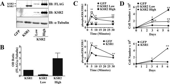
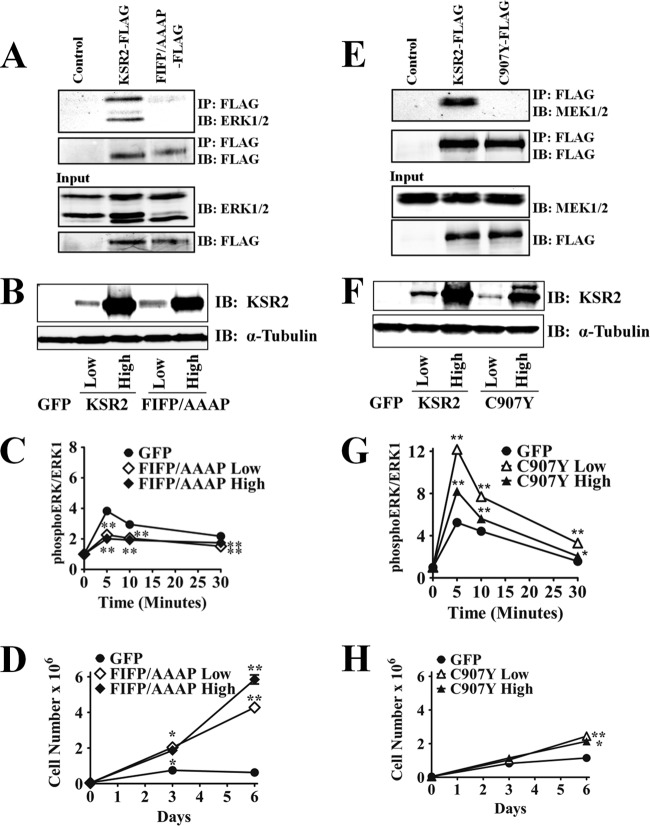
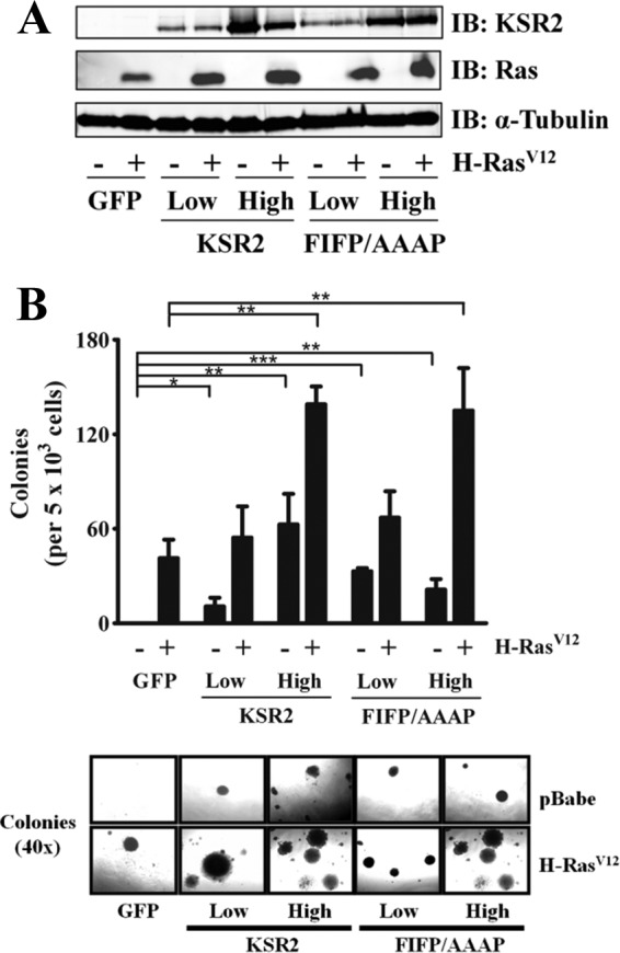
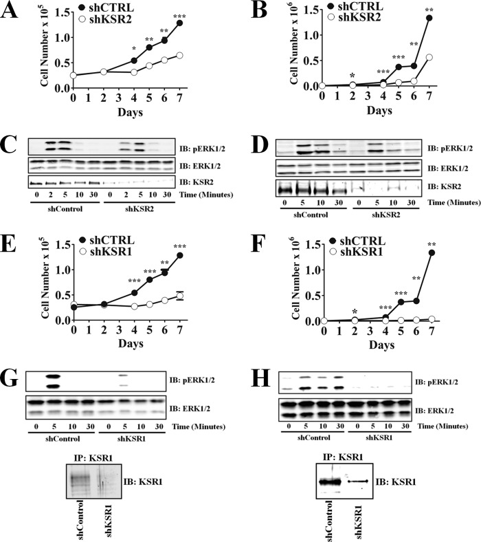
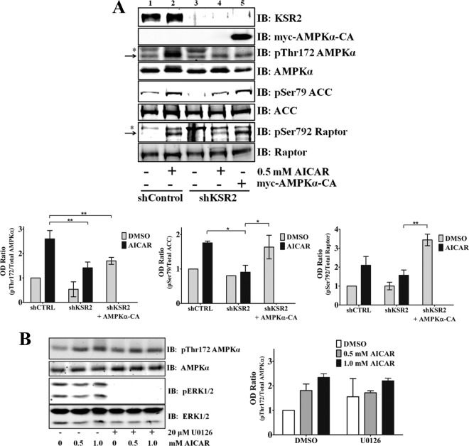
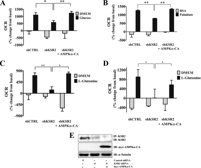
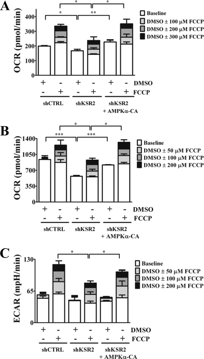
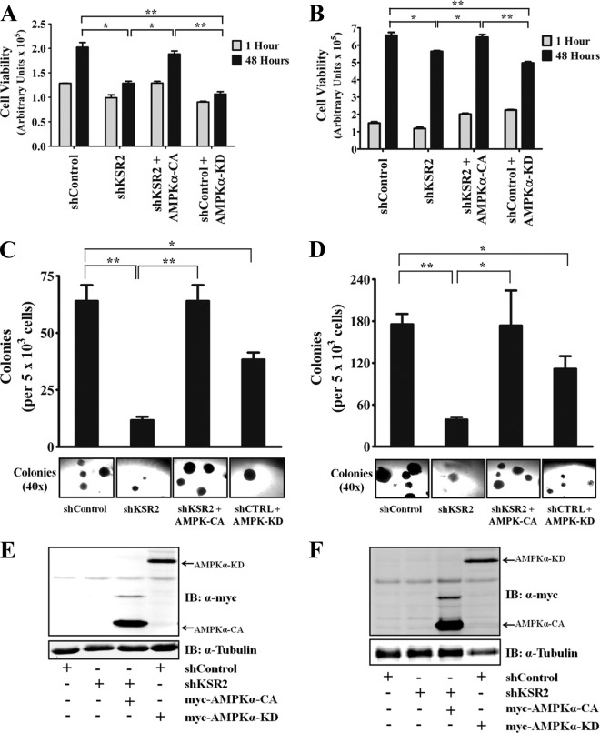
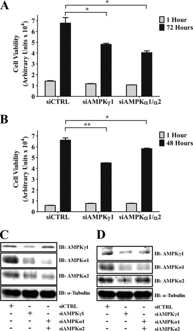
Kinase suppressor of Ras 1 (KSR1) and KSR2 are scaffolds that promote extracellular signal-regulated kinase (ERK) signaling but have dramatically different physiological functions. KSR2(-/-) mice show marked deficits in energy expenditure that cause obesity. In contrast, KSR1 disruption has inconsequential effects on development but dramatically suppresses tumor formation by activated Ras. We examined the role of KSR2 in the generation and maintenance of the transformed phenotype in KSR1(-/-) mouse embryo fibroblasts (MEFs) expressing activated Ras(V12) and in tumor cell lines MIN6 and NG108-15. KSR2 rescued ERK activation and accelerated proliferation in KSR1(-/-) MEFs. KSR2 expression alone induced anchorage-independent growth and synergized with the transforming effects of Ras(V12). Similarly, RNA interference (RNAi) of KSR2 in MIN6 and NG108-15 cells inhibited proliferation and colony formation, with concomitant defects in AMP-activated protein kinase (AMPK) signaling, nutrient metabolism, and metabolic capacity. While constitutive activation of AMPK was sufficient to complement the loss of KSR2 in metabolic signaling and anchorage-independent growth, KSR2 RNAi, MEK inhibition, and expression of a KSR2 mutant unable to interact with ERK demonstrated that mitogen-activated protein (MAP) kinase signaling is dispensable for the transformed phenotype of these cells. These data show that KSR2 is essential to tumor cell energy homeostasis and critical to the integration of mitogenic and metabolic signaling pathways.


Figures

Fig 1
Fig 1
KSR2 differentially mediates growth factor-induced ERK activation and cell proliferation. Bicistronic recombinant retroviruses that encompass genes for KSR2 and GFP, KSR1 and GFP, or GFP alone were expressed in KSR1−/− MEFs. (A) Whole-cell extracts (WCEs) were immunoblotted (IB) with the indicated antibodies to confirm the expression of KSR proteins after FACS. (B) The optical density (OD) values for FLAG and α-tubulin immunoreactive bands of three distinct membranes from the experiment described for panel A were quantified using the Li-COR Odyssey system. (C) In situ analysis of PDGF-induced ERK activation. (D) Proliferation of KSR1−/− MEFs expressing KSR2 at low or high levels, KSR1, or GFP only. All data shown are the results of triplicate measurements and representative of at least three independent experiments. Data represent means ± standard deviations. *, P ≤ 0.001; **, P ≤ 0.0001 (two-tailed Student's t test).
Fig 2
Fig 2
KSR2 mediates cell proliferation through an ERK1/2-independent pathway. (A and E) 293T cells transfected with the indicated plasmids were lysed and evaluated via immunoprecipitation (IP) using anti-FLAG-agarose to detect KSR2-ERK and KSR2-MEK association, respectively. Precipitates were eluted, resolved by electrophoresis, and probed on Western blots with the indicated antibodies. WCEs were analyzed to show the uniform expression of FLAG-tagged KSR2 proteins MEK and ERK. (B to D and F to H) Bicistronic recombinant retroviruses that encompass genes for KSR2 and GFP, KSR2.AAAP and GFP, KSR2.C907Y and GFP, or GFP alone were expressed in KSR1−/− MEFs. (B and F) Western blots showing the level of KSR2, KSR2.AAAP, and KSR2.C907Y protein expression in KSR1−/− MEFs after FACS. (C and G) In situ analysis of PDGF-induced ERK activation. (D and H) Proliferation of KSR1−/− MEFs expressing KSR2.AAAP, KSR2.C907Y or GFP only. IB, immunoblot. All data shown are the results of triplicate measurements and representative of at least three independent experiments. Data represent means ± standard deviations. *, P ≤ 0.0001; **, P ≤ 0.01 (two-tailed Student's t test).
Fig 3
Fig 3
KSR2 promotes anchorage-independent growth in an ERK-independent manner and synergizes H-RasV12-induced anchorage-independent growth of KSR1−/− MEFs. KSR1−/− MEFs were infected with a bicistronic recombinant retrovirus encompassing KSR2 and GFP, KSR2.AAAP and GFP, or GFP only, with either H-RasV12 or the respective vector alone. (A) A representative immunoblot (IB) using the indicated antibodies, illustrating the expression levels of KSR2, KSR2.AAAP, and H-RasV12 after sorting. (B) Anchorage-independent growth was examined by assessing the growth of MEFs expressing the indicated proteins in soft agar. Data are representative of three independent experiments and are illustrated as the number of colonies per 5 × 10³ cells present after 4 weeks. The lower panel shows representative photomicrographs of colonies for each sample. Data represent means ± standard deviations of the results determined with triplicate samples. *, P ≤ 0.05; **, P ≤ 0.01; ***, P ≤ 0.0001 (two-tailed Student's t test).
Fig 4
Fig 4
KSR2 knockdown results in decreased growth rates without affecting growth factor-mediated ERK activation. Tumor cell lines were infected with lentiviruses expressing a nontargeting short hairpin (shCTRL) or short hairpins that target KSR2 (shKSR2) or KSR1 (shKSR1). Cells expressing the indicated short hairpin were selected with puromycin. Growth rates of MIN6 (A and E) and NG108-15 (B and F) cell lines were assessed by seeding 2.5 × 10⁴ and 5 × 10³ cells, respectively, in 35-mm-diameter dishes and triplicate counts performed at the indicated time points. MIN6 (C and G) and NG108-15 (D and H) cells were stimulated with EGF for the indicated times, and the phosphorylation and total levels of ERK were assessed by immunoblotting (IB) with the indicated antibodies. KSR1 expression and knockdown were confirmed via KSR1 immunoprecipitations (IP). (A, B, E, and F) *, P ≤ 0.01; **, P ≤ 0.001; ***, P ≤ 0.0001 (two-tailed Student's t test). Data are representative of the results of three independent experiments.
Fig 5
Fig 5
ERK activity is not necessary for KSR2-mediated AICAR-induced AMPK activation in MIN6 cells. (A) MIN6 cells expressing the nontargeting short hairpin, the KSR2-specific short hairpin, or the KSR2-specific short hairpin and the constitutively active AMPKα2 (AMPKα-CA) were seeded in serum-free media for 4 h prior to stimulation with AICAR for 30 min (*, nonspecific bands). (B) MIN6 cells were subjected to serum starvation for 4 h and treated with the MEK inhibitor U0126 for 30 min prior to treatment with AICAR for 30 min at the indicated concentrations. For both panels, KSR2 knockdown, AMPKα, ACC, Raptor, and ERK activity were examined by immunoblotting (IB) with the indicated antibodies. Immunoblots were quantified using the Li-COR Odyssey system, and the optical densities are illustrated as means ± standard deviations. *, P ≤ 0.05; **, P ≤ 0.01 (two-tailed Student's t test). Data are representative of the results of at least three independent experiments.
Fig 6
Fig 6
KSR2 knockdown attenuates the ability of tumor cells to metabolize nutrients. Cells expressing the nontargeting short hairpin, the KSR2-specific short hairpin, or the KSR2-specific short hairpin and the constitutively active AMPKα2 (AMPKα-CA) were seeded in growth media on XF24 microplates 16 h prior to analysis. MIN6 (A to C) and NG108-15 (D) cells were stimulated with 22.5 mM glucose (A), 200 μM palmitate (B), or 10 mM l-glutamine (C and D), after incubation in bicarbonate-free low-buffered media containing low concentrations of nutrients as described in Materials and Methods. The rate of oxygen consumption (OCR) was measured prior to and after nutrient addition. (E) Representative immunoblots illustrating the expression of the indicated proteins represented in panel D. Data are represented as means ± standard errors of the means of percent change from basal values (n = 3 to 5 wells). *, P ≤ 0.05; **, P ≤ 0.01 (two-tailed Student's t test). Data are representative of the results of at least three independent experiments. IP, immunoprecipitation.
Fig 7
Fig 7
KSR2 knockdown affects the maximal metabolic capacity of tumor cells. Cells expressing the nontargeting short hairpin, the KSR2-specific short hairpin, or the KSR2-specific short hairpin and constitutively active AMPKα2 (AMPKα-CA) were seeded in growth media on XF24 microplates 16 h prior to analysis. MIN6 (A) and NG108-15 (B and C) cells were plated in bicarbonate-free low-buffered media with a full complement of nutrients as described in Materials and Methods. The rates of oxygen consumption (OCR) and extracellular acidification (ECAR) were measured prior to and 15 min after three successive stimulations with FCCP at the indicated concentrations. Data represent means ± standard errors of the means of the results determined for 3 or 4 wells. *, P ≤ 0.05; **, P ≤ 0.01; ***, P ≤ 0.001 (two-tailed Student's t test). Statistical comparison was performed on data collected after the third stimulation. Data are representative of the results of three independent experiments.
Fig 8
Fig 8
AMPK activity is required for KSR2-mediated anchorage-independent growth. MIN6 (A) and NG108-15 (B) cells expressing the nontargeting short hairpin, the KSR2-specific short hairpin, the KSR2-specific short hairpin and the constitutively active AMPKα2 (AMPKα-CA), or the nontargeting short hairpin and dominant-negative AMPKα2 (AMPKα-KD) were seeded in poly(HEMA)-coated plates, and the ability to grow in an anchorage-independent manner was examined by assessing cell viability using CellTiter-Glo at the indicated time points. Data representative of at least three independent experiments are illustrated as means ± standard errors of the means of the results determined for triplicate samples. Additionally, anchorage-independent growth of MIN6 (C) and NG108-15 (D) cells was examined after plating in soft agar. Data are illustrated as the number of colonies per 5 × 10³ cells present after 4 weeks (C) and 2 weeks (D). The lower panel shows representative photomicrographs of colonies for each sample. Data are representative of at least three independent experiments and illustrated as means ± standard deviations of the results determined with triplicate samples. Immunoblots (IB) demonstrating the expression of the myc-tagged AMPKα constructs in MIN6 (E) and NG108-15 (F) cells are shown. *, P ≤ 0.01; **, P ≤ 0.001 (two-tailed Student's t test).
Fig 9
Fig 9
AMPK knockdown attenuates tumor cell anchorage-independent growth. MIN6 cells (A) and NG108-15 cells (B) were transfected with the indicated siRNA and seeded in poly(HEMA)-coated plates. Anchorage-independent viability was examined at the indicated time points using CellTiter-Glo. Data representative of the results of at least three independent experiments are illustrated as means ± standard errors of the means of triplicate samples. *, P ≤ 0.01; **, P ≤ 0.001 (two-tailed Student's t test). (C and D) Representative immunoblots (IB) denoting the expression level of the indicated proteins in MIN6 and NG108-15 cells, respectively.

Similar articles

Cited by

References

    1. Abe Y, et al. 2010. Bioenergetic characterization of mouse podocytes. Am. J. Physiol. Cell Physiol. 299:C464–C476 - PMC - PubMed
    1. Buzzai M, et al. 2005. The glucose dependence of Akt-transformed cells can be reversed by pharmacologic activation of fatty acid beta-oxidation. Oncogene 24:4165–4173 - PubMed
    1. Cacace AM, et al. 1999. Identification of constitutive and ras-inducible phosphorylation sites of KSR: implications for 14-3-3 binding, mitogen-activated protein kinase binding, and KSR overexpression. Mol. Cell. Biol. 19:229–240 - PMC - PubMed
    1. Choi SW, Gerencser AA, Nicholls DG. 2009. Bioenergetic analysis of isolated cerebrocortical nerve terminals on a microgram scale: spare respiratory capacity and stochastic mitochondrial failure. J. Neurochem. 109:1179–1191 - PMC - PubMed
    1. Corton JM, Gillespie JG, Hawley SA, Hardie DG. 1995. 5-Aminoimidazole-4-carboxamide ribonucleoside. A specific method for activating AMP-activated protein kinase in intact cells? Eur. J. Biochem. 229:558–565 - PubMed

Publication types

Substances