. 2012 Sep;32(18):3743-55.
doi: 10.1128/MCB.00032-12. Epub 2012 Jul 16.

Hydrolase controls cellular NAD, sirtuin, and secondary metabolites

Affiliations

Hydrolase controls cellular NAD, sirtuin, and secondary metabolites

Motoyuki Shimizu et al. Mol Cell Biol. 2012 Sep.

Abstract

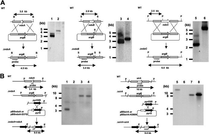
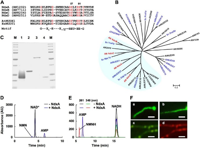
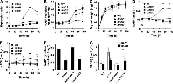
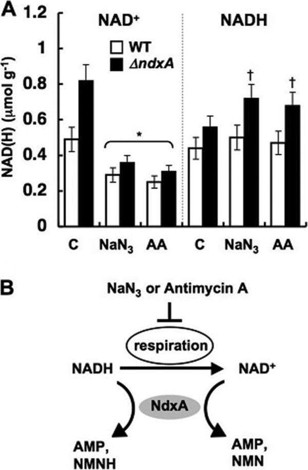
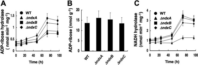
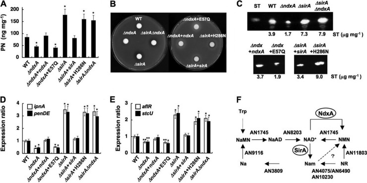
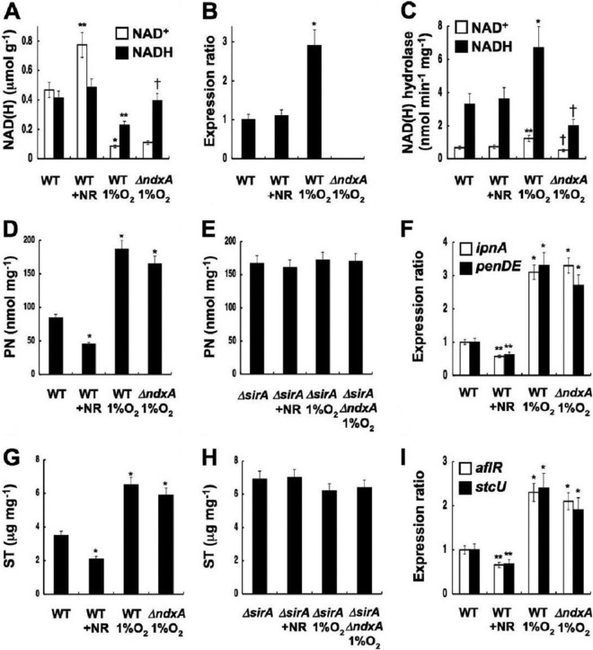
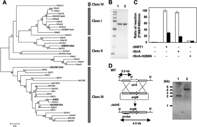
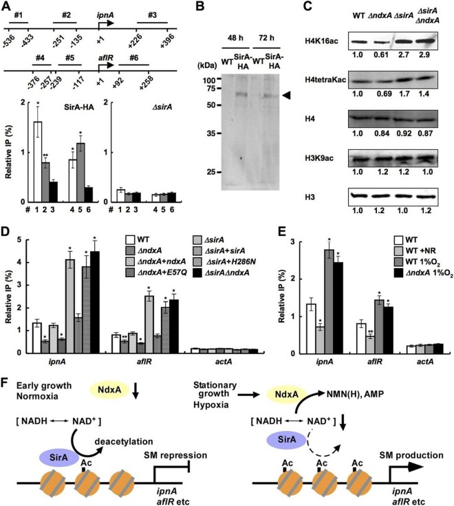
Cellular levels of NAD(+) and NADH are thought to be controlled by de novo and salvage mechanisms, although evidence has not yet indicated that they are regulated by NAD(+) degradation. Here we show that the conserved nudix hydrolase isozyme NdxA hydrolyzes and decreases cellular NAD(+) and NADH in Aspergillus nidulans. The NdxA-deficient fungus accumulated more NAD(+) during the stationary growth phase, indicating that NdxA maintains cellular NAD(+)/NADH homeostasis. The deficient strain also generated less of the secondary metabolites sterigmatocystin and penicillin G and of their gene transcripts than did the wild type. These defects were associated with a reduction in acetylated histone H4 on the gene promoters of aflR and ipnA that are involved in synthesizing secondary metabolites. Thus, NdxA increases acetylation levels of histone H4. We discovered that the novel fungal sirtuin isozyme SirA uses NAD(+) as a cosubstrate to deacetylate the lysine 16 residue of histone H4 on the gene promoter and represses gene expression. The impaired acetylation of histone and secondary metabolite synthesis in the NdxA-deficient strain were restored by eliminating functional SirA, indicating that SirA mediates NdxA-dependent regulation. These results indicated that NdxA controls total levels of NAD(+)/NADH and negatively regulates sirtuin function and chromatin structure.


Figures

Fig 1
Fig 1
Disruption of ndx genes in A. nidulans and introduction of ndxA and sirA alleles to A. nidulans. (A) Strategy for homologous recombination into ndxA, ndxB, and ndxC loci to construct ndxA, ndxB, and ndxC gene disruptants. Total DNA from strains was digested with BstXI (B), SphI (S), and PstI (P) before Southern blotting. Bars indicate positions and sizes of hybridization probes. Lanes 1, 3, and 5, A. nidulans wild type (WT); lane 2, ΔNdxA; lane 4, ΔNdxB; lane 6, ΔNdxC. (B) Strategy for introducing ndxA and sirA to ΔNdxA2 and ΔSirA2. Plasmids harboring wild-type or mutated ndxA and sirA were integrated to chromosomal pyrG regions by single crossover. Total DNA from strains was digested with PstI (P) before Southern blotting. Bars indicate positions and sizes of hybridization probes. Lanes 1 and 5, A. nidulans WT; lane 2, ΔNdxA2; lane 3, ΔNdxA2 plus pBSndxA; lane 4, ΔNdxA2 plus pBSndxA-E57Q; lane 6, ΔSirA2; lane 7, ΔSirA2 plus pBSsirA; lane 8, ΔSirA2 plus pBSsirA-H286N.
Fig 2
Fig 2
Identifying fungal nudix hydrolase isozymes. (A) Alignment of partial amino acid sequences among Ndx proteins of A. nidulans, A. thaliana AtNUDX1, and human HsNUDT1. Conserved residues are highlighted, and numbers indicate mutated residues. U, Ile, Leu, or Val. (B) Phylogenetic tree of nudix hydrolases of A. nidulans (prefixed with AN), S. cerevisiae (Sc), and A. thaliana (At). Proteins with reported enzyme activity are in blue. (C) SDS-PAGE of purified recombinant NdxA (lane 1), NdxB (lane 2), NdxC (lane 3), and NdxD (lane 4). Purified enzymes (1 μg) were resolved by SDS-PAGE and stained with Coomassie brilliant blue. Lanes M, Bio-Rad Precision protein standard kit. (D) Determination of reaction products of NAD+ hydrolysis catalyzed by recombinant NdxA. Reaction mixtures were analyzed by HPLC. (E) Similar experiments were performed using NADH as a substrate. (F) Fluorescence microscopy of visualized GFP-Ndx fusion proteins in A. nidulans: NdxA-GFP (a), NdxB-GFP (b), and GFP-NdxC (c). (d) DsRed-SKM produced in the same strain as that in subpanel c. Bars, 10 μm.
Fig 3
Fig 3
NdxA regulates cellular NAD(H). (A) Transcripts of ndxA, ndxB, and ndxC in A. nidulans wild type (WT) cultured at 30°C and quantified by PCR. (B) Intracellular NAD+ hydrolase activity. (C) Time-dependent changes in dry weight of cells. (D) Intracellular NAD+. (E) Intracellular NADH. (F and G) Intracellular NAD+ hydrolase activity and NAD(H) after a 72-h culture. WT, wild type; ΔndxA, ΔNdxA; ΔndxB, ΔNdxB; ΔndxC, ΔNdxC; ΔndxA+ndxA, ΔNdxA2 strain transformed with pNdxA; ΔndxA+E57Q, ΔNdxA2 strain transformed with pNdxA-E57Q. Data are means ± standard deviations (n = 3). *, P < 0.005; **, P < 0.03 versus WT.
Fig 4
Fig 4
NdxA affects cellular NAD(H) by respiratory activity. (A) NAD(H) in cells incubated with 4 mM NaN3 and 30 μM antimycin A (AA) for 24 h. Untreated controls (C) are shown. Data are means ± standard deviations (n = 3). *, P < 0.03 versus control. †, P < 0.03 versus wild type (WT). (B) Model for modulation of cellular NAD(H) levels by NdxA through respiratory activity.
Fig 5
Fig 5
Hydrolyzing activity of ADP-ribose and NADH and ADP-ribose contents in A. nidulans. (A) Time-dependent changes in intracellular ADP-ribose hydrolase activity. (B) ADP-ribose contents in the A. nidulans cells after a 72-h culture. (C) Time-dependent changes in intracellular NADH hydrolase activity. WT, wild type; ΔndxA, ΔNdxA; ΔndxB, ΔNdxB; ΔndxC, ΔNdxC. Data are means of three experiments. Error bars indicate standard deviations.
Fig 6
Fig 6
NdxA requirement for maximal secondary metabolite production. (A) Penicillin G (PN) production after 72 h in culture. (B) Bacterial growth inhibition assay shows penicillin production by A. nidulans. (C) Sterigmatocystin production after 72 h in culture. (D and E) Transcripts of secondary metabolite genes in cells quantified by PCR after 72 h in culture. WT, wild type; ΔndxA, ΔNdxA; ΔndxA+ndxA, ΔNdxA2 strain transformed with pNdxA; ΔndxA+E57Q, ΔNdxA2 strain transformed with pNdxA-E57Q; ΔsirA, ΔSirA; ΔsirA+sirA, ΔSirA2 strain transformed with pSirA; ΔsirA+H286N, ΔSirA2 strain transformed with pSirA-H286N; ΔsirAΔndxA, ΔSirA ΔNdxA. Data are means ± standard deviations (n = 3). *, P < 0.005 versus WT. (F) NAD+ synthesis in A. nidulans. Predicted genes for nicotinamide riboside (NR) salvage pathways for NAD+ synthesis and for de novo synthetic pathway of NAD+ are presented as gene identities. NaMN, nicotinic acid mononucleotide; NaAD, nicotinic acid adenine dinucleotide; NMN, nicotinamide mononucleotide; Na, nicotinic acid; Nam, nicotinamide. NdxA counteracts NMN adenylyltransferase (AN1745) in that they catalyze NMN+ and NAD+ interconversion.
Fig 7
Fig 7
Cellular NAD(H) regulates secondary metabolite synthesis. Aspergillus nidulans wild type (WT), ΔNdxA (ΔndxA), ΔSirA (ΔsirA), and ΔSirA ΔNdxA (ΔsirA ΔndxA) were cultured for 72 h under normoxic and hypoxic (1% O2) conditions. +NR, WT cultured with 0.3 mM nicotinamide riboside. (A) Intracellular NAD(H). (B) PCR-quantified ndxA transcripts. (C) Intracellular NAD(H) hydrolase activity. (D and E) Penicillin G production. (F) Penicillin G gene transcripts quantified by PCR. (G and H) Sterigmatocystin (ST) production. (I) Sterigmatocystin gene transcripts quantified by PCR. Data are means ± standard deviations (n = 3). *, P < 0.005; **, P < 0.03 (versus WT); †, P < 0.03 (versus WT with 1% O2).
Fig 8
Fig 8
Identification of sirtuin isozyme SirA from A. nidulans. (A) Phylogenetic relationship among sirtuin isozymes. All amino acid sequences of proteins similar to sirtuins collected from A. nidulans (prefixed with AN), S. cerevisiae (Sc), Schizosaccharomyces pombe (Sp), and human genes are shown as their corresponding gene identifier and/or gene names. SIRT and Hdac represent human proteins. Proteins described in the text are highlighted by boldface and underlining. Classes of HDAC are shown on the right. (B) SDS-PAGE of purified recombinant SirA (rSirA) (lane 1) and rSirA-H286N (lane 2) (1 μg each). (C) NAD+-dependent HDAC activity of rSirA with (solid bars) or without (open bars) 2 mM nicotinamide. Reactions proceeded with NAD+ (200 μM), rSIRT1, rSirA, and rSirA-H286N (25 μg each). Data are means of three experiments. Error bars indicate standard deviations. P < 0.005. (D) Strategy for gene disruption of sirA and Southern blot analysis of A. nidulans WT (lane 1) and ΔSirA2 (lane 2). Total DNA from strains was digested with HindIII (H) before blotting. The position and size of the hybridization probe are shown.
Fig 9
Fig 9
NdxA represses histone deacetylation. (A) ChIP analyses indicate SirA association with ipnA and aflR gene promoters in A. nidulans cells cultured for 48 h. Top panel, positions (1 to 6) of immunoprecipitated DNA. Data are means ± standard deviations (n = 3). *, P < 0.005, and **, P < 0.01, versus ΔsirA. (B) Western blot analysis of cell extracts of the strain producing SirA-HA. Anti-HA antibody detected levels of SirA-HA similar between early stationary (48-h) and stationary (72-h) growth phases. Arrowhead, SirA-HA. (C) Western blotting detected acetylated histones in nuclear fractions of cells cultured for 72 h. H3 and H4, total histones H3 and H4. Representative results of four repeated experiments are presented with relative signal intensity under bands. (D and E) ChIP findings of effects of ndxA and sirA (D) and of nicotinamide riboside and hypoxia (E) on H4K16 acetylation at ipnA, aflR, and actA gene promoters in strains cultured for 72 h. Relative IP represents signal ratio between precipitated and input DNA. WT, wild type; ΔndxA, ΔNdxA; ΔndxA+ndxA, ΔNdxA2 strain transformed with pNdxA; ΔndxA+E57Q, ΔNdxA2 strain transformed with pNdxA-E57Q; ΔsirA, ΔSirA; ΔsirA+sirA, ΔSirA2 strain transformed with pSirA; ΔsirA+H286N, ΔSirA2 strain transformed with pSirA-H286N; ΔsirAΔndxA, ΔSirA ΔNdxA. Data are means ± standard deviations (n = 3). *, P < 0.005, and **, P < 0.01, versus WT. (F) Model of negative epigenetic control by NdxA through NAD(H) hydrolysis.

Similar articles

Cited by

References

    1. AbdelRaheim SR, Cartwright JL, Gasmi L, McLennan AG. 2001. The NADH diphosphatase encoded by the Saccharomyces cerevisiae NPY1 nudix hydrolase gene is located in peroxisomes. Arch. Biochem. Biophys. 388: 18–24 - PubMed
    1. Abdelraheim SR, Spiller DG, McLennan AG. 2003. Mammalian NADH diphosphatases of the Nudix family: cloning and characterization of the human peroxisomal NUDT12 protein. Biochem. J. 374: 329–335 - PMC - PubMed
    1. Bayram O, et al. 2008. VelB/VeA/LaeA complex coordinates light signal with fungal development and secondary metabolism. Science 320: 1504–1506 - PubMed
    1. Belenky P, et al. 2007. Nicotinamide riboside promotes Sir2 silencing and extends lifespan via Nrk and Urh1/Pnp1/Meu1 pathways to NAD+. Cell 129: 473–484 - PubMed
    1. Bernreiter A, et al. 2007. Nuclear export of the transcription factor NirA is a regulatory checkpoint for nitrate induction in Aspergillus nidulans. Mol. Cell. Biol. 27: 791–802 - PMC - PubMed

Publication types

MeSH terms

LinkOut - more resources