. 2012 Oct;11(10):989-1007.
doi: 10.1074/mcp.M111.012138. Epub 2012 Jul 16.

Temporal profiling and pulsed SILAC labeling identify novel secreted proteins during ex vivo osteoblast differentiation of human stromal stem cells

Affiliations

Temporal profiling and pulsed SILAC labeling identify novel secreted proteins during ex vivo osteoblast differentiation of human stromal stem cells

Lars P Kristensen et al. Mol Cell Proteomics. 2012 Oct.

Abstract

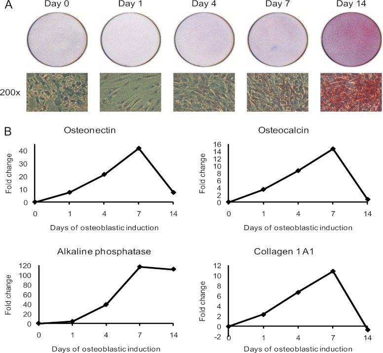
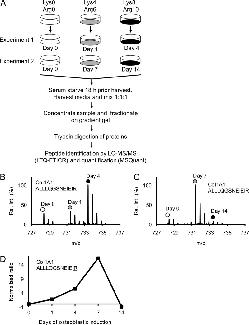
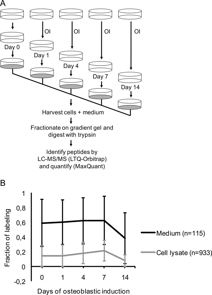
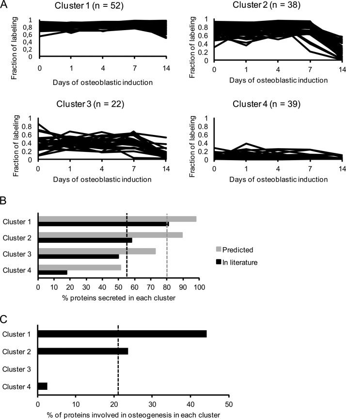
It is well established that bone forming cells (osteoblasts) secrete proteins with autocrine, paracrine, and endocrine function. However, the identity and functional role for the majority of these secreted and differentially expressed proteins during the osteoblast (OB) differentiation process, is not fully established. To address these questions, we quantified the temporal dynamics of the human stromal (mesenchymal, skeletal) stem cell (hMSC) secretome during ex vivo OB differentiation using stable isotope labeling by amino acids in cell culture (SILAC). In addition, we employed pulsed SILAC labeling to distinguish genuine secreted proteins from intracellular contaminants. We identified 466 potentially secreted proteins that were quantified at 5 time-points during 14-days ex vivo OB differentiation including 41 proteins known to be involved in OB functions. Among these, 315 proteins exhibited more than 2-fold up or down-regulation. The pulsed SILAC method revealed a strong correlation between the fraction of isotope labeling and the subset of proteins known to be secreted and involved in OB differentiation. We verified SILAC data using qRT-PCR analysis of 9 identified potential novel regulators of OB differentiation. Furthermore, we studied the biological effects of one of these proteins, the hormone stanniocalcin 2 (STC2) and demonstrated its autocrine effects in enhancing osteoblastic differentiation of hMSC. In conclusion, combining complete and pulsed SILAC labeling facilitated the identification of novel factors produced by hMSC with potential role in OB differentiation. Our study demonstrates that the secretome of osteoblastic cells is more complex than previously reported and supports the emerging evidence that osteoblastic cells secrete proteins with endocrine functions and regulate cellular processes beyond bone formation.


Figures

Fig. 1.
Fig. 1.
Induction of ex vivo osteoblastic differentiation of hMSC. Osteoblastic differentiation of the human marrow stromal (mesenchymal) stem cell line (hMSC-TERT) in SILAC medium was evaluated by the level of alkaline phosphatase activity and the expression of OB differentiation marker proteins. A, Cytochemical staining for alkaline phosphatase (upper part). The lower part shows the bright field images of the corresponding cells at 200× magnification. The red staining indicates alkaline phosphatase activity and the induction of OB differentiation in SILAC medium. B, Quantification of osteoblastic gene expression of osteonectin, osteocalcin, alkaline phosphatase, and collagen type I during the time course of OB differentiation using qRT-PCR. The data is expressed as fold changes using β-2-microglobulin as internal standard. All results are average of at least two independent experiments.
Fig. 2.
Fig. 2.
Schematic outline of the complete SILAC experiment to quantify the relative abundance changes of proteins secreted from MSC during osteoblastic differentiation. Mass spectrometry-based proteomics were used to quantify changes in the relative abundance of proteins secreted from MSC during osteoblast differentiation. A, Three populations of MSC were prelabeled with different isotope variants of lysine and arginine as indicated. The fully labeled cell populations were then stimulated with osteoblastic inducers for 1 and 4 days in experiment 1 and for 7 and 14 days in experiment 2. The unstimulated “day 0” cells were used as a common reference. Eighteen hours before collecting the conditioned medium with secreted proteins the cells were carefully washed and changed to serum free medium containing osteoblastic inducers. The medium collected from the three cell populations in each experiment was mixed in a ratio of 1:1:1 based on the total protein concentration. Each sample was then concentrated and fractionated on a 1D gradient gel and subjected to in-gel trypsin digestion. The resulting peptide mixtures were analyzed by reverse-phase liquid chromatography coupled on-line to an LTQ-FTICR instrument and quantified using the MSQuant software. B, The mass spectrum of a collagen 1 A1 (ALLLQGSNEIEIR) peptide from experiment 1 illustrates the relative abundance changes during OB differentiation where the same peptide originating from the three different cell populations can be distinguished by the isotope mass differences as indicated. C, The corresponding mass spectrum of a collagen 1 A1 (ALLLQGSNEIEIR) peptide from experiment 2. D, The signal intensity ratios of the collagen 1 A1 peptide from experiment 1 and 2 (Fig. 2B, 2C) were integrated into one temporal protein abundance profile over five time-points. All ratios were normalized to 0 (using day 0 as baseline) meaning that a normalized ratio of ±1 equals a twofold change in abundance.
Fig. 3.
Fig. 3.
Schematic outline of the pulsed SILAC experiment to quantify the fraction of labeling of proteins secreted from MSC during osteoblastic differentiation. To distinguish true secreted proteins from a potential background of intracellular proteins we measured the degree of heavy labeled amino acid incorporation following 18 h incubation of cells in SILAC medium and compared the results for secreted proteins collected from the medium with the results obtained for intracellular proteins from lysed cells. A, The osteoblastic induction (OI) was conducted in unlabeled medium containing 2% FBS for the indicated time-points. Eighteen hours prior to collection of the conditioned medium with secreted proteins and the harvest of cells, the nonlabeling medium was replaced with serum free SILAC medium containing D4 lysine and 13C614N4 arginine as well as osteoblastic inducers. Thus, the pulsed labeling time matches the time the cells secrete protein into the medium. B, Comparison of the average fraction of labeling during osteoblastic induction for proteins from the medium (n = 115) and cell lysate (n = 933), respectively. The standard deviation for each time point is indicated as error bars. The proteins in the medium have a significantly higher incorporation of labeled amino acids than proteins in the cell lysate at all time-points (p < 0.05).
Fig. 4.
Fig. 4.
Cluster analysis of secreted proteins during osteoblastic differentiation based on their fraction of labeling profile followed by enrichment analysis of proteins with a known role in OB differentiation. To evaluate the relevance of proteins with a high fraction of labeling during OB differentiation we clustered the proteins into four groups and compared the relative enrichment of protein predicted or validated to be secreted and proteins with a known role in OB differentiation for each of these clusters. A, Secreted proteins were clustered based on their fraction of labeling during osteoblastic differentiation considering only those quantified at all time-points in the pulsed SILAC labeling experiments and identified in the complete SILAC labeling experiment. The proteins in cluster 1 have a very high fraction of labeling in all time-points during the osteoblastic differentiation. Proteins in cluster 2 have a high fraction of labeling from day 0 to day 7 but are dramatically reduced at day 14. Proteins in cluster 3 have an overall intermediate fraction of labeling at all time-points. Proteins in cluster 4 have a very low fraction of labeling in all time-points during differentiation. B, The percentage of proteins in each of the four clusters that are predicted secreted (based on bioinformatics predication software) (gray) and validated secreted (through manual literature search) (black). The gray and black dashed lines represent the percentage of all the proteins before clustering annotated as predicted secreted (80%) and validated secreted (54%), respectively. Thus, the clustering based on fraction of labeling leads to enrichment for secreted proteins in cluster 1. C, The percentage of proteins known to be associated with OB differentiation in the four clusters. The dashed line indicates the percentage of all proteins before clustering directly involved in osteoblastic differentiation (22%). Thus, there is enrichment from 22% to 44% in cluster 1, while there are none or virtually none in cluster 3 and 4.
Fig. 5.
Fig. 5.
Searching for novel regulators of osteoblastic differentiation among those identified with a high fraction of labeling in the pulsed SILAC experiment and found to be regulated in the complete SILAC experiment. Functional annotation of secreted proteins from cluster 1 that exhibit more than twofold up- or down-regulation during early (day 1 or 4) or late (day 7 or 14) ex vivo OB differentiation of hMSC-TERT cells. The dashed lines between each of the four groups (early down-regulated, late down-regulated, early up-regulated, and late up-regulated) indicate the percentage expected if the distribution of the biological clusters is random. There is an enrichment for early up-regulated proteins among the growth factors, late down-regulated proteins among the proteases, and late up-regulated among the proteins related to both OB differentiation and extracellular matrix.
Fig. 6.
Fig. 6.
Direct comparison of the temporal expression profiles of significantly regulated candidate proteins as quantified by SILAC and qRT-PCR. The figure compares the temporal profiles of candidate proteins and their corresponding gene expression quantified by qRT-PCR. The early up-regulated candidate proteins are follistatin-like 1, cathepsin D, legumain, stanniocalcin 2, selectin-like osteoblast-derived protein, protein KIAA1199, plasminogen activator activator inhibitor-1 (PAI-1), and pentraxin-related protein PTX3. Complement factor H exhibits significantly late up-regulation during the course of ex vivo differentiation of hMSC-TERT cells. The data was expressed as a fold change normalized to the unstimulated day 0. All quantified mRNA values represent the average of at least two independent experiments. A significantly regulated protein ratio was indicated with red * (p < 0.05). The blue * indicate a significant regulation on mRNA level (p < 0.05).
Fig. 7.
Fig. 7.
Production of stanniocalcin 2 by hMSC. A, Direct comparison of the STC2 expression and secretion during osteoblast differentiation of hMSC as measured by qRT-PCR, SILAC (from conditioned medium), Western blot from cell lysates and Western blot from conditioned medium. All data were log2 normalized into a normalized ratio with respect to the values of unstimulated day 0. B, Western blot analysis of STC2 protein levels in cell lysates (upper lane, 50 μg/well) and conditioned medium (lower lane, 25 μg/well).
Fig. 8.
Fig. 8.
Human recombinant stanniocalcin 2 stimulates osteoblastic differentiation of hMSC. Cells were cultured in 24-well plate and induced to osteoblastic differentiation in absence or presence of different concentrations of recombinant human STC2 protein. A, Alkaline phosphatase (ALP) activity and staining were performed after 7 days induction. B, Mineralized matrix formation was demonstrated and quantified by Alizarin red S staining after 13 days treatment. C, Expression of specific osteoblastic marker genes as quantified by qRT-PCR. ALP: alkaline phosphatase; Col I: collagen type I; OC: osteocalcin; OPN: osteopontin. Data were represented as mean ± S.D. from four independent experiments. * indicate p < 0.05, ** indicate p < 0.001.

References

    1. Fukumoto S., Martin T. J. (2009) Bone as an endocrine organ. Trends Endocrinol. Metab. 20, 230–236 - PubMed
    1. Kobayashi Y., Udagawa N., Takahashi N. (2009) Action of RANKL and OPG for osteoclastogenesis. Crit. Rev. Eukaryot. Gene Exp. 19, 61–72 - PubMed
    1. Porter R. L., Calvi L. M. (2008) Communications between bone cells and hematopoietic stem cells. Arch. Biochem. Biophys. 473, 193–200 - PMC - PubMed
    1. Lorenzo J., Horowitz M., Choi Y. (2008) Osteoimmunology: interactions of the bone and immune system. Endocr. Rev. 29, 403–440 - PMC - PubMed
    1. Lee N. K., Sowa H., Hinoi E., Ferron M., Ahn J. D., Confavreux C., Dacquin R., Mee P. J., McKee M. D., Jung D. Y., Zhang Z., Kim J. K., Mauvais-Jarvis F., Ducy P., Karsenty G. (2007) Endocrine regulation of energy metabolism by the skeleton. Cell 130, 456–469 - PMC - PubMed

Publication types

MeSH terms