. 2012 Jul 26;487(7408):443-8.
doi: 10.1038/nature11314.

Oligodendroglia metabolically support axons and contribute to neurodegeneration

Affiliations

Oligodendroglia metabolically support axons and contribute to neurodegeneration

Youngjin Lee et al. Nature. .

Abstract

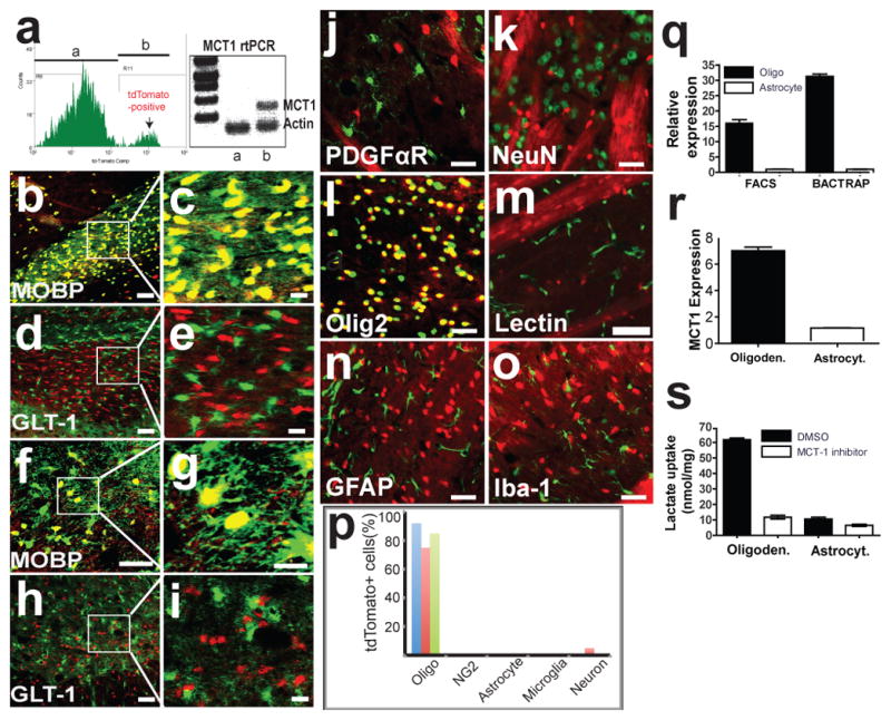
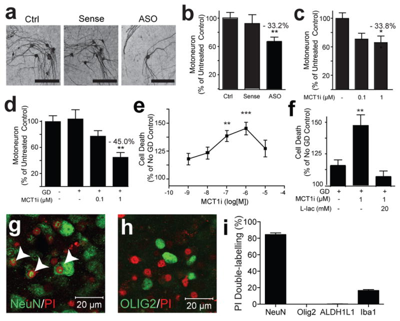
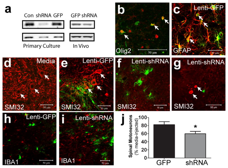
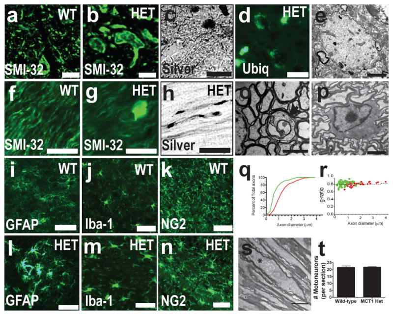
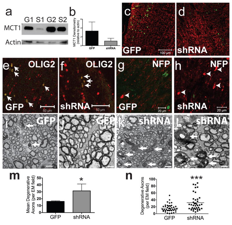
Oligodendroglia support axon survival and function through mechanisms independent of myelination, and their dysfunction leads to axon degeneration in several diseases. The cause of this degeneration has not been determined, but lack of energy metabolites such as glucose or lactate has been proposed. Lactate is transported exclusively by monocarboxylate transporters, and changes to these transporters alter lactate production and use. Here we show that the most abundant lactate transporter in the central nervous system, monocarboxylate transporter 1 (MCT1, also known as SLC16A1), is highly enriched within oligodendroglia and that disruption of this transporter produces axon damage and neuron loss in animal and cell culture models. In addition, this same transporter is reduced in patients with, and in mouse models of, amyotrophic lateral sclerosis, suggesting a role for oligodendroglial MCT1 in pathogenesis. The role of oligodendroglia in axon function and neuron survival has been elusive; this study defines a new fundamental mechanism by which oligodendroglia support neurons and axons.


Conflict of interest statement

Competing interests statement: Authors have no competing interests.

Figures

Figure 1
Figure 1. MCT1 expressed primarily within oligodendroglia in the CNS
(a) Histogram and rtPCR for MCT1 mRNA from FAC sorted cells with (b) or without (a) tdTomato expression. (b–e) Corpus callosum of tdTomato MCT1 reporter mice crossed with BAC-MOBP eGFP (b,c) or BAC-GLT1 eGFP (d,e) mice. (f–i) Spinal cord of tdTomato MCT1 reporter mice crossed with BAC-MOBP eGFP (f,g) or BAC-GLT1 eGFP (h,i) mice (Scale bars = 50 and 14 μm for low and high power, respectively). (j–o) TdTomato MCT1 reporter mouse brain immunostained with cell-specific markers (scale bars = 12.5 μm, except 50 μm for lectin). (p) Percentage of tdTomato cells co-labelled with CNS cell markers in the spinal cord (blue), cortex (red), and corpus callosum (green). (q,r) Real-time rtPCR of MCT1 mRNA isolated from oligodendroglia and astroglia by FACS and BacTRAP techniques (q; means of 4 replicates) and human oligodendroglioma (M03.13) and astrocytoma (U87) cell lines (r). (s) Lactate transport and blockade by selective MCT1 inhibitor in oligodendroglioma and astrocytoma.
Figure 2
Figure 2. MCT1 required for neuronal survival in vitro.
(a–d) Photomicrograph (a) and quantification of motoneurons in spinal cord slice cultures treated with media only (Ctrl), MCT1 sense oligonucleotides (Sense), or MCT1 antisense oligonucleotides (ASO) for 3 weeks (b; n ≥ 55 sections per group), following 3 weeks treatment with MCT1i (c; n ≥ 27 sections per group), or 2 hrs of glucose deprivation (GD) ± MCT1i (d; n ≥ 30 sections per group). (e,f) Propidium iodide (PI) uptake in slice cultures treated with 2 hrs GD + MCT1i (e; n ≥ 10 sections per group) or 2 hrs GD, ± MCT1i, ± 20 mM lactate (f; n = 15 for all groups). (g,h) PI uptake in slices treated with GD + MCT1i labelled with neuronal (g; co-localized cells marked with arrowheads) or oligodendroglia (h) marker. (i) Percentage of PI-labelled cells co-localizing cell-specific markers (n 10 sections per group, Error bars = ± S.E.M. *p < 0.05, **p < 0.01, ***p < 0.001).
Figure 3
Figure 3. Lentiviral MCT1 shRNA is toxic to motoneurons
(a) Lenti-shRNA (shRNA) downregulates MCT1 in primary astrocyte cultures and in vivo compared to untreated (Con) or lenti-GFP (GFP). (b,c) Olig2 (b) and glial fibrillary acidic protein (c; GFAP) in lenti-GFP injected spinal cords. (d–f) Non-phosphorylated neurofilament (SMI-32) (red) in spinal cords injected with media (d), Lenti-GFP (e), or Lenti-MCT1 shRNA (f, arrows indicate motoneurons). (g) SMI-32 labels aberrant axonal swellings in lenti-shRNA injected spinal cords. (h,i) Iba1 positive microglia in spinal cords injected with lenti-GFP (h) and lenti-MCT1 shRNA (i). (j) Quantification of cervical spinal motoneurons injected with lenti-GFP (n = 8) or lenti-MCT1 shRNA (n = 10), relative to contralateral media injection (Scale bars = 50 μm. Error bars = ± S.E.M. *p < 0.05).
Figure 4
Figure 4. MCT1 heterozygote null mice develop widespread axonopathy
(a–d) Spinal cords of wild-type (WT) and MCT1+/− mice (HET) immunostained for non-phosporylated neurofilament (SMI-32) (a,b), silver stained (c), or immunostained for ubiquitin (d; Scale bars = 20 um). (e) Electron microscopy of axonal spheroid in spinal cord (scale bar = 2 μm). (f–h) Optic nerves of WT and HET mice immunostained for SMI-32 (f,g) or silver stained (h; scale bars = 20 um). (i–n) GFAP (i.l), Iba-1 (j,m), and NG2 (k,n) in WT (i–k) and HET mice (l–n ; Scale bars = 50 um). (o,p) Degenerating axon (o; indicated by arrow, scale bar = 2 μm) and intact oligodendrocyte (p; scale bar = 6 μm) in optic nerve. (q,r) Axon diameter (q) and g ratio (r) in HET (green line/dots ; n=158) and WT (red line/dots ; n=78) mice. (s) Distended mitochondrion (astrerisk) in HET optic nerve. (t) Number of spinal motorneurons per section from WT and HET mice.
Figure 5
Figure 5. Selective downregulation of MCT1 in oligodendroglia produces axonal injury
(a,b) Western blot of optic nerves injected with lentivirus expressing MBP-GFP (G) or MBP-MCT1shRNA (S). (c,d) Glial fibrillary acidic protein (GFAP) immunoreactivity in GFP-expressing (GFP) and MCT1shRNA-GFP expressing (shRNA) viruses (scale bars = 100 μm). (e,f) GFP and shRNA viruses co-localize exclusively with Olig2 (scale bars = 50 μm). (g,h) Axon swellings (arrowheads) labelled with SMI-32 following injection of GFP (g) and shRNA (h) viruses (scale bars = 20 μm). (i–l) Low (i,k ; scale bars = 1 μm) and high (j,l; scale bars = 0.5 μm) magnification EM photomicrographs following injection of GFP and shRNA viruses (arrows indicate myelin ovoids and asterisks indicate dark degenerating axons). (m,n) Mean number (m ; n=4 nerves/group) and scatterplot (n ; n=40 fields/group) of degenerative axons per EM field in GFP- and shRNA-injected optic nerves (*p<0.05, ***p<0.001).
Figure 6
Figure 6. MCT1 reduced in ALS patients and SOD1G93A mice
(a) Immunoblots of MCT1 and myelin related proteins from sporadic ALS (sALS) and non-ALS patients. (b– c) (N ≥ 8 for ALS patients and ≥ 6 for non-ALS patients). (d–g) Immunofluorescence of MCT1 reporter alone (d,f) and double-labelled (red) with CC-1 (e,g; green) in endstage SOD1G93A transgenic mice (d,e) and littermate controls (f,g; scale bars = 100 μm). Dashed lines delineate boundary of ventral horn gray matter.

Comment in

Similar articles

Cited by

References

    1. Trapp BD, et al. Axonal transection in the lesions of multiple sclerosis. N Engl J Med. 1998;338:278–285. - PubMed
    1. Garbern JY, et al. Patients lacking the major CNS myelin protein, proteolipid protein 1, develop length-dependent axonal degeneration in the absence of demyelination and inflammation. Brain. 2002;125:551–561. - PubMed
    1. Griffiths I, et al. Axonal swellings and degeneration in mice lacking the major proteolipid of myelin. Science. 1998;280:1610–1613. - PubMed
    1. Lappe-Siefke C, et al. Disruption of Cnp1 uncouples oligodendroglial functions in axonal support and myelination. Nat Genet. 2003;33:366–374. - PubMed
    1. Funfschilling U, et al. Glycolytic oligodendrocytes maintain myelin and long-term axonal integrity. Nature. 2012;485:517–521. - PMC - PubMed

Publication types

MeSH terms