. 2012 Aug 23;488(7412):499-503.
doi: 10.1038/nature11280.

Mutations in the profilin 1 gene cause familial amyotrophic lateral sclerosis

Affiliations

Mutations in the profilin 1 gene cause familial amyotrophic lateral sclerosis

Chi-Hong Wu et al. Nature. .

Abstract

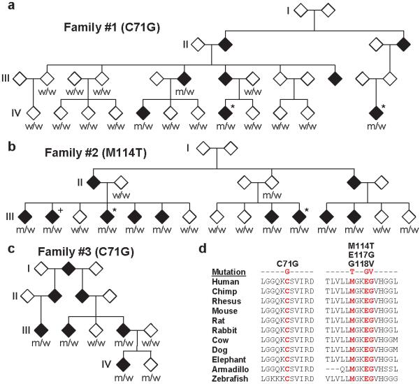
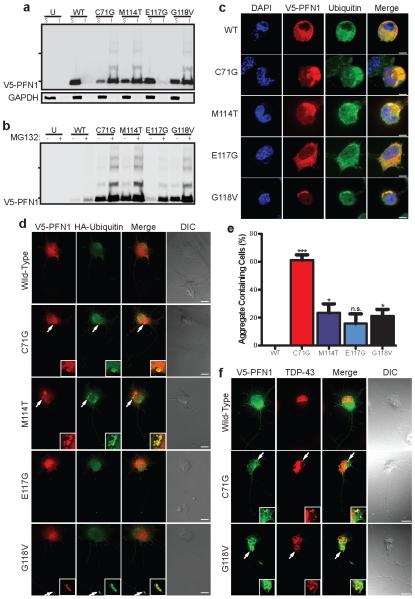
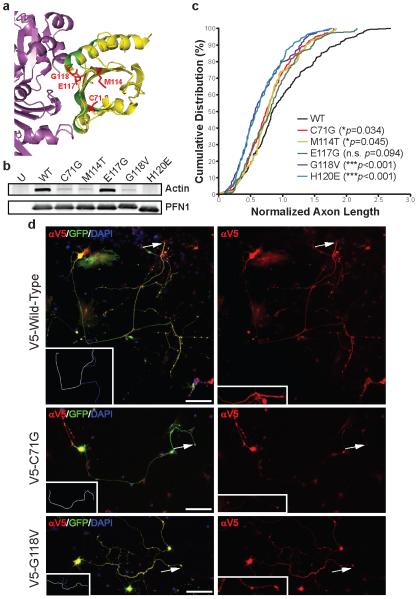
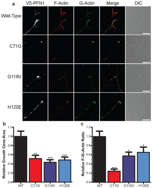
Amyotrophic lateral sclerosis (ALS) is a late-onset neurodegenerative disorder resulting from motor neuron death. Approximately 10% of cases are familial (FALS), typically with a dominant inheritance mode. Despite numerous advances in recent years, nearly 50% of FALS cases have unknown genetic aetiology. Here we show that mutations within the profilin 1 (PFN1) gene can cause FALS. PFN1 is crucial for the conversion of monomeric (G)-actin to filamentous (F)-actin. Exome sequencing of two large ALS families showed different mutations within the PFN1 gene. Further sequence analysis identified 4 mutations in 7 out of 274 FALS cases. Cells expressing PFN1 mutants contain ubiquitinated, insoluble aggregates that in many cases contain the ALS-associated protein TDP-43. PFN1 mutants also display decreased bound actin levels and can inhibit axon outgrowth. Furthermore, primary motor neurons expressing mutant PFN1 display smaller growth cones with a reduced F/G-actin ratio. These observations further document that cytoskeletal pathway alterations contribute to ALS pathogenesis.


Figures

Figure 1
Figure 1. Exome sequencing identifies PFN1 gene mutations in familial ALS
a-c, Familial ALS pedigrees harboring PFN1 mutations are shown. Asterisks indicate samples subjected to exome sequencing. To prevent identification of individual family members, the gender of each subject and information on the lower generation are withheld. Genotypes of available DNA samples for the indicated PFN1 mutation are shown (w=wild-type, m=mutant). The genotype of sample III:2 in Family #2 (+) was inferred from the genotypes of spouse and progeny (not shown). d, The evolutionary conservation of PFN1 mutations is shown. For each, the mutated amino acid is colored in red.
Figure 2
Figure 2. Mutant PFN1 produces ubiquitinated insoluble aggregates
a, Western blot analysis of transfected N2A cells subject to NP-40-soluble (S) and insoluble (I) fractionation. b, Transfected cells were treated with MG132 and processed as in (a). Hash marks indicate 25 kDa and 37 kDa markers. Transfected N2A cells (c) and PMNs (d) were stained with V5, HA (PMNs) and ubiquitin (N2A) antibodies. Example aggregates are enlarged in the inset in (d). e, Transfected N2A cells displaying insoluble aggregates were counted and analyzed using one-way ANOVA testing with Dunnett’s multiple test comparison (n=127-135 transfected cells from 3 independent experiments). *P<0.05, ***P<0.001, n.s. P>0.05. Error bars indicate SEM. f, Transfected PMNs stained with V5 and TDP-43 antibodies. Scale bars: 5 μm (c), 10 μm (d, f)
Figure 3
Figure 3. Mutant PFN1 inhibits axon outgrowth
a, PFN1-actin interaction region (PDB accession: 2BTF) using the PyMOL Molecular Graphics System (v. 1.4). Magenta: actin; Yellow: PFN1; Green: actin-binding PFN1 residues; Red: ALS-linked mutated PFN1 residues. b, Transfected HEK293 cells were immunoprecipitated with a V5 antibody and then immunoblotted with antibodies for either V5 or actin. c, PMNs transfected with wild-type or mutant V5-PFN1 and a GFP expressing construct were stained to detect V5-PFN1. d, Cumulative distribution of axon lengths relative to the mean of wild-type PFN1 transfected cells was plotted. The axon tip, indicated by arrows, is enlarged in the inset in (d), right panel. P values are given in the legend (n=104-161 cells from 4 independent experiments). Scale bar: 100 μm.
Figure 4
Figure 4. Mutant PFN1 reduces growth cone size and F-/G-actin expression
a, PMNs were transfected with either wild-type or mutant V5-PFN1. At 3 days post-transfection, cells were fixed and stained to detect V5-PFN1, F-actin (Phalloidin, red) and G-actin (DNase I, green). The growth cone region of representative cells is shown. Scale bar: 10 μm. The growth cone area (b) and F-/G-actin expression (c) of transfected cells was determined and plotted. Comparisons to the wild-type V5-PFN1 transfected cells were made using one-way ANOVA testing. *P<0.05, **P<0.01, ***P<0.001 (n=27-35 cells from 3 independent experiments). Error bars indicate SEM.

Similar articles

Cited by

References

    1. Kabashi E, et al. TARDBP mutations in individuals with sporadic and familial amyotrophic lateral sclerosis. Nat Genet. 2008;40:572–574. - PubMed
    1. Sreedharan J, et al. TDP-43 mutations in familial and sporadic amyotrophic lateral sclerosis. Science. 2008;319:1668–1672. - PMC - PubMed
    1. Kwiatkowski TJ, Jr., et al. Mutations in the FUS/TLS gene on chromosome 16 cause familial amyotrophic lateral sclerosis. Science. 2009;323:1205–1208. - PubMed
    1. Vance C, et al. Mutations in FUS, an RNA processing protein, cause familial amyotrophic lateral sclerosis type 6. Science. 2009;323:1208–1211. - PMC - PubMed
    1. Johnson JO, et al. Exome sequencing reveals VCP mutations as a cause of familial ALS. Neuron. 2010;68:857–864. - PMC - PubMed

Publication types

MeSH terms