. 2013 Jan;70(1):137-52.
doi: 10.1007/s00018-012-1075-5. Epub 2012 Jul 17.

Pyk2 cytonuclear localization: mechanisms and regulation by serine dephosphorylation

Affiliations

Pyk2 cytonuclear localization: mechanisms and regulation by serine dephosphorylation

Camille Faure et al. Cell Mol Life Sci. 2013 Jan.

Abstract

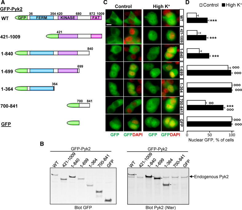
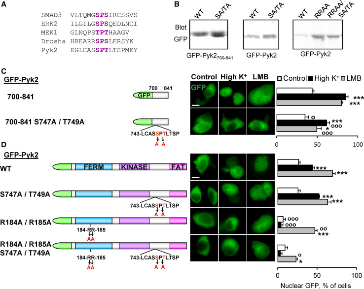
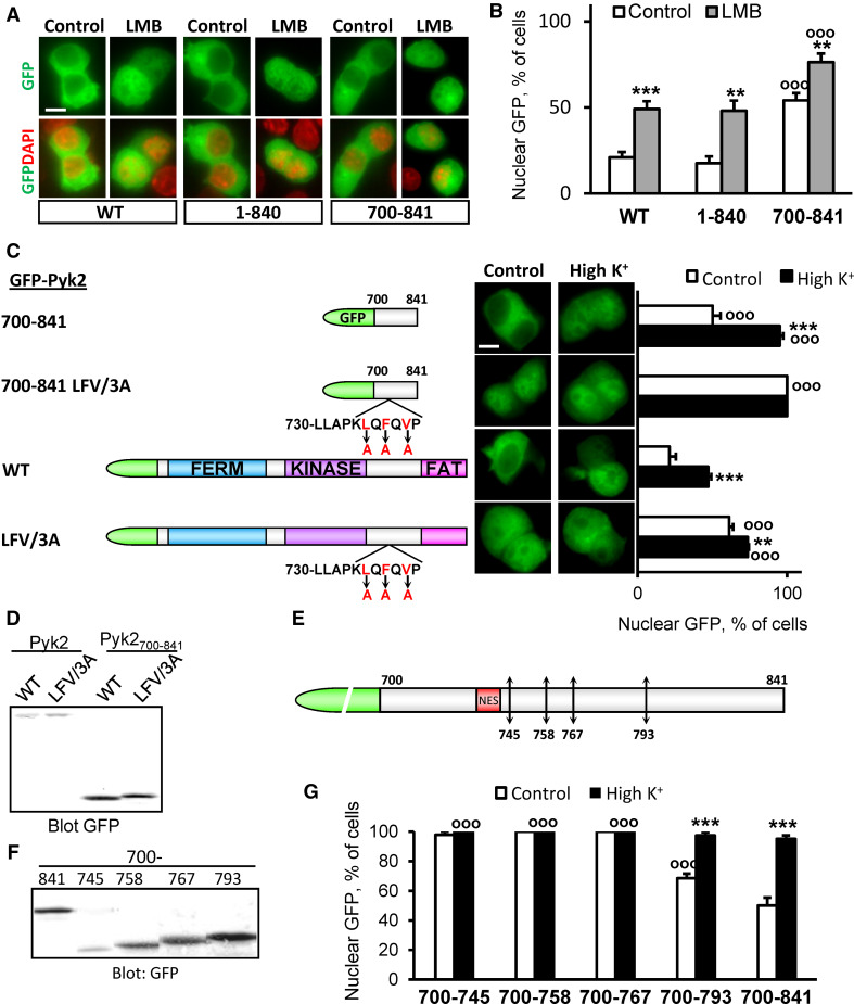
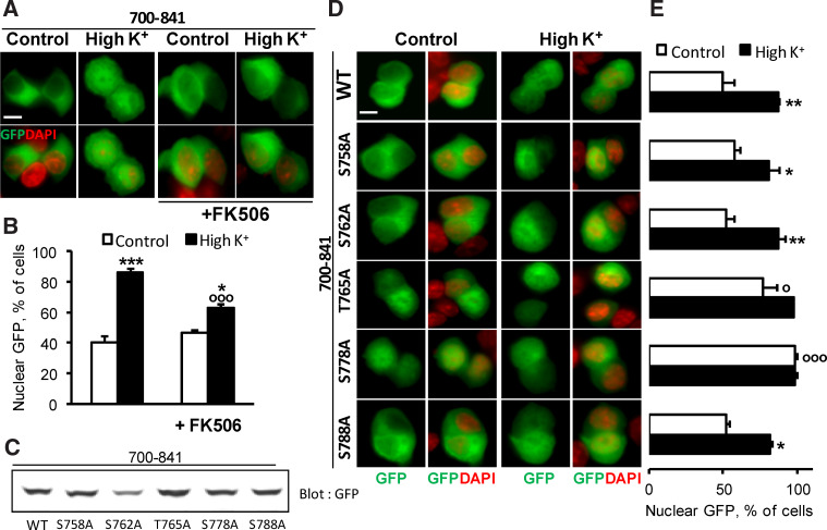
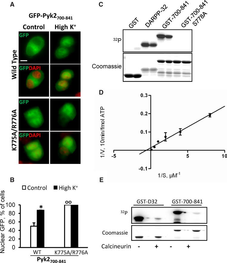
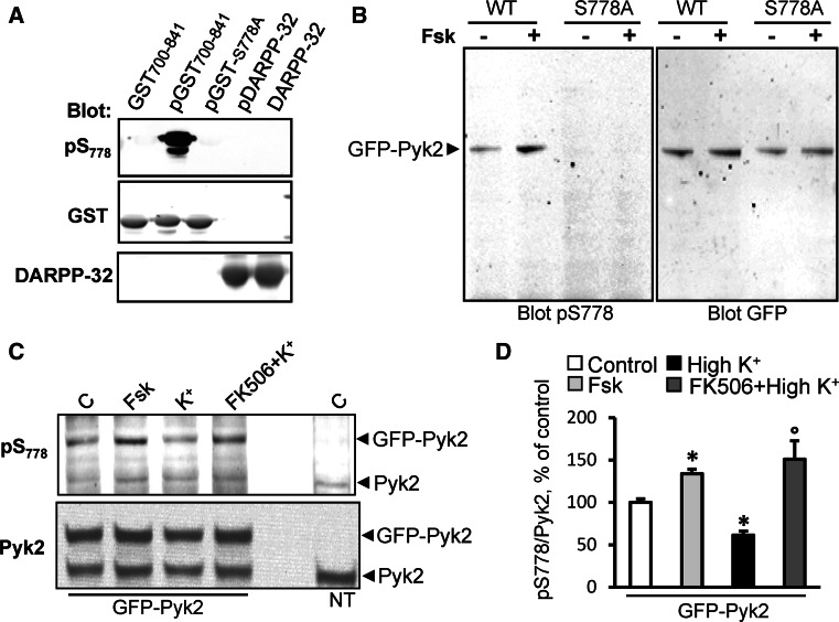
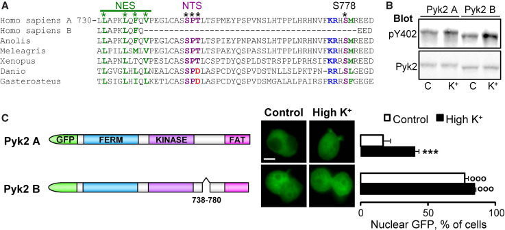
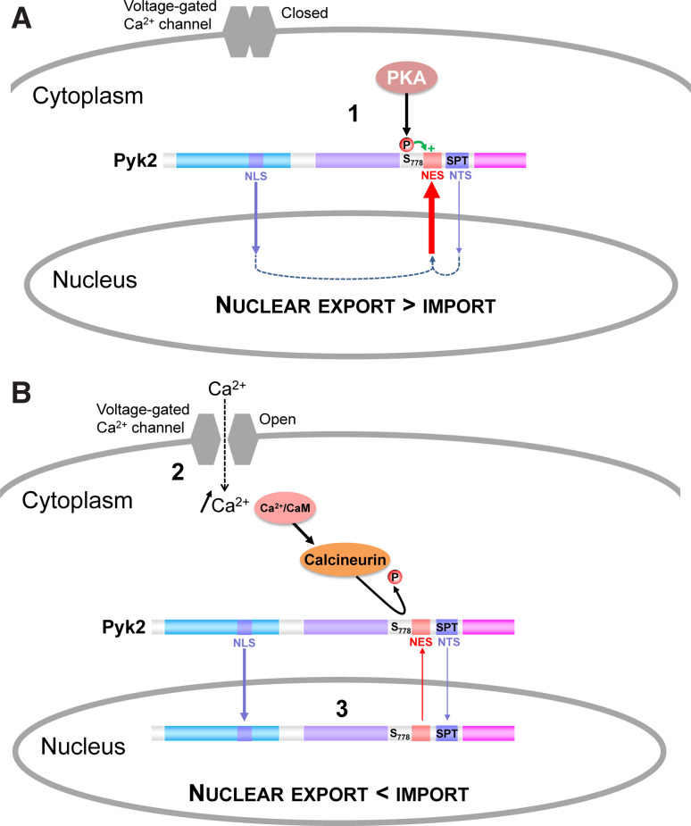
Cytonuclear signaling is essential for long-term alterations of cellular properties. Several pathways involving regulated nuclear accumulation of Ser/Thr kinases have been described but little is known about cytonuclear trafficking of tyrosine kinases. Proline-rich tyrosine kinase 2 (Pyk2) is a cytoplasmic non-receptor tyrosine kinase enriched in neurons and involved in functions ranging from synaptic plasticity to bone resorption, as well as in cancer. We previously showed the Ca(2+)-induced, calcineurin-dependent, nuclear localization of Pyk2. Here, we characterize the molecular mechanisms of Pyk2 cytonuclear localization in transfected PC12 cells. The 700-841 linker region of Pyk2 recapitulates its depolarization-induced nuclear accumulation. This region includes a nuclear export motif regulated by phosphorylation at residue S778, a substrate of cAMP-dependent protein kinase and calcineurin. Nuclear import is controlled by a previously identified sequence in the N-terminal domain and by a novel nuclear targeting signal in the linker region. Regulation of cytonuclear trafficking is independent of Pyk2 activity. The region regulating nuclear localization is absent from the non-neuronal shorter splice isoform of Pyk2. Our results elucidate the mechanisms of Ca(2+)-induced nuclear accumulation of Pyk2. They also suggest that Pyk2 nuclear accumulation is a novel type of signaling response that may contribute to specific long-term adaptations in neurons.


Conflict of interest statement

None.

Figures

Fig. 1
Fig. 1
Pyk2 700–841 region recapitulates cytonuclear regulation. a Schematic representation of Pyk2 constructs. b Expression levels of the various GFP-Pyk2 constructs in PC12 cells. Left panel anti-GFP immunoblot of PC12 cells transfected with GFP-Pyk2 (WT), deletion mutants (GFP-Pyk2421–1009, GFP-Pyk21–840, GFP-Pyk21–699, GFP-Pyk21–364, GFP-Pyk2700–841, as indicated) or GFP. Right panel immunoblot with an antibody reacting with the N-terminal region of the protein. GFP-Pyk2 is indicated by an asterisk when visible (i.e. N-terminus present). c Transfected PC12 cells were treated with high K+ (High K+) or control solution for 3 min. GFP fluorescence and nuclei stained with DAPI were analyzed with a fluorescence microscope. d Quantification of the number of cells in c with n ≥ c GFP fluorescence. Values are means + SEM, two-way ANOVA: depolarization effect F (6,42) = 9.93, p < 0.0001, deletion effect F (6.42) = 175.95, p < 0.0001, interaction F (1,42) = 73.71, p < 0.0001. Newman–Keuls test: ***p < 0.001 versus control. °°p < 0.01, °°°p < 0.001 versus WT. Scale bar 5 μm
Fig. 2
Fig. 2
Pyk2700–841 contains an atypical nuclear import motif. a Sequence alignment showing the NTS identified in SMAD3, ERK2, MEK1, Drosha, and the putative Pyk2 NTS. b Anti-GFP immunoblotting of PC12 cells transfected with GFP-Pyk2700–841 or GFP-Pyk2, with the indicated mutations (S747A, T749A, R184A, and R185A). PC12 cells transfected with GFP-Pyk2700–841 (c) or GFP-Pyk2 (d) with the indicated mutations (scheme on the left) were treated with high K+ (High K+, 40 mM, 3 min), LMB (LMB, 11 ng/ml, 3 h), or control solution. GFP fluorescence and nuclei stained with DAPI (middle) were analyzed and the number of cells with n ≥ c GFP quantified (right). Values are means + SEM. c Two-way ANOVA: mutation effect F (1,23) = 26.92, p < 0.0001, treatment effect F (1,23) = 118.57, p < 0.0001, interaction F (1,23) = 5.48, p < 0.05. Newman–Keuls test: *p < 0.05, **p < 0.01, ***p < 0.001 versus control. °p < 0.05, °°p < 0.01, °°°p < 0.001 versus WT. d Two-way ANOVA: p < 0.0001, mutation effect F (3,44) = 63.13, p < 0.0001, treatment effect F (1,44) = 14.55, p < 0.0005, interaction F (3,44) = 11.29. Newman–Keuls test: *p < 0.05, **p < 0.01, ***p < 0.001 versus control. °p < 0.05, °°p < 0.01, °°°p < 0.001 versus WT. Scale bar 5 μm
Fig. 3
Fig. 3
Pyk2700–841 contains a nuclear export motif. a PC12 cells transfected with GFP-Pyk2 (WT), 1–840 or 700–841 were incubated in the absence or presence of LMB (11 ng/ml, 3 h). GFP-Pyk2 fluorescence and nuclei stained with DAPI were analyzed. b Percentage of cells with nuclear GFP. Values are means + SEM, two-way ANOVA: deletion effect F (2,20) = 31.09, p < 0.0001, LMB effect F (1,20) = 48.88, p < 0.0001, interaction F (2,20) = 0.39, p > 0.05. Newman–Keuls test: **p < 0.01, ***p < 0.001 versus control. °°°p < 0.001 compared to WT. c PC12 cells transfected with WT or LFV/3A GFP-Pyk2 or GFP-Pyk2700–841 (scheme on the left) were treated with high K+ (High K+) or control solution for 3 min. GFP fluorescence and nuclei stained with DAPI were analyzed (middle) and the percentage of cells with n ≥ c GFP quantified (right). Values are means + SEM, two-way ANOVA: deletion effect F (4,30) = 158.77, p < 0.0001, depolarization effect F (1,30) = 113.22, p < 0.0001, interaction F (4,30) = 21.71, p < 0.0001. Newman–Keuls test: **p < 0.01, ***p < 0.001 versus control. °°°p < 0.001 versus WT. d Immunoblot analysis of constructs as in c with anti-GFP or anti-Pyk2 antibody. e Positions of stop codons inserted in GFP-Pyk2700–841 (745, 758, 767 and 793). f PC12 cells transfected with the constructs presented in e were analyzed by immunoblotting with anti-GFP. g Percentage of transfected cells with n ≥ c GFP. Values are means + SEM, two-way ANOVA: deletions effect F (4,25) = 47.88, p < 0.0001, depolarization effect F (1,25) = 82.58, p < 0.0001, interaction F (4,25) = 32.49, p < 0.0001. Newman–Keuls test: ***p < 0.001 versus control. °°°p < 0.001 versus 700–841. Scale bar 5 μm
Fig. 4
Fig. 4
Pyk2700–841 cytonuclear localization is regulated by calcineurin. a PC12 cells were transfected with GFP-Pyk2700–841 and treated with high K+ (High K+ 40 mM) or control solution for 3 min in the absence or presence of FK506 (2 μM, 20 min before high K+). GFP fluorescence and nuclei stained with DAPI were analyzed. b Quantification of the number of cells in a with n ≥ c GFP. Values are means + SEM, two-way ANOVA: depolarization effect F (1,14) = 80.92, p < 0.0001, FK506 effect F (1,14) = 5.88, p < 0.05, interaction F (1,14) = 17.49, p < 0.001. Newman–Keuls test: *p < 0.05, ***p < 0.001 versus control. °°°p < 0.001 versus high K+. c Anti-GFP immunoblotting of PC12 cells transfected with GFP-Pyk2700–841, S758A, S762A, T765A, S778A or S788A. d GFP fluorescence and DAPI nuclear staining of PC12 cells transfected as in c and treated with high K+ (High K+ 40 mM) or control solution for 3 min. e Percentage of cells in d with n ≥ c GFP. Values are means + SEM, two-way ANOVA: p < 0.05, mutation effect F (5,12) = 14.33, p < 0.0001, depolarization effect F (1,12) = 72.7, p < 0.0001, interaction F (5,12) = 3.82. Newman–Keuls test: *p < 0.05 versus control. °p < 0.05, °°°p < 0.001 versus 700–841 WT. Scale bar 5 μm
Fig. 5
Fig. 5
S778 is a substrate of PKA and is dephosphorylated by calcineurin in vitro. a PC12 cells transfected with WT or K775A/R776A GFP-Pyk2700–841 were treated with high K+ (High K+ 40 mM) or control solution for 3 min. GFP fluorescence and nuclei stained with DAPI were analyzed. b Percentage of cells in a with n ≥ c GFP. Values are means + SEM. Two-way ANOVA: mutation effect F (1,4) = 63.45, p < 0.005, depolarization effect F (1,4) = 22.64, p < 0.01, interaction F (1,4) = 22.64, p < 0.01. Newman–Keuls test: **p < 0.01 versus control. °°p < 0.01 versus 700–841 WT. c Purified GST, DARPP-32, WT or S778A GST700–841 were incubated with PKA catalytic subunit and [γ-32P]ATP. 32P incorporation was analyzed with phosphorimager (upper panel) and proteins with Coomassie staining (lower panel). d Lineweaver–Burk plot of 32P incorporation with increasing concentrations (0.25–10 μM) of GST700–841. f Purified GST, alone or fused to DARPP-32 (GST-DARPP-32) or Pyk2700–841 (GST-700–841) were phosphorylated on glutathione-Sepharose beads with PKA and [γ-32P]ATP as in a, washed and incubated for 30 min in the absence (−) or presence (+) of calcineurin (PP2B). Scale bar 5 μm
Fig. 6
Fig. 6
Regulation of S778 phosphorylation by PKA and calcineurin in cells. a The pS778 antibody reacts specifically with Pyk2 phosphorylated by PKA. Wild type GST-Pyk2700–841 incubated in the absence of PKA and ATP (lane 1), WT or S778A GST-Pyk2700–841 incubated in the presence of PKA and ATP (lane 2 pGST700–841, lane 3 pGST-S778A). DARPP-32 incubated in the presence (lane 4 pDARPP-32) or absence of PKA and ATP (lane 5 DARPP-32) was used as a specificity control. Samples were analyzed by immunoblotting with anti-pS778, (upper panel), anti-GST (middle) or anti-DARPP-32 antibody (lower). b COS7 cells transfected with WT or S778A GFP-Pyk2 and treated or not with forskolin (Fsk, 10 μM, 10 min) were analyzed by immunoblotting with anti-pS778 antibody (left panel) or anti-GFP antibody (right panel). c PC12 cells were transfected with GFP-Pyk2 and incubated in the absence (control, C) or presence of forskolin (Fsk, 10 μM, 10 min) or high K+ (40 mM, 5 min) with or without calcineurin inhibitor FK506 (2 μM, 15 min before K+) were analyzed by immunoblotting with anti-pS778 antibody (upper panel) or anti-Pyk2 antibody (bottom panel). Non-transfected cells (NT) are also shown. d Quantification of the results in c for Pyk2-GFP. Values are means + SEM. One-way ANOVA: F (3,8) = 10.41 (GFP), p < 0.05. Newman–Keuls test: *p < 0.05 versus control, °p < 0.05 versus high K+
Fig. 7
Fig. 7
S778 phosphorylation controls Pyk2 nuclear export. a GFP fluorescence, pS778 immunoreactivity and DAPI staining in PC12 cells transfected with WT or S778A GFP-Pyk2 and incubated in the presence of a control or depolarizing (High K+ 40 mM) solution for 3 min. b Percentage of c > n GFP-positive cells in a. Values are means + SEM. Two-way ANOVA: mutation effect F (3,8) = 128.58, p < 0.0001, depolarization effect F (1,8) = 23.99, p < 0.005, interaction F (3,8) = 6.01, p < 0.05. Newman–Keuls test: ***p < 0.001 versus control, °°°p < 0.001 versus WT. c Class distribution of GFP-Pyk2-transfected cells in a depending on cytoplasmic pS778 immunoreactivity and GFP localization with schematic representation of each class (cytoplasm and nucleus: green, compartment with highest GFP concentration; red pS778 immunoreactivity, yellow colocalization). Values are means + SEM. One-way ANOVA: F (7,8) = 27.96, p < 0.0001. Newman–Keuls test: ***p < 0.001, **p < 0.01 high K+ versus control. d GFP fluorescence, pS778 immunoreactivity, and DAPI staining of PC12 cells transfected with GFP-Pyk2 and incubated in the presence of vehicle (control), or H89 (100 μM, 30 min), or myrPKI14–22 (10 μM, 10 min), or a depolarizing solution (High K+ 40 mM, 3 min). e Quantification of cells with GFP fluorescence stronger in the nucleus than in the cytoplasm in d. Values are means + SEM. One-way ANOVA: F (5,12) = 43.41, p < 0.0001. Newman–Keuls test: ***p < 0.001 versus control, °°p < 0.01 versus High K+. f PC12 cells transfected with WT or S778A GFP-Pyk2 were treated with LMB (LMB, 11 ng/ml, 3 h) as in Fig. 3a. Results are expressed as % increase in nuclear/cytoplasmic fluorescence ratio (n/c) in response to LMB-treatment. Values are means + SEM. Student t test: ***p < 0.01. g PC12 cells transfected with GFP-Pyk2 WT or S778A treated with high K+ (High K+ 40 mM) or control solution for 5 min were analyzed by immunoblotting with anti-pY402 antibody (upper) or anti-GFP antibody (lower). h Values are means + SEM. Two-way ANOVA: mutation effect F (1,6) = 1.94, p > 0.05, treatment effect F (2,6) = 88.37, p < 0.0001, interaction F (2,6) = 0.32, p > 0.05. Newman–Keuls test: **p < 0.01 versus control, ***p < 0.001 versus control. Scale bar 5 μm
Fig. 8
Fig. 8
The NES, NTS, and S778 are conserved between species and absent in Pyk2 isoform B. a Alignment of Pyk2 protein sequences from Homo sapiens, Anolis carolinensis, Meleagris gallopavo, Xenopus silurana, Danio rerio, Gasterosteus aculeatus together with the Homo sapiens isoform B lacking one exon. The NES, NTS, and S778 are shown with asterisk. The residues in the PKA consensus site are indicated in blue (−3, −2 basic) and green (+1 hydrophobic). b PC12 cells transfected with GFP-Pyk2 isoform A (long) or B (short) were treated with high K+ (K+, 40 mM) or control solution (C) for 5 min and analyzed by immunoblotting with anti-pY402 antibody (upper panel) or anti-Pyk2 antibody (lower panel). c PC12 cells transfected with GFP-Pyk2 isoform A or B (scheme on the left) were treated with high K+ (High K+) or control solution for 3 min. The percentage of cells with n ≥ c GFP fluorescence (middle) was quantified (right panel). Values are means + SEM, two-way ANOVA: isoform effect F (1,12) = 261.83, p < 0.0001, K+ effect F (1,12) = 118.57, p < 0.0005 interaction F (1,12) = 6.42, p < 0.05. Newman–Keuls test: ***p < 0.001 versus control, °°°p < 0.001 versus WT. Scale bar 5 μm
Fig. 9
Fig. 9
Working model of Pyk2 intracellular localization regulation. a In basal conditions, Pyk2 is phosphorylated on S778 by PKA, the NES in the linker region between the kinase and the FAT domain, is active, and Pyk2 is predominantly cytoplasmic. b After a rise of Ca2+, for example through opening of voltage-sensitive Ca2+ channels, calcineurin is activated and dephosphorylates S778 leading to the inactivation of the NES. Pyk2 then accumulates in the nucleus due to the predominant activity of the NLS and possibly the NTS

Similar articles

Cited by

References

    1. Lev S, et al. Protein tyrosine kinase Pyk2 involved in Ca(2+)-induced regulation of ion channel and MAP kinase functions. Nature. 1995;376(6543):737–745. doi: 10.1038/376737a0. - DOI - PubMed
    1. Salter MW, Kalia LV. Src kinases: a hub for NMDA receptor regulation. Nat Rev Neurosci. 2004;5(4):317–328. doi: 10.1038/nrn1368. - DOI - PubMed
    1. Girault JA, et al. FAK and PYK2/CAK in the nervous system, a link between neuronal activity, plasticity and survival? Trends Neurosci. 1999;22:257–263. doi: 10.1016/S0166-2236(98)01358-7. - DOI - PubMed
    1. Hsin H, et al. Proline-rich tyrosine kinase 2 regulates hippocampal long-term depression. J Neurosci. 2010;30(36):11983–11993. doi: 10.1523/JNEUROSCI.1029-10.2010. - DOI - PMC - PubMed
    1. Bartos JA, et al. Postsynaptic clustering and activation of Pyk2 by PSD-95. J Neurosci. 2010;30(2):449–463. doi: 10.1523/JNEUROSCI.4992-08.2010. - DOI - PMC - PubMed

Publication types

MeSH terms

LinkOut - more resources