Review
. 2012 Oct;29(10):1074-98.
doi: 10.1039/c2np20025b. Epub 2012 Jul 17.

Explorations of catalytic domains in non-ribosomal peptide synthetase enzymology

Review

Explorations of catalytic domains in non-ribosomal peptide synthetase enzymology

Gene H Hur et al. Nat Prod Rep. 2012 Oct.

Abstract

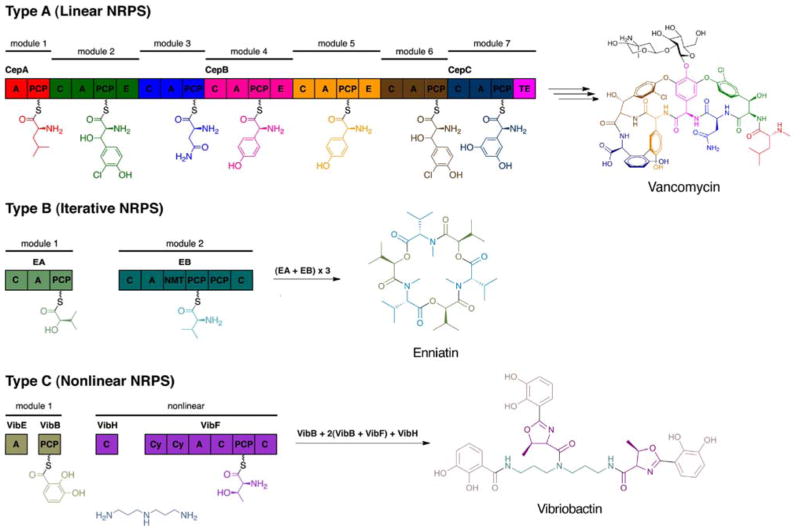
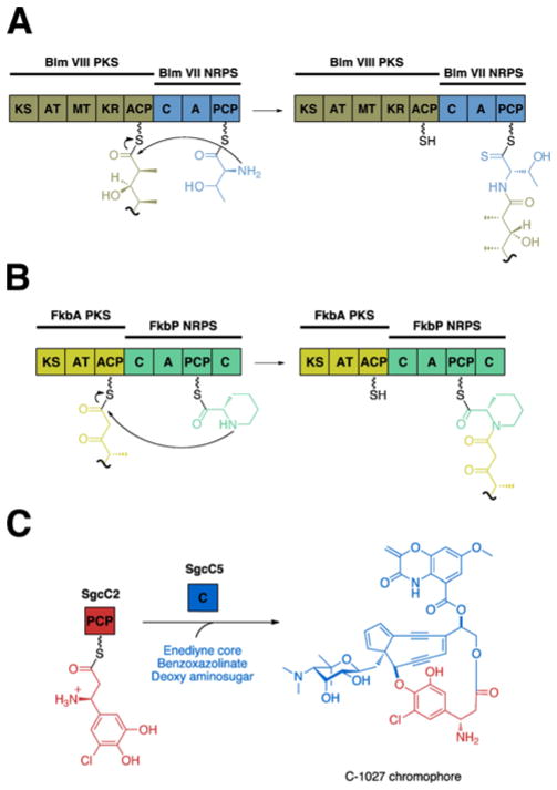
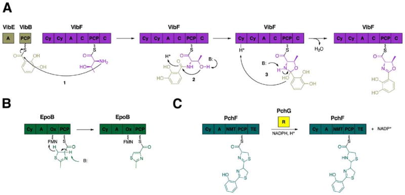
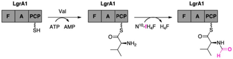
Many pharmaceuticals on the market today belong to a large class of natural products called nonribosomal peptides (NRPs). Originating from bacteria and fungi, these peptide-based natural products consist not only of the 20 canonical L-amino acids, but also non-proteinogenic amino acids, heterocyclic rings, sugars, and fatty acids, generating tremendous chemical diversity. As a result, these secondary metabolites exhibit a broad array of bioactivity, ranging from antimicrobial to anticancer. The biosynthesis of these complex compounds is carried out by large multimodular megaenzymes called nonribosomal peptide synthetases (NRPSs). Each module is responsible for incorporation of a monomeric unit into the natural product peptide and is composed of individual domains that perform different catalytic reactions. Biochemical and bioinformatic investigations of these enzymes have uncovered the key principles of NRP synthesis, expanding the pharmaceutical potential of their enzymatic processes. Progress has been made in the manipulation of this biosynthetic machinery to develop new chemoenzymatic approaches for synthesizing novel pharmaceutical agents with increased potency. This review focuses on the recent discoveries and breakthroughs in the structural elucidation, molecular mechanism, and chemical biology underlying the discrete domains within NRPSs.


Figures

Fig. 1
Fig. 1
A variety of nonribosomal peptides with structural features that confer their bioactivity (highlighted).
Fig. 2
Fig. 2
Biosynthetic strategies for assembling nonribosomal peptides (NRPS).
Fig. 3
Fig. 3
(A) Crystal structure of the Bacillus subtilis Sfp protein (red and yellow) complexed with CoA (purple). Residues D107, E109, and E151 coordinate with Mg2+ ion (blue sphere) and H90 binds to CoA (purple). Mutational analysis of residues K112, E117, and K120 (orange) determined the loop region (cyan) that forms the binding pocket for the PCP. (B) Coordination diagram depicting the proposed mechanism of the phosphopantetheinylation reaction.
Fig. 4
Fig. 4
(A) Structure of Bacillus subtilis SrfTEII (red and yellow) in complex with TycC3–PCP (blue), a PCP from the tyrocidine system. Residues S86, D190, and H216 of SrfTEII form the catalytic triad. Key residues (purple), including the ‘lid’ region (orange) of SrfTEII that interact with TycC3–PCP are highlighted. (B) Structure of RifR. Residues S94, D200, H228 form the catalytic triad of the RifR active site and the flexible linker region (purple) as well as the ‘lid’ region (orange) are indicated.
Fig. 5
Fig. 5
(A) Adenylation reaction in NRPS. (B) Crystal structure of the adenylation domain, consisting of the large N-terminal domain (red and yellow) and the smaller C-terminal domain (gray), from the gramicidin S synthetase, GrsA, complexed with AMP (purple) in the presence of Mg2+ion (blue sphere). The 10 catalytic residues (orange) termed ‘codons’ are highlighted. (C) Crystal structure of DltA, consisting of the large N-terminal domain (red and yellow), the linker region (orange) and the smaller C-terminal domain (gray), bound to ATP (purple). Invariant residues K492, E298, and R397 stabilize ATP in the presence of Mg2+ ion (light blue sphere).
Fig. 6
Fig. 6
Structures of MbtH-like proteins. (A) Crystal structure of PA2412. (B) NMR structure of PA2412. Disordered residues at the N-terminus have been omitted for clarity. Flexible region between the two α-helices is shown in purple. (C) NMR solution structure of MbtH. Disordered residues at the N-terminus have been omitted for clarity. Flexible C-terminus is shown in purple.
Fig. 7
Fig. 7
Ribbon diagrams of the NMR solution structures of TycC3–PCP in three different conformations (A) the A-state, (B) the A/H-state and (C) the H-state. Helices αI (blue), αII (red), αIII/loop III(orange), and αIV (purple) undergo conformational changes in each state.
Fig. 8
Fig. 8
Structure of the PCP–TE didomain of the Escherichia coli enterobactin synthetase, EntF. Active site residues S180 of the TE domain (red and yellow) and S48A of the PCP (blue) are 17.5 Å apart. F42 stabilizes the interactions between the PCP and TE and helices α4TE-α5TE form the ‘lid’ region (purple).
Fig. 9
Fig. 9
(A) Crystal structure of the TycC5–6 PCP–C bidomain from the tyrocidine NRPS. The active site residues H224 of the C domain (red and yellow) and S43 from the PCP (blue) are positioned 47 Å apart. Residues (orange) responsible for proper interaction between the PCP and C domain are highlighted. (B) Crystal structure of the termination module from the Bacillus subtilis surfactin NRPS, SrfA–C. Linker regions (green) connecting the C domain (orange), A domain (gray and purple), PCP (blue), and TE domain (red) are indicated. Active site residues H147 of the C domain and S1003 from the PCP are 16.4 Å apart. Residues (black) determined to be responsible for proper interaction between the PCP and C domain are highlighted.
Fig. 10
Fig. 10
(A) Peptide bond formation catalyzed by the C domain. (B) X-ray crystal structure of the stand-alone C domain, VibH, from the Vibrio cholerae vibrioactin synthetase. The N-terminal (red) and C-terminal (blue) subdomains are connected by a linker region (purple), forming a V-shaped canyon. The ‘His’ motif (black), consisting of the catalytic residue H126, marks the active site, which is located at the junction of these two subdomains.
Fig. 11
Fig. 11
Different condensation reactions catalyzed by the C domain. (A) The C domain in the bleomycin NRPS subunit, BlmVII, condenses an aminoacyl substrate (blue) with a ketide unit (gold). (B) The C domain in the FK520 NRPS subunit, FkbP, catalyzes condensation between pipecolate (green) and a ketide unit (yellow). (C) The free-standing C domain, SgcC5, from the C-1027 NRPS catalyzes ester bond formation.
Fig. 12
Fig. 12
Crystal structure of the Epimerization domain from Tyrocidine synthetase A (TycA).
Fig. 13
Fig. 13
Enzymatic reactions of the tailoring domains. (A) The cyclization (Cy) domain from the Vibrio cholerae vibriobactin NRPS subunit, VibF, catalyze cyclization of threonine to form the oxazoline ring in three steps. (B) The oxidation (Ox) domain from the epothilone synthetase B, EpoB, oxidizes the thiazoline ring to the thiazole in the presence of the cofactor flavin mononucleotide (FMN). (C) The reduction (R) domain from the pyochelin synthetase, PchF, reduces the thiazoline ring to the thiazolidine in the presence of NADPH.
Fig. 14
Fig. 14
N- and C-methylation of amionacyl substrates in NRP biosynthesis. (A) The N-methyltransferase (NMT) domain from the pyochelin NRPS, PchF, transfers a methyl group from S-adenosyl methionine (SAM) to the amine group of the substrate tethered on the PCP. (B) Crystal structure of NMT, MtfA, from the chloroeremomycin synthetase complexed with SAM (purple stick model) with one monomer designated in gray. (C) The C-methyltransferase (CMT), GlmT, from the CDA producer Streptomyces coelicolor transfers the methyl group from SAM to the β-carbon of α-ketoglutarate in a stereospecific manner.
Fig. 15
Fig. 15
The formylation (F) domain of the Bacillus brevis linear gramicidin NRPS subunit, LgrA1, catalyzes formylation of valine in the presence of N-formyltetrahydrofolate.
Fig. 16
Fig. 16
(A) Proposed mechanism of flavin-dependent halogenases. (B) Crystal structure of the chondrochloren halogenase, CndH (red and yellow), from the myxobacterium Chondromyces crocatus Cm c5 complexed with FAD (purple) and in the presence of Cl ion (blue sphere). Active site residue K76 reacts with HOCl to form the chlorinating reagent and E387 (black) acts as the base to complete halogenation.
Fig. 17
Fig. 17
Structure and mechanism of αKG-dependent halogenases. (A) Crystal structure of the halogenase domain, SyrB2, from the syringomycin E synthetase complexed with αKG (purple) in the presence of Fe(II) ion (blue sphere). Residues N123, T143, and R254 (orange) form hydrogen bonds with the Cl ion (green sphere) and residues A118, F121, and S231 (gray) form the hydrophobic pocket. (B) Crystal structure of the halogenase domain, CytC3, from the γ,γ-dichloroaminobutyrate synthetase complexed with αKG (purple) in the presence of Fe2+ ion (blue sphere). Corresponding residues from SyrB2 are indicated as well. (C) Proposed mechanism of non-heme Fe(II)/αKG-dependent halogenases.
Fig. 18
Fig. 18
(A) The thioesterase (TE) domain from the vancomycin NRPS catalyzes release of the linear peptide through hydrolysis after three crosslinking reactions. (B) The TE domain from the daptomycin NRPS catalyzes the release of the peptidic product through an intramolecular macrocyclization.
Fig. 19
Fig. 19
Crystal structure of the surfactin thioesterase domain, SrfTEI, depicted as an asymmetric dimer. The monomer on the left is in the closed ‘C’ conformation with the ‘lid’ region (purple) covering the active site, while the other is in the open ‘O’ conformation with the ‘lid’ flipped back. Residues S80, D107, and H207 (black) form the catalytic triad and A81 and V27 (blue) form the oxyanion hole.
Fig. 20
Fig. 20
Crystal structure of the fengycin thioesterase domain, FenTEI. The ‘lid’ region (purple) is flipped back designating FenTEI in the open conformation. Residues S84, D111, and H201 (black) form the catalytic triad and A85 and I30 (blue) form the oxyanion hole.
Fig. 21
Fig. 21
The thioesterase domain from the thiocoraline synthetase, TioS, catalyzes macrothiolactonization.
Fig. 22
Fig. 22
(A) COM domains from the tyrocidine system regulate the protein interactions between the first two modules, TycA and TycB1, for proper processing of the aminoacyl substrates, L-Phe and L-Pro, to form the dipeptidyl intermediate. (B) Pantetheine azide and difluorocyclooctyne pantetheine modified the PCPs of TycA and TycB1, respectively, and displayed sensitivity to the protein interactions governed by the COM domains of these cognate partner NRPSs through crosslinking.
Fig. 23
Fig. 23
Ribbon diagram depicting the COM hand interaction between two SrfA-C monomers. The myc-His6 tag (purple) interacts not only with the putative COMA sequence (blue) of SrfA-C, but also two additional βsheets (red) located on the C domain.
Fig. 24
Fig. 24
Ribbon diagram of the tubulysin docking domain, TubCdd (red and yellow). A new αββαα-fold is displayed with residues R20, E26, R27, R29, Q31 and V36 (black) making up the docking code.

Similar articles

Cited by

References

    1. Finking R, Marahiel MA. Annu Rev Microbiol. 2004;58:453–488. - PubMed
    1. Cane DE, Walsh CT, Khosla C. Science. 1998;282:63–68. - PubMed
    1. Stein T, Vater J, Kruft V, Otto A, Wittmann-Liebold B, Franke P, Panico M, McDowell R, Morris HR. J Biol Chem. 1996;271:15428–15435. - PubMed
    1. Weber G, Schorgendorfer K, Schneider-Scherzer E, Leitner E. Curr Genet. 1994;26:120–125. - PubMed
    1. Jirakkakul J, Punya J, Pongpattanakitshote S, Paungmoung P, Vorapreeda N, Tachaleat A, Klomnara C, Tanticharoen M, Cheevadhanarak S. Microbiology. 2008;154:995–1006. - PubMed

Publication types