. 2012 Oct;56(10):5031-9.
doi: 10.1128/AAC.00891-12. Epub 2012 Jul 16.

Existence of separate domains in lysin PlyG for recognizing Bacillus anthracis spores and vegetative cells

Affiliations

Existence of separate domains in lysin PlyG for recognizing Bacillus anthracis spores and vegetative cells

Hang Yang et al. Antimicrob Agents Chemother. 2012 Oct.

Abstract

As a potential antimicrobial, the bacteriophage lysin PlyG has been reported to specifically recognize Bacillus anthracis vegetative cells only and to kill B. anthracis vegetative cells and its germinating spores. However, how PlyG interacts with B. anthracis spores remains unclear. Herein, a 60-amino-acid domain in PlyG (residues 106 to 165), located mainly in the previously identified catalytic domain, was found able to specifically recognize B. anthracis spores but not vegetative cells. The exosporium of the spores was found to be the most probable binding target of this domain. This is the first time that a lysin for spore-forming bacteria has been found to have separate domains to recognize spores and vegetative cells, which might help in understanding the coevolution of phages with spore-forming bacteria. Besides providing new biomarkers for developing better assays for identifying B. anthracis spores, the newly found domain may be helpful in developing PlyG as a preventive antibiotic to reduce the threat of anthrax in suspected exposures to B. anthracis spores.


Figures
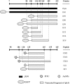
Fig 1
Fig 1
Schematic positions of the truncated proteins and synthetic peptides used in the study.
Fig 2
Fig 2
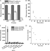
Binding characteristics of the recombinant proteins for B. anthracis vegetative cells and spores. (A) Changes in residual fluorescence intensities of EP9, EP3, and EP0 after reaction with B. anthracis cells for 30 min. (B) Time gradient of fluorescence changes of the supernatant during reaction of EP0 with B. anthracis cells. (C) Specificity of EP0 for B. anthracis spores. After staining with EP0, spores were analyzed by the fluorescence microscope system under the same instrument conditions. (D) Time gradient of fluorescence changes of the supernatant during reaction of EP0 with B. anthracis spores.
Fig 3
Fig 3
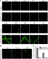
Binding features of the truncated proteins and the peptide for B. anthracis spores, heat-treated vegetative cells, and sonicated spores. (A) Binding profiles with B. anthracis spores and vegetative cells. (B) Binding profiles with sonicated spores. (C) Relative fluorescence intensities of intact spores and sonicated spores after staining with EP0. All images were taken under the same instrument conditions. Bar = 2 μm.
Fig 4
Fig 4
Structure-based analysis of the truncated fragments within the SBD. (A) Structure of the PlyG SBD based on the structure of the catalytic domain of PlyG (PDB 2L47_A; GI 350610326). All of the truncated sites are labeled with different colors. (B) Summarized binding results for the truncated fragments within the SBD.
Fig 5
Fig 5
Characteristics of EC6 association with B. anthracis (B. A) spores. (A) Differences in binding of the truncated proteins to B. anthracis spores with the same concentrations and conditions. (B) Kinetics of EC6 association with B. anthracis spores at concentrations of 674.6 nM (black) and 337.3 nM (dark gray). EGFP (304.4 nM; gray) and PBS (light gray) were used as the control and the blank, respectively. (C) Saturation binding curve of EC6, shown by the molar concentration of Cf relative to Cb. (D) Simulated Scatchard plot for EC6 association with B. anthracis spores (adjusted r2 = 0.9918).
Fig 6
Fig 6
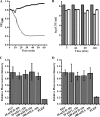
PlyG-mediated spore binding and killing. (A) Lysin activity of PlyG on B. anthracis vegetative cells. One unit of two-step-purified PlyG (open squares) displays high activity against B. anthracis cells, in contrast to the buffer control (solid squares). (B) Effect of PlyG on B. anthracis spores. Heat-activated B. anthracis spores were treated with PlyG (10 U) in the presence of either 100 mM l-alanine (light gray) or d-alanine (white) for different times. The resulted CFU were counted and are contrasted with results for the blank control (gray). EG1 can bind to inactivated (C) or live (D) B. anthracis spores in the presence of either l-alanine or d-alanine, and the relative fluorescence intensity is not obviously different from that of the buffer control.
Fig 7
Fig 7
Transmission electron microscopy pictures of B. anthracis spores, sonicated spores, and the detached exosporium labeled with bare AuNPs and N21-AuNPs, respectively. Bar = 500 nm.

Similar articles

Cited by

References

    1. Acharya G, et al. 2007. Label-free optical detection of anthrax-causing spores. J. Am. Chem. Soc. 129:732–733 - PubMed
    1. Brumlik MJ, et al. 2001. Use of long-range repetitive element polymorphism-PCR to differentiate Bacillus anthracis strains. Appl. Environ. Microbiol. 67:3021–3028 - PMC - PubMed
    1. Clery-Barraud C, Gaubert A, Masson P, Vidal D. 2004. Combined effects of high hydrostatic pressure and temperature for inactivation of Bacillus anthracis spores. Appl. Environ. Microbiol. 70:635–637 - PMC - PubMed
    1. Daniel A, et al. 2010. Synergism between a novel chimeric lysin and oxacillin protects against infection by methicillin-resistant Staphylococcus aureus. Antimicrob. Agents Chemother. 54:1603–1612 - PMC - PubMed
    1. Dixon TC, Meselson M, Guillemin J, Hanna PC. 1999. Anthrax. N. Engl. J. Med. 341:815–826 - PubMed

Publication types

MeSH terms

Substances