

Virilising ovarian tumour in a woman with an adrenal nodule
- PMID: 22802276
- PMCID: PMC3029143
- DOI: 10.1136/bcr.07.2010.3139
Virilising ovarian tumour in a woman with an adrenal nodule
Abstract
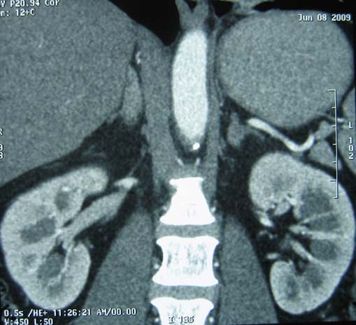
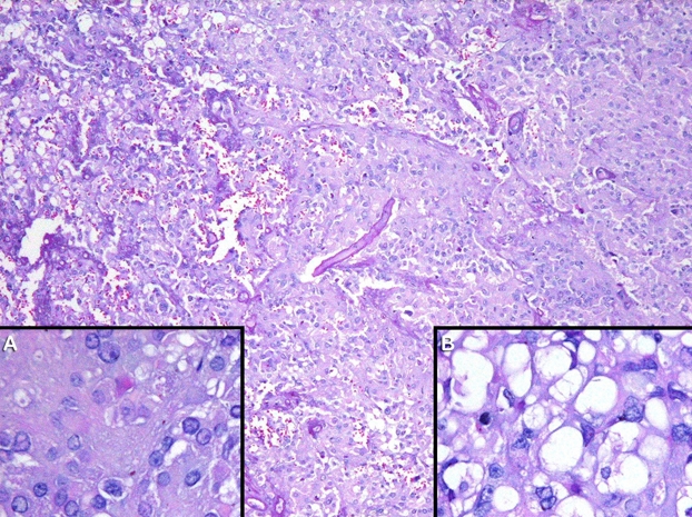
Androgen secreting tumours are the least commonly encountered androgen excess disorders, having a prevalence of 0.2%. Androblastomas of the ovary comprise less than 0.5% of all ovarian tumours. Pure Leydig cell tumours are very rare and almost always show secretion of male sex hormones. A 41-year-old multipara Filipino woman presented with a 2-year history of amenorrhoea and virilisation characterised by hirsutism, androgenic alopecia, masculine habitus and clitoromegaly. Diagnostic evaluation showed markedly elevated serum testosterone and normal dehydroepiandrosterone sulfate. Normal ovaries were seen on initial transvaginal ultrasound. A low dose dexamethasone suppression test suggested an ovarian source. A left adrenal nodule was seen on CT scan. Doppler transvaginal ultrasound revealed a solid lobulated structure in the right ovary. The patient underwent surgery and histopathology showed a Leydig cell tumour, hilar type. Serum testosterone levels normalised 3 days after surgery. Specific clinical and biochemical investigation of androgen secreting neoplasms is very important for correct diagnosis of these rare tumours.
Conflict of interest statement
Figures


References
-
- Azziz R, Sanchez LA, Knochenhauer ES, et al. Androgen excess in women: experience with over 1000 consecutive patients. J Clin Endocrinol Metab 2004;89:453–62 - PubMed
-
- Carmina E, Rosato F, Jannì A, et al. Extensive clinical experience relative prevalence of different androgen excess disorders in 950 women referred because of clinical hyperandrogenism. J Clin Endocrinol Metab 2006; 91:2–6 - PubMed
-
- Roth LM, Anderson MC, Govan AD, et al. Sertoli-Leydig cell tumors: a clinicopathologic study of 34 cases. Cancer.1981; 48:187–197 - PubMed
-
- Wenham RM, Berchuck A. The molecular biology of Sertoli-leydig cell tumors of the ovary. CME Journal of Gynecol Oncol.2002;7:121–125
-
- Hyperandrogenic Disorders Task Force AACE Medical Guidelines for Clinical Practice for the Diagnosis and Treatment of Hyperandrogenic Disorders. Endocr Pract 2001;7:121–134 - PubMed
Publication types
MeSH terms
Supplementary concepts
LinkOut - more resources
Full Text Sources
Medical