Review
. 2012 Jul;9(3):102-10.
doi: 10.1513/pats.201201-005AW.

Role of the lysophospholipid mediators lysophosphatidic acid and sphingosine 1-phosphate in lung fibrosis

Affiliations
Review

Role of the lysophospholipid mediators lysophosphatidic acid and sphingosine 1-phosphate in lung fibrosis

Barry S Shea et al. Proc Am Thorac Soc. 2012 Jul.

Abstract

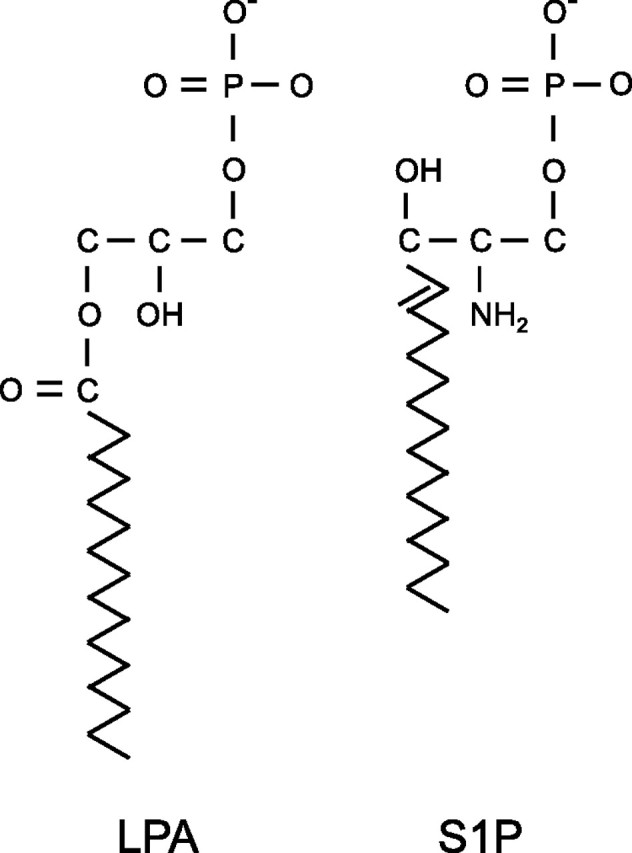
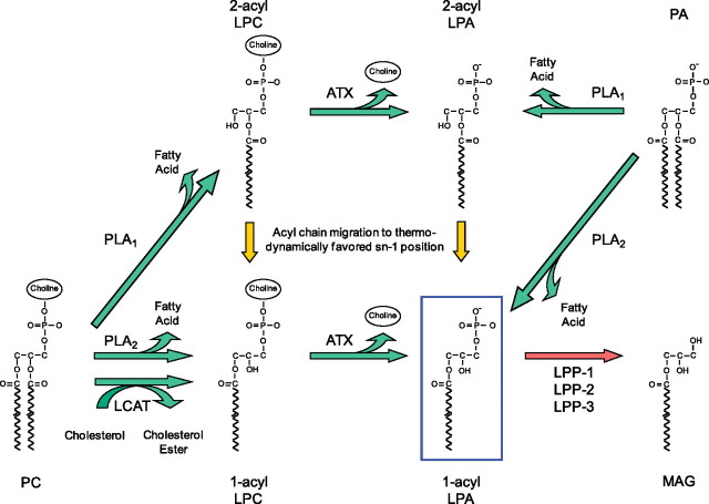
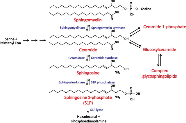
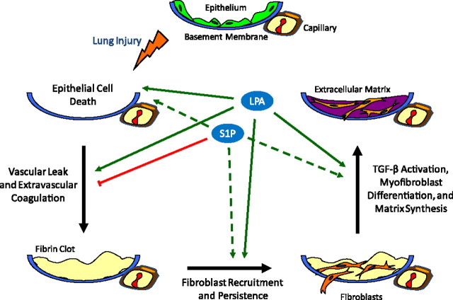
Aberrant wound healing responses to lung injury are believed to contribute to fibrotic lung diseases, such as idiopathic pulmonary fibrosis (IPF). The lysophospholipids lysophosphatidic acid (LPA) and sphingosine 1-phosphate (S1P), by virtue of their ability to mediate many basic cellular functions, including survival, proliferation, migration, and contraction, can influence many of the biological processes involved in wound healing. Accordingly, recent investigations indicate that LPA and S1P may play critical roles in regulating the development of lung fibrosis. Here we review the evidence indicating that LPA and S1P regulate pulmonary fibrosis and the potential mechanisms through which these lysophospholipids may influence fibrogenesis induced by lung injury.


Figures

Figure 1.
Figure 1.
Chemical structures of lysophosphatidic acid (LPA) and sphingosine 1-phosphate (S1P).
Figure 2.
Figure 2.
Lysophosphatidic acid (LPA) metabolism. LPA can be produced by several different pathways. Phospholipids such as phosphatidylcholine (PC) can be converted to lysophospholipids such as lysophosphatidylcholine (LPC) by members of the phospholipase A1 (PLA1) and phospholipase A2 (PLA2) families of enzymes. These lysophospholipids can be converted to LPA by the lysophospholipase D activity of autotaxin (ATX). Alternatively, LPA can be generated from phospholipids by the sequential actions of lecithin cholesterol acyltransferase (LCAT) and ATX. Last, PLA1 and PLA2 enzymes can produce LPA directly by hydrolysis of phosphatidic acid (PA). Several PLA1 and PLA2 enzymes have been implicated in extracellular LPA production, including mPA-PLA1α/Lipase H (LIPH), mPA-PLA1β, PS-PLA1, and sPLA2-IIA. LPA can also be degraded in several ways. Of these, hydrolysis of LPA to monoacylglycerol (MAG) by lipid phosphate phosphatases (LPP-1, LPP-2, and LPP-3) is likely the major pathway for clearance of LPA in vivo.
Figure 3.
Figure 3.
Sphingosine 1-phosphate (S1P) metabolism. Ceramide is considered to be the central molecule in sphingolipid metabolism. It can be generated de novo from serine and palmitoyl-CoA through several intermediate steps, or it can be formed from the catabolism of sphingomyelin and complex glycosphingolipids. Ceramide can be metabolized via several different pathways, one of which is the conversion by ceramidases to sphingosine, which can in turn be phosphorylated by sphingosine kinases (Sphk) to synthesize S1P. S1P can either be converted back to sphingosine by the action of S1P phosphatase or the lipid phosphate phosphatases, or it can be degraded by S1P lyase.
Figure 4.
Figure 4.
Lysophosphatidic acid (LPA) and sphingosine 1-phosphate (S1P) regulation of lung fibrosis. Schematic representation of the biological processes implicated in lung fibrosis that appear to be regulated by LPA and S1P. Evidence reviewed in this article indicates that LPA contributes to the development of fibrosis after lung injury through multiple mechanisms (solid green arrows), including the induction of: (1) epithelial cell apoptosis; (2) increased vascular permeability, resulting in increased intraalveolar coagulation; (3) fibroblast recruitment into the injured airspaces and their resistance to apoptosis; and (4) activation of latent TGF-β. Mechanisms 1 to 3 appear to be mediated by LPA signaling through LPA1, whereas mechanism 4 appears to be mediated by LPA signaling through LPA2. S1P may have both anti- and profibrotic effects in the lung. Evidence reviewed in this article indicates that S1P inhibits the development of pulmonary fibrosis through attenuation of lung vascular leak (red line). This mechanism appears to be mediated by S1P signaling through S1P1 expressed on endothelial cells. Potential profibrotic activities of S1P, which have not yet been investigated in the lung (dashed green arrows), include the induction of: (1) epithelial cell apoptosis, (2) fibroblast recruitment, and (3) myofibroblast differentiation.

References

    1. Ley B, Collard HR, King TE. Clinical course and prediction of survival in idiopathic pulmonary fibrosis. Am J Respir Crit Care Med 2010;183:431–440. - PubMed
    1. Selman M, Pardo A. Role of epithelial cells in idiopathic pulmonary fibrosis: from innocent targets to serial killers. Proc Am Thorac Soc 2006;3:364–372. - PubMed
    1. Gharaee-Kermani M, Hu B, Phan SH, Gyetko MR. Recent advances in molecular targets and treatment of idiopathic pulmonary fibrosis: focus on TGFbeta signaling and the myofibroblast. Curr Med Chem 2009;16:1400–1417. - PubMed
    1. Antoniou KM, Pataka A, Bouros D, Siafakas NM. Pathogenetic pathways and novel pharmacotherapeutic targets in idiopathic pulmonary fibrosis. Pulm Pharmacol Ther 2007;20:453–461. - PubMed
    1. Coward WR, Saini G, Jenkins G. The pathogenesis of idiopathic pulmonary fibrosis. Ther Adv Respir Dis 2010;4:367–388. - PubMed

Publication types

MeSH terms