. 2012 Oct;80(10):3721-32.
doi: 10.1128/IAI.00442-12. Epub 2012 Jul 16.

Abscess formation and alpha-hemolysin induced toxicity in a mouse model of Staphylococcus aureus peritoneal infection

Affiliations

Abscess formation and alpha-hemolysin induced toxicity in a mouse model of Staphylococcus aureus peritoneal infection

Sabine Rauch et al. Infect Immun. 2012 Oct.

Abstract

Staphylococcus aureus is a frequent cause of skin infection and sepsis in humans. Preclinical vaccine studies with S. aureus have used a mouse model with intraperitoneal challenge and survival determination as a measure for efficacy. To appreciate the selection of protective antigens in this model, we sought to characterize the pathological attributes of S. aureus infection in the peritoneal cavity. Testing C57BL/6J and BALB/c mice, >10(9) CFU of S. aureus Newman were needed to produce a lethal outcome in 90% of animals infected via intraperitoneal injection. Both necropsy and histopathology revealed the presence of intraperitoneal abscesses in the vicinity of inoculation sites. Abscesses were comprised of fibrin as well as collagen deposits and immune cells with staphylococci replicating at the center of these lesions. Animals that succumbed to challenge harbored staphylococci in abscess lesions and in blood. The establishment of lethal infections, but not the development of intraperitoneal abscesses, was dependent on S. aureus expression of alpha-hemolysin (Hla). Active immunization with nontoxigenic Hla(H35L) or passive immunization with neutralizing monoclonal antibodies protected mice against early lethal events associated with intraperitoneal S. aureus infection but did not affect the establishment of abscess lesions. These results characterize a mouse model for the study of intraperitoneal abscess formation by S. aureus, a disease that occurs frequently in humans undergoing continuous ambulatory peritoneal dialysis for end-stage renal disease.


Figures
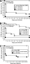
Fig 1
Fig 1
Challenge of mice by intraperitoneal injection of S. aureus. Cohorts of 6-week-old BALB/c (A) or C57BL/6 (B) mice (n = 20) were infected by intraperitoneal injection with the indicated doses of S. aureus Newman, and animal survival was recorded over 15 days. (C) Six-week-old BALB/c mice were infected by intraperitoneal injection with 9 × 10⁸ CFU of S. aureus Newman either with or without (mock) prior heat treatment at 55°C for 20 min. As determined by CFU enumeration on agar plates, heat treatment reduced colony formation by 97.5%. Animal survival was monitored over 15 days. Statistical significance was calculated by the log-rank test (P < 0.0001).
Fig 2
Fig 2
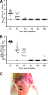
Fate of staphylococci in the peritoneal cavities of mice. Six-week-old BALB/c mice were infected by intraperitoneal injection with 5 × 10⁸ CFU of S. aureus Newman. (A) At timed intervals (3 h or 1, 2, 3, 6, or 10 days) after inoculation, blood from cohorts of mice (n = 7) was collected by cardiac puncture and plated on tryptic soy agar (TSA), and the staphylococcal load was enumerated as CFU. Black bars indicate means of observations. (B) At timed intervals (3 h or 1, 3, 6, 10, or 15 days) after inoculation, the peritoneal cavity of mice was rinsed with 1 ml of sterile PBS, and the staphylococcal load was enumerated as CFU on agar plates. (C) Animals that had been infected by intraperitoneal injection with 5 × 10⁸ CFU of S. aureus Newman were euthanized and necropsied, and peritoneal abscesses on the abdominal walls (black arrowheads) were excised and analyzed for histopathology or staphylococcal load (see subsequent figures).
Fig 3
Fig 3
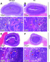
Staphylococcal lesions associated with the kidney. (A) Six-week-old BALB/c mice were infected by intraperitoneal injection with 5 × 10⁸ CFU of S. aureus Newman. At timed intervals, animals (n = 15) were euthanized and necropsied, and the kidneys were excised, fixed in formalin, thin sectioned, and analyzed for histopathology of H&E-stained tissues. The percentages of kidneys with surface abscesses or parenchymal abscesses were recorded. (B and C) Example of an H&E-stained surface abscess associated with the peritoneal lining of the kidney. (D and E) Example of an H&E-stained abscess positioned within the kidney parenchyma.
Fig 4
Fig 4
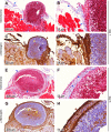
Fibrin and collagen deposits sequester staphylococci within the peritoneal cavity. Animals that had been infected via intraperitoneal injection with 5 × 10⁸ CFU of S. aureus Newman for 1 or 3 days were euthanized and necropsied, and peritoneal abscesses on the abdominal wall were excised and analyzed for histopathology. Thin-sectioned samples were either stained with Masson's trichrome (ABEF) or with antibody against mouse fibrinogen, followed by immunohistochemical detection (brown), and counterstained with H&E (CDGH). Arrowheads highlight the presence of collagen (blue), granulocytes (blue), staphylococci (yellow), and fibrin (brown).
Fig 5
Fig 5
Persistence of S. aureus lesions in the peritoneal cavity. Animals were infected with 5 × 10⁸ CFU of S. aureus Newman via intraperitoneal injection and were euthanized and necropsied on days 1, 3, 6, and 15. Peritoneal abscesses on the abdominal wall excised and analyzed for histopathology. Abdominal tissues were fixed in formalin, embedded, thin sectioned, and stained with H&E. Enlarged segments of the lesion include the outer layers (black box) and the staphylococcal community (red box). Arrowheads indicate granulocytes (blue), fibrin (red), staphylococci (yellow), pseudocapsule of staphylococcal abscess communities (white), and macrophages (green).
Fig 6
Fig 6
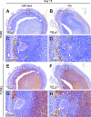
Macrophages and apoptotic immune cells in peritoneal abscesses caused by S. aureus. BALB/c mice were infected by intraperitoneal injection with 5 × 10⁸ CFU of S. aureus Newman wild-type or hla mutant bacteria. Animals were euthanized 15 days after infection and necropsied, and peritoneal abscesses on the abdominal wall were excised, fixed in formalin, thin sectioned, and processed for histopathology. (A to D) Murine macrophages in peritoneal abscesses were detected in serial thin sections with an antibody against F4/80. (E to H) Apoptotic immune cells were identified by using TUNEL. Arrowheads identify staphylococci (yellow), the pseudocapsule of staphylococcal abscess communities (white), macrophages (green), and apoptotic cells (red).
Fig 7
Fig 7
Staphylococcal genes required for the early lethal outcome associated with intraperitoneal S. aureus infections. (A) Cohorts of 6-week-old BALB/c mice (n = 20) were infected with 5 × 10⁸ CFU of S. aureus Newman (wild type) or mutants with insertional lesions in saeR, agrA, mgrA, or srtA. Animal survival was monitored over 15 days, and the statistical significance determined as follows: wild type versus saeR mutant, P < 0.0001; wild type versus agrA mutant, P < 0.0001; wild type versus mgrA mutant, P = 0.0018; and wild type versus srtA mutant, P = 0.071. (B) Cohorts of 6-week-old BALB/c mice (n = 20) were infected with 5 × 10⁸ CFU of S. aureus Newman (wild type), a mutant with an insertional lesion in hla, or the hla mutant with the complementing plasmid (phla). Animal survival was monitored over 15 days, and the statistical significance was determined as follows: wild type versus hla mutant, P < 0.0001; and hla mutant versus hla (phla) mutant, P = 0.1439 (C) Cohorts of 6-week-old BALB/c mice (n = 15) were infected with 5 × 10⁸ CFU of S. aureus MW2, Newman, or USA300 LAC. Animal survival was monitored over 15 days, and the statistical significance determined as follows: MW2 versus USA300 LAC strains, P < 0.0001; MW2 versus Newman strains, P = 0.0004; and Newman versus USA300 LAC strains, P = 0.1553. Statistical significance was determined by the log-rank test.
Fig 8
Fig 8
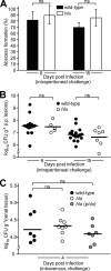
Alpha-hemolysin is dispensable for the formation of S. aureus abscess lesions in the peritoneal cavity or in kidney parenchyma. Cohorts of 6-week-old BALB/c mice (n = 20) were infected by intraperitoneal injection with 5 × 10⁸ CFU of S. aureus Newman wild type or hla mutant. Animals were euthanized 6 or 15 days after infection. Peritoneal abscess lesions on the abdominal walls were identified during necropsy and recorded as the percentages of animals presenting with lesions: wild type versus hla mutant, P = 0.7000 (day 6); and wild type versus hla mutant, P = 1.0 (day 15). (B) Peritoneal abscesses were excised from the abdominal wall, and staphylococcal load in the lesions was determined by plating homogenized tissue on agar plates and enumerating the CFU: wild type versus hla mutant, P = 0.7107 (day 6); and wild type versus hla mutant, P = 1.0 (day 15). (C) BALB/c mice were infected by intravenous injection of 107 CFU of S. aureus Newman into the periorbital plexus. Animals were euthanized 4 days after challenge, and the kidneys were removed during necropsy and either analyzed for abscess formation via histopathology or the staphylococcal load was determined by plating homogenized tissue on agar plates and enumerating the CFU: wild type versus hla mutant, P = 0.4009; and wild type versus hla mutant (phla), P = 0.7308. Differences in staphylococcal load were examined for statistical significance with the nonparametric Mann-Whitney test.
Fig 9
Fig 9
Histopathology of peritoneal abscesses caused by hla mutant S. aureus. BALB/c mice were infected by intraperitoneal injection with 5 × 10⁸ CFU of S. aureus Newman wild-type (A, C, E, and G) or hla mutant (B, D, F, and H) bacteria. Animals were euthanized either at 6 or 15 days after infection and necropsied, and peritoneal abscesses on the abdominal wall were excised, fixed in formalin, thin sectioned, and analyzed for the histopathology of H&E-stained tissues. Enlarged images highlight staphylococcal communities within each lesion (C, D, G, and H). Arrowheads identify staphylococci (yellow), fibrin (red), the pseudocapsule of staphylococcal abscess communities (white), and macrophages (green).
Fig 10
Fig 10
Hla-neutralizing antibodies cannot prevent the establishment of S. aureus abscesses in the peritoneal cavity. (A) Three-week-old BALB/c mice were immunized by intramuscular injection with either PBS (mock) or 20 μg of purified HlaH35L emulsified in complete Freund adjuvant. Animals were boosted with the same antigen dose emulsified in incomplete Freund adjuvant on day 12. Antibody titers were analyzed by ELISA on day 20: anti-HlaH35L in mock samples, <1:100; and anti-HlaH35L in HlaH35L immune samples, 1:2,500. On day 21, cohorts of animals (n = 15) were challenged by intraperitoneal injection of 5 × 10⁸ CFU of S. aureus Newman, and survival was monitored over 15 days. (B) Six-week-old BALB/c mice were immunized by intraperitoneal injection with either 5 mg kg−1 (body weight) of purified Hla-neutralizing MAb 7B8 (αHla) (48) or of mouse IgG2a isotype control (IgG2a). Four hours later, cohorts of animals (n = 15) were challenged by intraperitoneal injection of 5 × 10⁸ CFU of S. aureus Newman, and survival was monitored over 15 days. (C) BALB/c mice (n = 15) that had been immunized by intramuscular injection with either PBS (mock) or 20 μg of purified HlaH35L and then challenged by intraperitoneal injection of 5 × 10⁸ CFU of S. aureus Newman were euthanized 15 days after challenge. Peritoneal abscess lesions on the abdominal walls were identified during necropsy and recorded as the percentages of animals presenting with these lesions: wild type versus hla mutant, P = 1.0 (day 15). (D) BALB/c mice (n = 15) that had been immunized by intraperitoneal injection with 5 mg kg−1 of MAb 7B8 (αHla) or of mouse IgG2a isotype-control (IgG2a) were challenged by intraperitoneal injection of 5 × 10⁸ CFU of S. aureus Newman. Animals were euthanized 15 days after challenge. Peritoneal abscess lesions on the abdominal wall were identified during necropsy and recorded as the percentages of animals presenting with these lesions: wild type versus hla mutant, P = 1.0 (day 15).

References

    1. Baba T, Bae T, Schneewind O, Takeuchi F, Hiramatsu K. 2007. Genome sequence of Staphylococcus aureus strain Newman and comparative analysis of staphylococcal genomes. J. Bacteriol. 190:300–310 - PMC - PubMed
    1. Bae T, et al. 2004. Staphylococcus aureus virulence genes identified by bursa aurealis mutagenesis and nematode killing. Proc. Natl. Acad. Sci. U. S. A. 101:12312–12317 - PMC - PubMed
    1. Bagdasarian N, Heung M, Malani PN. 2012. Infectious complications of dialysis access devices. Infect. Dis. Clin. N. Am. 26:127–141 - PubMed
    1. Bernheimer AW. 1965. Staphylococcal alpha toxin. Ann. N. Y. Acad. Sci. 128:112–123 - PubMed
    1. Bubeck-Wardenburg J, Patel R, Schneewind O. 2007. Surface proteins and exotoxins are required for the pathogenesis of Staphylococcus aureus pneumonia. Infect. Immun. 74:1040–1044 - PMC - PubMed

Publication types

MeSH terms