. 2012 Nov;206(9):1473-8.
doi: 10.1093/infdis/jis464. Epub 2012 Jul 16.

New features of invasive candidiasis in humans: amyloid formation by fungi and deposition of serum amyloid P component by the host

Affiliations

New features of invasive candidiasis in humans: amyloid formation by fungi and deposition of serum amyloid P component by the host

Kevin B Gilchrist et al. J Infect Dis. 2012 Nov.

Abstract

Background: Invasive candidiasis occurs in the gastrointestinal tract, especially in neutropenic patients. We were interested in determining whether invasive fungi formed amyloid in humans as they are known to do in vitro. We also sought to characterize the consequence(s) of such amyloid formation.

Methods: Tissue from 25 autopsy patients with invasive candidiasis of the gastrointestinal tract was stained with amyloidophilic dyes and for the presence of serum amyloid P component (SAP). Confirmation of the interaction of SAP and Candida was demonstrated using Candida albicans and mutants for amyloid formation.

Results: Amyloid was present on the cellular surface of fungi invading gut tissue. Moreover, SAP bound to the fungal cell walls, confirming the presence of amyloid. In vitro observations showed SAP bound avidly to fungi when amyloid formed in fungal cell walls. An unexpected result was the lack of host neutrophils in response to the invading fungi, not only in neutropenic patients but also in patients with normal or increased white blood counts.

Conclusions: We report the first demonstration of functional fungal amyloid in human tissue and the binding of SAP to invading fungi. It is postulated that fungal amyloid, SAP, or a complex of the proteins may inhibit the neutrophil response.


Figures

Figure 1.
Figure 1.
Invasion of gastrointestinal tissue by Candida. A, Hematoxylin-eosin stain of intestinal tissue (×200). At the top of the figure, yeasts and filamentous forms of fungi staining reddish-blue (arrows) are present in an ulcerating lesion of the gastrointestinal mucosa with little inflammatory response. B, Gomori methenamine silver stain of intestinal tissue (×200). Yeast and filamentous forms of Candida stain black (arrows) and are seen within tissue, on the ulcerated mucosal tissue, and in the lumen of the gastrointestinal tract. C, Submucosal tissue of the gastrointestinal tract with invasive candidiasis stained for serum amyloid P component (×200). The protein is present on yeasts and filamentous forms of fungi, staining a reddish-brown (arrows).
Figure 2.
Figure 2.
Binding of serum amyloid P (SAP) component to yeast in vitro. SAP, 25 µg/ml, was incubated with (AD) C. albicans; (EH) S. cerevisiae expressing Als5p on its surface; or (IL) S. cerevisiae transformed with empty vector only. The cells were washed and incubated with fluorescent antibody to SAP component. A, D, E, H, I, and L are brightfield images, and B, C, F, G, J, and K are the respective corresponding fluorescence images. There was no fluorescence in preparations of C. albicans or Als5p-expressing S. cerevisiae when SAP or primary antibody was omitted (data not shown).
Figure 3.
Figure 3.
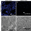
Thioflavin T staining of tissue samples. Infected gut tissue (A) and uninfected spleen sample (B) were stained with 100 nM thioflavin T and imaged via confocal microscopy. Lower panels are bright light without stain.
Figure 4.
Figure 4.
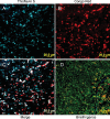
Amyloid dye staining and birefringence of infected gastrointestinal tissue. Tissue was costained with 0.1% thioflavin S (A) and 0.1% Congo red (B) and analyzed for birefringence. Part (C) shows the 2 stains merged. Note apple-green birefringence denoting amyloid in (D).

Comment in

References

    1. Otzen D, Nielsen PH. We seek them here, we find them there: functional bacterial amyloid. Cell Mol Life Sci. 2008;65:910–27. - PMC - PubMed
    1. Olsen A, Jonsson A, Normark S. Fibronectin binding mediated by a novel class of surface organelles on Eschereichia coli. Nature. 1989;338:652–5. - PubMed
    1. Dranginis A, Rauceo J, Coronado J, Lipke PN. A biochemical guide to yeast adhesins: glycoproteins for social and antisocial occasions. Microbiol Molec Biol Rev. 2007;71:282–94. - PMC - PubMed
    1. Finkel J, Mitchell A. Genetic control of Candida albicans biofilm development. Nat Rev Microbiol. 2011;9:109–18. - PMC - PubMed
    1. Otoo H, Lee K, Qiu W, Lipke PN. Candida albicans Als adhesins have conserved amyloid-forming sequences. Eukaryot Cell. 2007;7:776–82. - PMC - PubMed

MeSH terms

Substances