Review
. 2010 Dec 29:2010:bcr1020103385.
doi: 10.1136/bcr.10.2010.3385.

Duodenal adenocarcinoma arising from a pyloric gland adenoma with a brief review of the literature

Affiliations
Review

Duodenal adenocarcinoma arising from a pyloric gland adenoma with a brief review of the literature

T S Khor et al. BMJ Case Rep. .

Abstract

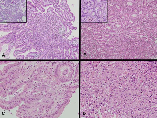
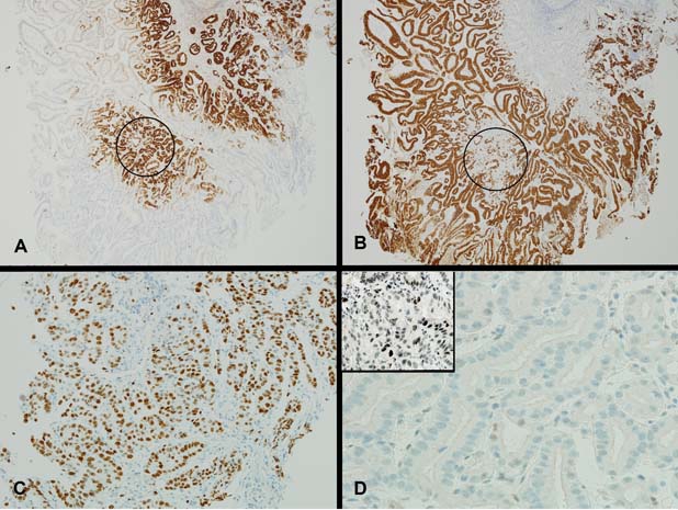
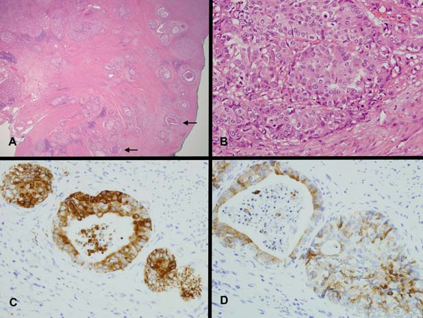
Pyloric gland-type adenoma of the duodenum with documented malignant progression is rare. A case is presented of an 87-year-old man with bloating and nausea, who on investigation was found to have a polyp on the anteroinferior wall of the duodenal cap. Histologic examination of the polyp showed features of a pyloric gland adenoma (PGA) demonstrating the full spectrum of progression from low- to high-grade dysplasia and finally invasive adenocarcinoma. The carcinoma showed gastric-type differentiation highlighted by its mucin immunohistochemistry profile and was of advanced stage with lymph node metastasis. The literature on PGAs and the little documentations on progression to carcinoma in duodenal PGAs are reviewed.


Conflict of interest statement

Competing interests None.

Figures

Figure 1
Figure 1
(A) Sections demonstrating a polypoid lesion arising among distorted duodenal villi composed of tubular glands lined by columnar epithelium with basal nuclei and pale to oeosinophilic ground glass cytoplasm (H&E ×100, inset PAS with Diastase, ×200). No apical mucin cap is demonstrated within the epithelium of the glands (inset). (B) Sections demonstrating abrupt transition of typical pyloric type glands to glands with low-grade dysplasia showing irregularity of gland outline with elongated, pseudostratified and mildly hyperchromatic nuclei (H&E ×100, inset H&E ×400). Higher magnification showing two glands in the centre with abrupt transition within the same gland from typical bland cytology of pyloric type epithelium to low-grade dysplasia with mildly hyperchromatic and pseudostratified nuclei (inset). (C) High-grade dysplasia with irregular papillary structures and fused irregular glands lined by epithelium showing enlarged nuclei with loss of polarity, nuclear pleomorphism and enlarged nucleoli (H&E ×400). Increasing numbers of mitotic figures are seen. (D) Intramucosal carcinoma with syncytial growth of back-to-back and fused poorly formed microglands and solid clusters of malignant cells with increasing nuclear pleomorphism, loss of polarity and nucleolar prominence (H&E ×400).
Figure 2
Figure 2
(A) There is strong expression of MUC6 in the glands of the pyloric gland adenoma (MUC6 ×40). In some areas of the polyp, the glands positive for MUC6 also show patchy co-expression of MUC5AC (circled area, compare with circled area in figure 2B). (B) There is expression of MUC5AC either restricted to surface epithelium with lack of staining within the glands of the pyloric gland adenoma or with patchy co-expression within the glands of the pyloric gland adenoma (circled area) (MUC5AC ×40). (C) A high proliferation index is shown within areas of high-grade dysplasia (MIB-1 ×200). (D) Typical glands of pyloric gland adenoma negative for p53 (p53 pyloric gland adenoma ×400, inset p53 pyloric gland adenoma high-grade dysplasia/intramucosal adenocarcinoma ×400). Area of high-grade dysplasia/intramucosal adenocarcinoma with moderate to strong nuclear staining for p53 (inset).
Figure 3
Figure 3
(A) Invasive adenocarcinoma (H&E ×20). Infiltrating adenocarcinoma with cribriform or cystically dilated glands, cords and solid nests. Some glands show central comedo-like necrosis (arrows). (B) The cells show vesicular nuclei with prominent nucleoli (H&E ×400). The cytoplasm is pale to oeosinophilic with a ground glass quality similar to the cells of the pyloric gland adenoma. (C) There is strong cytoplasmic staining for MUC6 in the cells of the adenocarcinoma (MUC6 ×400). (D) Patchy co-expression of MUC5AC is seen within cells of the adenocarcinoma (MUC5AC ×400).

References

    1. Chen ZM, Scudiere JR, Abraham SC, et al. Pyloric gland adenoma: an entity distinct from gastric foveolar type adenoma. Am J Surg Pathol 2009;33:186–93 - PubMed
    1. Vieth M, Kushima R, Borchard F, et al. Pyloric gland adenoma: a clinico-pathological analysis of 90 cases. Virchows Arch 2003;442:317–21 - PubMed
    1. Kushima R, Rüthlein HJ, Stolte M, et al. ‘Pyloric gland-type adenoma’ arising in heterotopic gastric mucosa of the duodenum, with dysplastic progression of the gastric type. Virchows Arch 1999;435:452–7 - PubMed
    1. Kushima R, Vieth M, Mukaisho K, et al. Pyloric gland adenoma arising in Barrett's esophagus with mucin immunohistochemical and molecular cytogenetic evaluation. Virchows Arch 2005;446:537–41 - PubMed
    1. Vieth M, Kushima R, de Jonge JP, et al. Adenoma with gastric differentiation (so-called pyloric gland adenoma) in a heterotopic gastric corpus mucosa in the rectum. Virchows Arch 2005;446:542–5 - PubMed