. 2012 Jul 15;26(14):1626-42.
doi: 10.1101/gad.191338.112.

Ptbp2 represses adult-specific splicing to regulate the generation of neuronal precursors in the embryonic brain

Affiliations

Ptbp2 represses adult-specific splicing to regulate the generation of neuronal precursors in the embryonic brain

Donny D Licatalosi et al. Genes Dev. .

Abstract

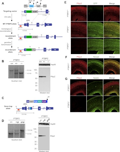
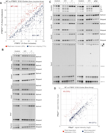
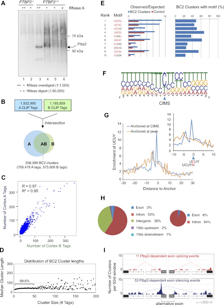
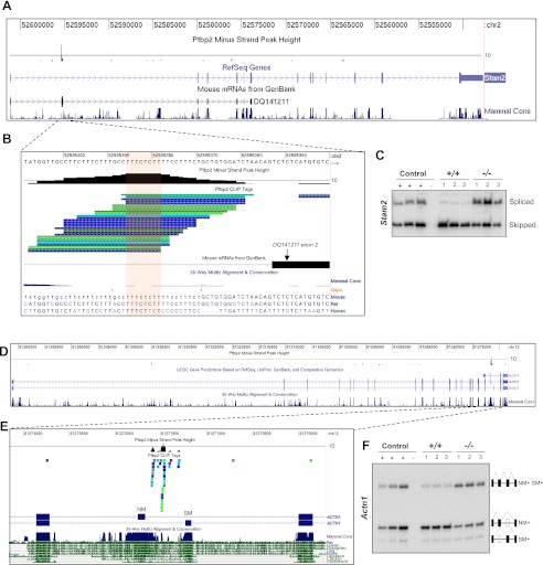
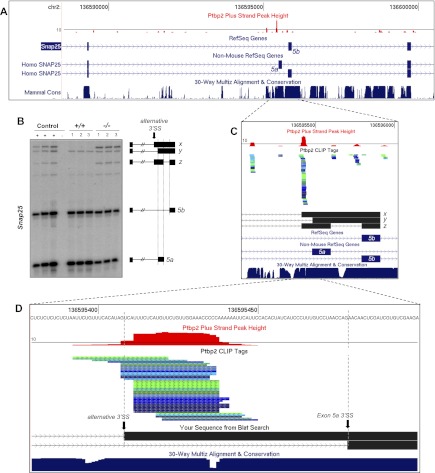
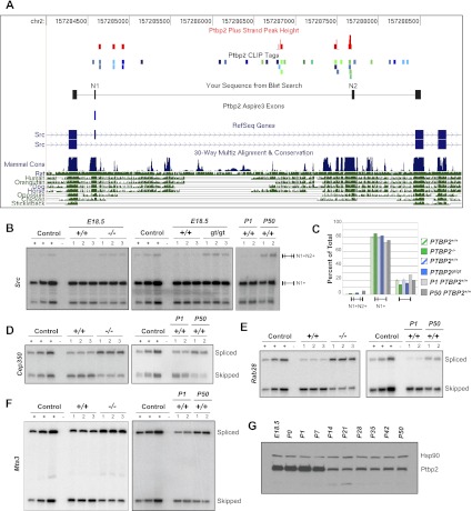
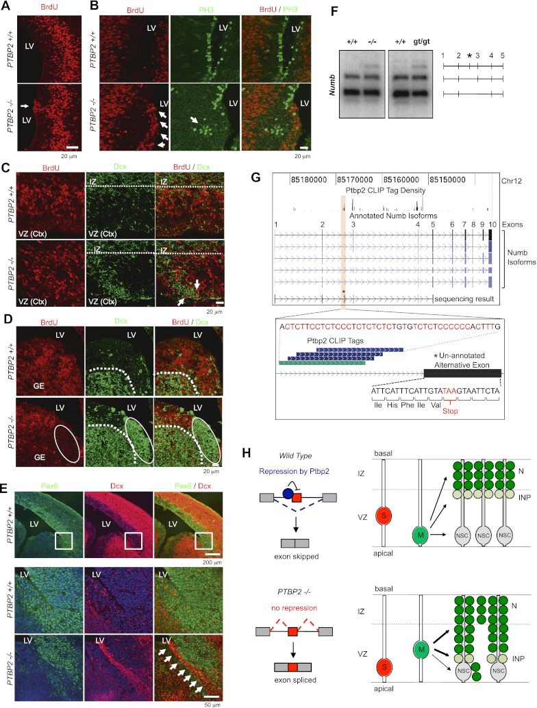
Two polypyrimidine tract RNA-binding proteins (PTBs), one near-ubiquitously expressed (Ptbp1) and another highly tissue-restricted (Ptbp2), regulate RNA in interrelated but incompletely understood ways. Ptbp1, a splicing regulator, is replaced in the brain and differentiated neuronal cell lines by Ptbp2. To define the roles of Ptbp2 in the nervous system, we generated two independent Ptbp2-null strains, unexpectedly revealing that Ptbp2 is expressed in neuronal progenitors and is essential for postnatal survival. A HITS-CLIP (high-throughput sequencing cross-linking immunoprecipitation)-generated map of reproducible Ptbp2-RNA interactions in the developing mouse neocortex, combined with results from splicing-sensitive microarrays, demonstrated that the major action of Ptbp2 is to inhibit adult-specific alternative exons by binding pyrimidine-rich sequences upstream of and/or within them. These regulated exons are present in mRNAs encoding proteins associated with control of cell fate, proliferation, and the actin cytoskeleton, suggesting a role for Ptbp2 in neurogenesis. Indeed, neuronal progenitors in the Ptbp2-null brain exhibited an aberrant polarity and were associated with regions of premature neurogenesis and reduced progenitor pools. Thus, Ptbp2 inhibition of a discrete set of adult neuronal exons underlies early brain development prior to neuronal differentiation and is essential for postnatal survival.


Figures

Figure 1.
Figure 1.
Ptbp2 is broadly expressed in the developing mouse brain, including neuronal precursors. (A) Gene targeting strategy to generate Ptbp2-null mice, including schematic of the targeting vector, wild-type (WT) allele, and recombinant allele before and after Cre-mediated recombination at loxP sites in the male germline. (B) Southern blot confirmation of genotypes (position of probe indicated) and Western blot analysis of animals derived from the gene targeting strategy (whole brain E18.5) with polyclonal anti-Ptbp2 antibody raised against full-length protein (Polydorides et al. 2000) and anti-Hsp90. The position of the Southern blot probe is indicated, as are NcoI sites (N) used to digest genomic tail DNA. (C) Schematic of the gene trap Ptbp2 allele from ES cell line NPX210, bearing an intronic cassette consisting of a splice acceptor (SA), neomycin resistance (neoR) and β-galactosidase (β-gal) genes, an internal ribosome entry site (IRES), and coding sequence for placental alkaline phosphatase (PLAP). (D) Southern blot and Western blot analysis of animals derived from gene trap ES cells, as in B. (E) Sections of Ptbp2 wild-type (+/+), heterozygous (+/−), and Ptbp2 knockout (KO [−/−]) E18.5 brain are stained with antibodies to Ptbp2 and GFP. Ptpb2 is nuclear, while GFP is cytoplasmic, and cells with red nuclei and green cytoplasm are evident in heterozygotes. (F) Costaining of Ptbp2 and Nova2 in wild-type E18.5 brains. (G) Costaining of Ptbp2 and Nestin in wild-type and knockout E18.5 brains.
Figure 2.
Figure 2.
Ptbp2-dependent alternative splicing in the brain. (A) Relative exon inclusion levels in wild-type (WT) and Ptbp2 knockout (KO) brains (ASPIRE3, P < 0.01, ΔIrank > 5). Red dots indicate alternative exons with higher inclusion in wild-type compared with knockout brains, and blue dots represent exons with increased inclusion in knockout compared with wild-type brains. (B) RT–PCR validation of seven ASPIRE candidates (additional candidates in are shown in the Supplemental Figures). In all cases, the control sample consists of an equal-parts mixture of wild-type and knockout cDNA amplified at different cycle numbers (see the Materials and Methods). (C) Comparison of alternative splicing in both Ptbp2-null strains (−/−, gene targeting; gt/gt, gene trap) (for additional examples, see Fig. 6). (D) Probeset signal intensities from MoEx 1.0ST analysis of wild-type and Ptbp2-null brains.
Figure 3.
Figure 3.
Ptbp2 HITS-CLIP. (A) Autoradiogram of nitrocellulose membrane containing protein–RNA complexes purified from Ptbp2-null (lanes 1,2) and wild-type (lanes 36) E18.5 mouse brains treated with a high (++) or low (+) concentration RNase A. (B) Summary of CLIP tag filtering and clustering to identify BC2 clusters. (C) Analysis of cortex A and B CLIP tag counts in each BC2 cluster. (D) Comparison of the length and numbers of CLIP tags for each BC2 cluster. (E) Top 10 tetramers present in BC2 sequences as determined by differences between the observed and expected frequency in BC2 compared with the shuffled control sequences. (F) CIMS-based identification of UCUY as a consensus binding site for Ptbp2. (G) Enrichment of UCUY near CIMS. (H) Genomic distribution of BC2 clusters (Ptbp2-binding events). (I) Distribution of BC2 clusters near alternative exons that are misspliced in Ptbp2-null brains. The total number (Y-axis) of BC2 clusters centered in each 50-nt window relative to alternative and flanking constitutive splice sites is shown for exons that exhibit increased skipping (top panel) and splicing (bottom panel) in Ptbp2 knockout compared with wild-type brains. Only BC2 clusters residing within the regions specified in the figure are displayed.
Figure 4.
Figure 4.
Ptbp2 binding upstream of alternative exons silences exon inclusion in vivo. Examples of high-density BC2 CLIP tag clusters predicting Ptbp2 action to repress exon inclusion. (A) Distribution of Ptbp2 CLIP tags to the Stam2 gene. Tag density is represented by a histogram where the black line indicates a CLIP tag density of 10 overlapping tags. (B) Zoom-in view of a high-density Ptbp2 CLIP cluster immediately upstream of the Ptbp2-dependent Stam2 alternative exon. (C) RT–PCR confirmation of aberrant (increased) splicing of Stam2 exon 2 in Ptbp2-null brains compared with wild-type littermate controls. (D) Distribution of Ptbp2 CLIP tags to the Actn1 gene. (E) Zoom-in view of Ptbp2 binding in the intron flanking the alternative NM and SM exons. (F) RT–PCR confirmation of a role for Ptbp2 in the regulation of Actn1 NM/SM splicing.
Figure 5.
Figure 5.
Ptbp2 represses aberrant alternative 3′ splice site selection. Ptbp2 silences alternative 3′ splice site utilization in Snap25 pre-mRNA. (A) Distribution of Ptbp2 CLIP tags in a region of Snap25 containing the alternative 5a and 5b exons. CLIP tag density is represented by a histogram where the black line indicates a CLIP tag density of 10 overlapping tags. (B) RT–PCR analysis of Snap25 5a/5b splicing in wild-type and Ptbp2-null brains. (C) Zoom-in of a region of Snap25 showing Ptbp2 CLIP tags and splice site utilization and intron retention identified from PCR products x, y, and z. (D) Zoom-in of C showing the position of the Ptbp2 CLIP cluster relative to the alternative 3′ splice site up-regulated in Ptbp2-null brains.
Figure 6.
Figure 6.
Ptbp2 inhibits developmentally regulated adult-enriched alternative exons. (A) Distribution of Ptbp2 binding in a region of the c-src gene containing the alternative N1 and N2 exons. Also shown is a BLAT search result following Sanger sequencing of the N1+N2+ isoform purified from acrylamide gels shown in B. (B) RT–PCR analysis of c-src splicing in both Ptbp2-null strains (and wild-type littermates, left and center panels) and in neonatal (P1) and adult (P50) wild-type brains (right panel). Control lanes correspond to cDNA mixtures amplified at different cycle numbers (see the Materials and Methods). (C) Quantification of c-src RT–PCR products from B. (DF) Analysis of Ptbp2-regulated exons in Cep350, Rab28, and Mta3 in wild-type and Ptbp2-null E18.5 brains (left) and wild-type neonatal and adult brains (right). (G) Western blot analysis of Ptbp2 and Hsp90 in wild-type brains at the indicated embryonic and postnatal days.
Figure 7.
Figure 7.
Ptbp2 deletion results in aberrant neural stem cell polarity and premature neurogenesis. Immunohistochemical analysis of E14.5 wild-type (+/+) and Ptbp2-null (−/−) brains using BrdU (A), BrdU and PH3 (B), BrdU and Dcx (C,D), and Pax6 and Dcx (E) antibodies. (LV) Lateral ventricle; (IZ) intermediate zone; (VZ) ventricular zone; (GE) ganglionic eminence. (F) RT–PCR analysis of Numb pre-mRNA splicing in wild-type (+/+) and Ptbp2-null (−/−, gt/gt) brains to monitor splicing of an alternative exon (exon 3, using primers corresponding to exons 1 [forward] and 5 [reverse]) indicates expression of aberrantly processed Numb mRNA in both Ptbp2-null strains at E18.5. The identity of RT–PCR products is shown on the right, including an unannotated isoform (top band) containing an additional exon (asterisk, confirmed by Sanger sequencing). (G, top panel) Genome browser view of Ptbp2 CLIP tag density along the Numb gene, including annotated alternative mRNA isoforms and the unannotated isoform identified by sequencing RT–PCR products. (Bottom panel) Zoom-in view of a 79-nt region of Numb showing the unannotated Ptbp2-repressed alternative exon and a BC2 CLIP cluster centered ∼20 nt upstream of the 3′ splice site. (H) Model summarizing a role for Ptbp2 in exon repression and the consequences to neural stem cell polarity and cell cycle. The results of a HITS-CLIP analysis of Ptbp2 targets and the morphology of Ptbp2−/− mice suggest a model in which Ptbp2 normally acts on a regulatory cascade in neural progenitors cycling between S and M phase and differentiating into neural stem cells (NSCs) and indirect neural pathway (INP) progenitors in the ventricular zone (VZ). (Top panel) Ptbp2's immediate action is primarily to repress inclusion of alternative exons; in aggregate, these mediate changes in proteins (cell cycle, cell cytoskeleton, and cell fate) that inhibit aberrant cell cycle entry in periventricular progenitors. For example, Ptbp2 may inhibit “activating” exons in transcripts encoding cell cycle proteins, restrict cytoskeletal proteins to those that align the mitotic spindle orthogonal to the ventricular surface, and inhibit the function of regulators of basal cell division. (Bottom panel) In Ptbp2−/− mice brains, neural progenitors show increased cell cycle entry in the ventricular zone (A–D) and increased indirect neural pathway progenitors (E) that may result from skewed mitotic spindle orientation and/or actions on regulators of basal cell cycle.

References

    1. Barash Y, Calarco JA, Gao W, Pan Q, Wang X, Shai O, Blencowe BJ, Frey BJ 2010. Deciphering the splicing code. Nature 465: 53–59 - PubMed
    1. Bark C, Bellinger FP, Kaushal A, Mathews JR, Partridge LD, Wilson MC 2004. Developmentally regulated switch in alternatively spliced SNAP-25 isoforms alters facilitation of synaptic transmission. J Neurosci 24: 8796–8805 - PMC - PubMed
    1. Baye LM, Link BA 2008. Nuclear migration during retinal development. Brain Res 1192: 29–36 - PMC - PubMed
    1. Boutz PL, Stoilov P, Li Q, Lin CH, Chawla G, Ostrow K, Shiue L, Ares MJ, Black DL 2007. A post-transcriptional regulatory switch in polypyrimidine tract-binding proteins reprograms alternative splicing in developing neurons. Genes Dev 21: 1636–1652 - PMC - PubMed
    1. Brocke L, Srinivasan M, Schulman H 1995. Developmental and regional expression of multifunctional Ca2+/calmodulin-dependent protein kinase isoforms in rat brain. J Neurosci 15: 6797–6808 - PMC - PubMed

Publication types

MeSH terms

Substances