

A distinct de novo expression of Nav1.5 sodium channels in human atrial fibroblasts differentiated into myofibroblasts
- PMID: 22802584
- PMCID: PMC3473287
- DOI: 10.1113/jphysiol.2012.233593
A distinct de novo expression of Nav1.5 sodium channels in human atrial fibroblasts differentiated into myofibroblasts
Abstract
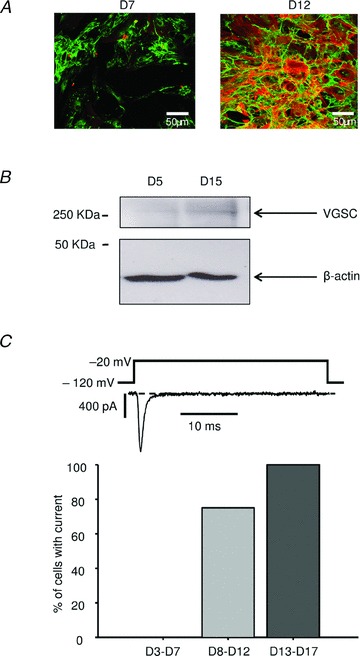
Fibroblasts play a major role in heart physiology. They are at the origin of the extracellular matrix renewal and production of various paracrine and autocrine factors. In pathological conditions, fibroblasts proliferate, migrate and differentiate into myofibroblasts leading to cardiac fibrosis. This differentiated status is associated with changes in expression profile leading to neo-expression of proteins such as ionic channels. The present study investigates further electrophysiological changes associated with fibroblast differentiation focusing on the activity of voltage-gated sodium channels in human atrial fibroblasts and myofibroblasts. Using the patch clamp technique we show that human atrial myofibroblasts display a fast inward voltage gated sodium current with a density of 13.28 ± 2.88 pA pF(-1) whereas no current was detectable in non-differentiated fibroblasts. Quantitative RT-PCR reveals a large amount of transcripts encoding the Na(v)1.5 α-subunit with a fourfold increased expression level in myofibroblasts when compared to fibroblasts. Accordingly, half of the current was blocked by 1 μm of tetrodotoxin and immunocytochemistry experiments reveal the presence of Na(v)1.5 proteins. Overall, this current exhibits similar biophysical characteristics to sodium currents found in cardiac myocytes except for the window current that is enlarged for potentials between -100 and -20 mV. Since fibrosis is one of the fundamental mechanisms implicated in atrial fibrillation, it is of great interest to investigate how this current could influence myofibroblast properties. Moreover, since several Na(v)1.5 mutations are related to cardiac pathologies, this study offers a new avenue on the fibroblasts involvement of these mutations.
Figures


Comment in
-
Voltage-gated Na⁺ channels: novel players in fibroblast-to-myofibroblast transition with a potential role in atrial arrhythmogenesis?J Physiol. 2012 Oct 15;590(20):4975. doi: 10.1113/jphysiol.2012.241455. J Physiol. 2012. PMID: 23082023 Free PMC article. No abstract available.
References
-
- Allessie M, Ausma J, Schotten U. Electrical, contractile and structural remodeling during atrial fibrillation. Cardiovasc Res. 2002;54:230–246. - PubMed
-
- Amin AS, Tan HL, Wilde AA. Cardiac ion channels in health and disease. Heart Rhythm. 2010;7:117–126. - PubMed
-
- Ancey C, Corbi P, Froger J, Delwail A, Wijdenes J, Gascan H, Potreau D, Lecron JC. Secretion of IL-6, IL-11 and LIF by human cardiomyocytes in primary culture. Cytokine. 2002;18:199–205. - PubMed
-
- Andrikopoulos P, Fraser SP, Patterson L, Ahmad Z, Burcu H, Ottaviani D, Diss JK, Box C, Eccles SA, Djamgoz MB. Angiogenic functions of voltage-gated Na+ channels in human endothelial cells: modulation of vascular endothelial growth factor (VEGF) signaling. J Biol Chem. 2011;286:16846–16860. - PMC - PubMed
-
- Armstrong CM, Hille B. Voltage-gated ion channels and electrical excitability. Neuron. 1998;20:371–380. - PubMed
Publication types
MeSH terms
Substances
LinkOut - more resources
Full Text Sources
Other Literature Sources
Miscellaneous