. 2012 Jul 31;109(31):12485-90.
doi: 10.1073/pnas.1207821109. Epub 2012 Jul 16.

Dopamine receptor D3 regulates endocytic sorting by a Prazosin-sensitive interaction with the coatomer COPI

Affiliations

Dopamine receptor D3 regulates endocytic sorting by a Prazosin-sensitive interaction with the coatomer COPI

Xin Zhang et al. Proc Natl Acad Sci U S A. .

Abstract

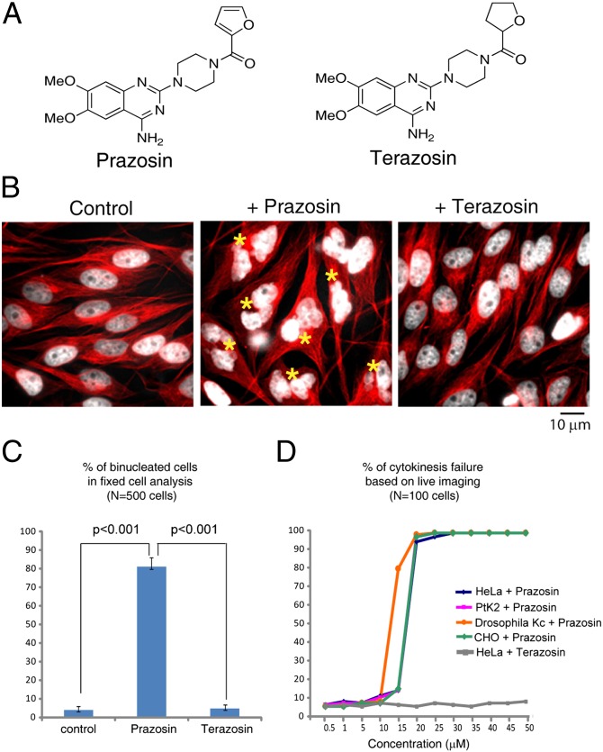
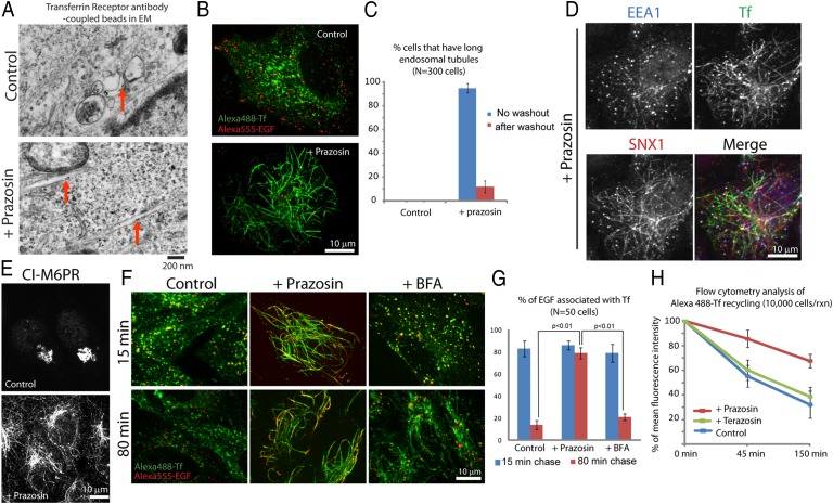
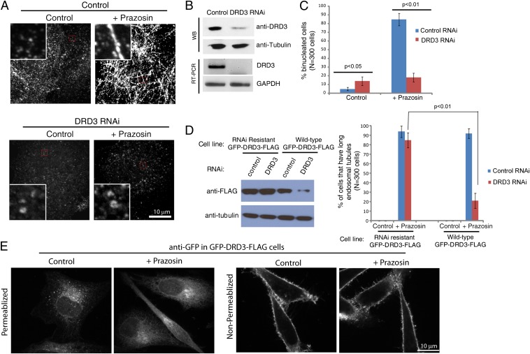
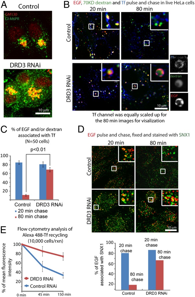
Macromolecules enter cells by endocytosis and are sorted to different cellular destinations in early/sorting endosomes. The mechanism and regulation of sorting are poorly understood, although transitions between vesicular and tubular endosomes are important. We found that the antihypertensive drug Prazosin inhibits endocytic sorting by an off-target perturbation of the G protein-coupled receptor dopamine receptor D(3) (DRD3). Prazosin is also a potent cytokinesis inhibitor, likely as a consequence of its effects on endosomes. Prazosin stabilizes a normally transient interaction between DRD3 and the coatomer COPI, a complex involved in membrane transport, and shifts endosomal morphology entirely to tubules, disrupting cargo sorting. RNAi depletion of DRD3 alone also inhibits endocytic sorting, indicating a noncanonical role for a G protein-coupled receptor. Prazosin is a powerful tool for rapid and reversible perturbation of endocytic dynamics.


Conflict of interest statement

The authors declare no conflict of interest.

Figures

Fig. 1.
Fig. 1.
Prazosin inhibits cytokinesis. (A) Chemical structures of Prazosin and its inactive analog Terazosin. (B) Fixed cell analysis shows that Prazosin induces cytokinesis failure and binucleated cell formation, whereas Terazosin does not. HeLa cells were incubated with DMSO, 20 μM Prazosin, or Terazosin for 36 h before being fixed and stained for microtubules (red) and DNA (white). Binucleated cells are indicated by an asterisk. (Scale bar: 10 μm.) (C) Quantification of B. (D) Quantification of cytokinesis failure in different cell lines after Prazosin treatment shows that Prazosin-induced cytokinesis inhibition is conserved. Terazosin-treated HeLa cells are also included (SI Appendix, SI Materials and Methods).
Fig. 2.
Fig. 2.
Prazosin induces endosomal tubulation and inhibits sorting. (A) Electron micrographs of Prazosin-induced transferrin receptor positive tubules. Transferrin receptor antibody-coupled gold beads were added to HeLa cells for 10 min before control DMSO, or 30 μM Prazosin was added for 1 additional h. Red arrows show gold beads in vesicles or tubules. (Scale bar: 200 nm.) (B) Prazosin-treated HeLa cells exhibit robust transferrin receptor-positive endosomal tubules, whereas lysosomes are not affected. Alexa488-Transferrin and Alexa555-EGF were added to cells for 45 min. Then, cells were chased in the presence of DMSO or 30 μM Prazosin for 30 min in marker-free medium to allow EGF to reach the lysosomes and clear from earlier pathways. (C) Quantification of the endosomal tubules in Prazosin-treated cells and washout cells using fixed cell analysis. Cells were treated with DMSO or 30 μM Prazosin for 30 min followed by washing out with drug-free medium for 30 min. Endosomal tubules longer than 5 μm are classified as long endosomal tubules. (D) Prazosin induces an array of endosomal tubules that contain transferrin (and transferrin receptor and Rab11) (SI Appendix, Fig. S2B), SNX1 (and SNX2), and EEA1 (and Rab5) (SI Appendix, Fig. S2B). Representative micrographs show transferrin, SNX1, and EEA1 localization in a Prazosin-treated HeLa cell. Alexa 488-Transferrin (green) was added to HeLa cells for 10 min before 30 μM Prazosin was added for an additional 1 h. Cells were washed and fixed before staining with anti-SNX1 (red) and anti-EEA1 (blue) antibodies. (E) Prazosin-treated HeLa cells form tubules that contain CI-M6PR. (F) Endosomal sorting is disrupted in endosomal tubules induced by Prazosin but not Brefeldin A. Pulse and chase experiments in HeLa cells show that both transferrin and EGF are trapped in endosomal tubules during sorting. HeLa cells were treated with DMSO control, 30 μM Prazosin, or 10 μg/mL BFA for 1 h. Then, Alexa 488-Transferrin and Alexa 555-EGF were added for 10 min. Images were taken at indicated times after washing out with marker- and phenol red-free medium. (G) Quantification of results in F. (H) Flow cytometry analysis of transferrin recycling in control, 30 μM Prazosin, or Terazosin-treated HeLa cells.
Fig. 3.
Fig. 3.
DRD3 is involved in endocytic sorting and mediates the effects of Prazosin. (A) DRD3 RNAi prevents Prazosin-induced endosomal tubule formation. Transferrin receptor staining is shown. HeLa cells were treated with control or DRD3 siRNA for 3 d before DMSO or 30 μM Prazosin was added for 1 h. (B) Western blot and semiquantitative RT-PCR show the expression and knockdown of DRD3 in HeLa cells. (C) DRD3 RNAi causes an increase in binucleated cells and prevents strong cytokinesis inhibition induced by Prazosin. (D) Western blots show the specificity and efficiency of DRD3 RNAi knockdown. Cells with long endosomal tubules (longer than 5 μm) were counted in three independent experiments. (E) The localization of DRD3. GFP-DRD3-FLAG-HeLa cells were treated with control DMSO or 30 μM Prazosin for 1 h before being fixed in PBS + formaldehyde. Cells were processed with anti-GFP antibody staining using two different conditions. Left shows cells that were permeabilized using 0.1% Triton X-100, and Right shows unpermeablized cells. Live GFP-DRD3-FLAG-HeLa cells show similar localizations to the anti-GFP staining in permeablized cells, indicating that the membrane population of DRD3 is relatively low compared with its intracellular population, which is similar to transferrin receptor. The commercially available anti-DRD3 antibody shows highly unspecific staining and is not suitable for immunofluorescence. (Scale bar: 10 μm.)
Fig. 4.
Fig. 4.
DRD3 is involved in endocytic sorting. (A) DRD3 RNAi causes increased CI-M6PR localization to vesicles in addition to the Golgi. Control or DRD3 RNAi-treated cells were stained for CI-M6PR (green) and a resident Golgi marker GM130 (red). (B) Pulse and chase experiments show sorting defects in DRD3 RNAi cells. HeLa cells were treated with control or DRD3 RNAi for 3 d before Alexa 555-EGF, FITC-70 kD-dextran, and Alexa 647-Transferrin were added for 10 min followed by washing out with marker- and phenol red-free medium. Pictures were taken at indicated time points in live cells. (C) Quantification of EGF and/or dextran that colocalize or partially colocalize with transferrin; (D) Endocytic cargoes are trapped in SNX1-positive endosomes after DRD3 RNAi. HeLa cells were treated with control or DRD3 RNAi for 3 d before Alexa 555-EGF (red) was added for 10 min followed by washing out with marker-free medium for indicated time points. Cells were than washed, fixed, and stained with anti-SNX1 (green). EGF that colocalizes or partially colocalizes with SNX1 was quantified. For each condition, around 50 cells were counted. Mean values from two independent experiments are shown. (Scale bar: 10 μm.) (E) Flow cytometry analysis of transferrin recycling in control or DRD3 RNAi knockdown HeLa cells.
Fig. 5.
Fig. 5.
The COPI complex interacts with DRD3 in the presence of Prazosin and is involved in its effects on endocytosis. (A) Pull-down experiments using anti-FLAG antibody in GFP-DRD3-FLAG-HeLa cells treated with DMSO, Prazosin, or Terazosin show that the interaction of DRD3 with COPI subunits COPB, COPC, and COPG is increased after Prazosin treatment. (B) COPB, COPD, and COPG localizations are disrupted by Prazosin treatment. HeLa cells were treated with DMSO or 30 μM Prazosin for 1 h before fixing and staining with COP antibodies. (C) COPB1 or COPB2 RNAi prevents endosomal tubule formation in Prazosin-treated HeLa cells. Transferrin receptor staining is shown. HeLa cells were treated with siRNAs for 3 d before DMSO or 30 μM Prazosin was added for 1 h. (D) Pulse and chase experiments show sorting defects in COPB RNAi cells. HeLa cells were treated with control or COPB1 + COPB2 RNAi for 3 d before Alexa 488-Tf and FITC-70 kD-dextran were added for 10 min followed by washing out with marker- and phenol red-free medium. Individual COPB1 or COPB2 RNAi resulted in similar phenotypes. Pictures were taken at indicated time points without fixing the cells. (Scale bar: 10 μm.) (E) Flow cytometry analysis of transferrin recycling in control or COPB1 or COPB2 RNAi knockdown HeLa cells.

Similar articles

Cited by

References

    1. Lappano R, Maggiolini M. G protein-coupled receptors: Novel targets for drug discovery in cancer. Nat Rev Drug Discov. 2011;10:47–60. - PubMed
    1. Rosenbaum DM, Rasmussen SG, Kobilka BK. The structure and function of G-protein-coupled receptors. Nature. 2009;459:356–363. - PMC - PubMed
    1. Platta HW, Stenmark H. Endocytosis and signaling. Curr Opin Cell Biol. 2011;23:393–403. - PubMed
    1. Hanyaloglu AC, von Zastrow M. Regulation of GPCRs by endocytic membrane trafficking and its potential implications. Annu Rev Pharmacol Toxicol. 2008;48:537–568. - PubMed
    1. Cavalli V, Corti M, Gruenberg J. Endocytosis and signaling cascades: A close encounter. FEBS Lett. 2001;498:190–196. - PubMed

Publication types

MeSH terms

LinkOut - more resources