. 2012 Jul 31;109(31):12698-703.
doi: 10.1073/pnas.1207219109. Epub 2012 Jul 16.

Unexpected role for MHC II-peptide complexes in shaping CD8 T-cell expansion and differentiation in vivo

Affiliations

Unexpected role for MHC II-peptide complexes in shaping CD8 T-cell expansion and differentiation in vivo

Jeong-su Do et al. Proc Natl Acad Sci U S A. .

Abstract

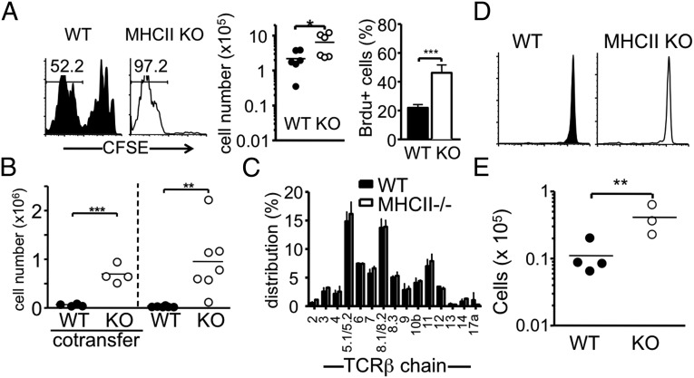
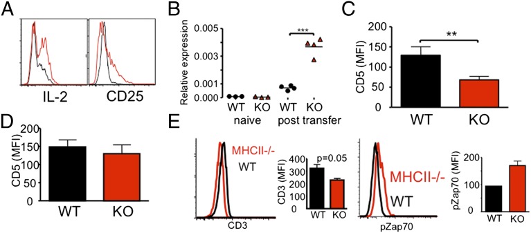
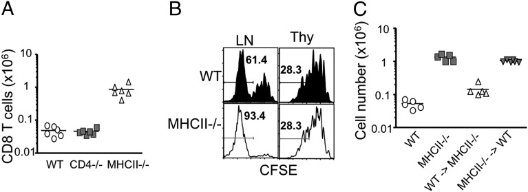
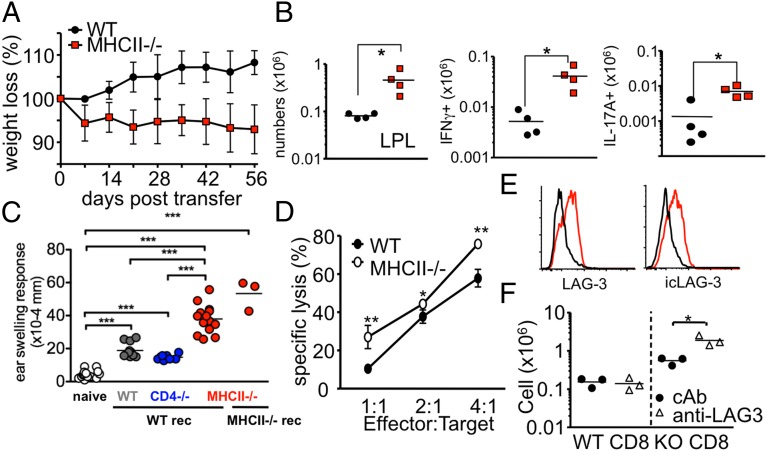
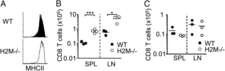
Here we report a unique role for MHC II-peptide complexes in controlling immune responses of naïve CD8 T cells. Compared with CD8 T cells from WT mice, CD8 T cells isolated from MHC II(-/-) mice hyperproliferated under lymphopenic conditions, differentiated into effector cells producing proinflammatory cytokines, and mediated more severe tissue inflammation. The elevated responses of MHC II(-/-) CD8 T cells were due to the absence of MHC II, but not CD4, T cells. The hyperreactivity appeared to be a feature of mature T cells, given its absence in CD8 single positive thymocytes derived from MHC II(-/-) mice. Expression of the MHC II ligand LAG3 was markedly enhanced during in vivo activation of MHC II(-/-) CD8 T cells, and blockade of MHC II-LAG3 interactions further enhanced T-cell expansion. Importantly, CD8 T cells isolated from H-2M(-/-) mice expressing WT levels of MHC II also displayed hyperresponsiveness similar to that of MHC II(-/-) CD8 T cells, suggesting that peptides presented on MHC II are involved in the control of CD8 T-cell responses. Our results uncover a previously undefined MHC II-dependent regulation that tunes CD8 T-cell reactivity and may have implications for an improved understanding of CD8 T-cell homeostasis and functions.


Conflict of interest statement

The authors declare no conflict of interest.

Figures

Fig. 1.
Fig. 1.
Expansion of MHCII−/− CD8 T cells in different lymphopenic recipients. (A) CFSE-labeled 1 × 10⁶ naive Thy1.1 WT or MHC II−/−CD8 T cells were transferred into groups of Rag1−/− recipients. CFSE profiles, absolute numbers, and BrdU incorporation of Thy1.1+ cells in the pLN were examined at 7 d after the transfer. The results shown are representative of six individually tested recipients. (B) FACS-sorted naïve Ly5.1 WT and Thy1.1 MHC II−/− CD8 T cells (5 × 10⁵ cells per recipient) were cotransferred or transferred separately into TCR- β/δ−/− recipients. The absolute numbers of donor cells in the pLN were examined at 7 d after the transfer. Each symbol represents an individually tested recipient. (C) Distribution of TCR- Vβ expression was compared between WT and MHC II−/− naïve CD8 T cells. Data shown are the mean ± SD of individually tested mice (n = 3). (D) CFSE-labeled 1 × 10⁶ naive Thy1.2 WT or Thy1.2 MHC II−/− CD8 T cells were transferred into Thy1.1 WT recipients. The CFSE profile was examined at 7 d after the transfer. (E) WT or MHCII−/− CD8 T cells were transferred into MHC II−/− Rag−/− recipients. The results shown are representative of three individually tested recipients. *P < 0.05; **P < 0.01; ***P < 0.001.
Fig. 2.
Fig. 2.
Activation phenotypes of MHC II−/− CD8 T cells in vivo. FACS-sorted naive Thy1.1 WT or MHC II−/− CD8 T cells were transferred into TCR-β−/− recipients. (A and B) CD25 and intracellular IL-2 expression (A) and CD8 T-cell IL-2 mRNA expression (B) were measured at 7 d after the transfer. Results shown are representative of between three and six individually tested recipients. (C and D) Surface CD5 expression was examined at 7 d posttransfer (C) or before transfer (D). Graphs show mean ± SD values of between four and six individually tested mice from two independent experiments. (E) Surface CD3ε expression on WT and MHC II−/− CD8 T cells was examined at 16 h posttransfer. The graph shows the mean ± SD of three individually tested mice. **P < 0.01; ***P < 0.001.
Fig. 3.
Fig. 3.
MHC II deficiency is responsible for the enhanced response of MHC II−/− CD8 T cells. (A) 1 × 10⁶ naive Thy1.2 WT, CD4−/−, and MHC II−/− CD8 T cells were transferred into Rag−/− recipients. CFSE profiles and the absolute numbers of donor T cells in the pLN were examined at 7 d after the transfer. The results shown are representative of six individually tested recipients in two independent experiments. (B) Thy1.1 WT or MHC II−/− thymic CD8+CD4CD44 or peripheral CD8+44− cells were transferred into Rag−/− recipients (1 × 10⁶ per recipient). CFSE profiles of the donor cells in the pLN were examined at 7 d after transfer. Similar results were observed from two independent experiments. (C) Lethally irradiated WT and MHC II−/− mice received BM cells from MHC II−/− and WT mice, respectively. After 6 wk of BM reconstitution, naive CD8 T cells were sorted and transferred into TCR-β−/− recipients. Donor cell recovery in the pLN was analyzed at 7 d after the transfer. The graph shows the mean ± SD of two independent experiments.
Fig. 4.
Fig. 4.
MHC II−/− CD8 T cells induced severe colitis and CHS. (A and B) 1 × 10⁶ naive Thy1.1 WT and MHC II KO CD8 T cells were transferred into TCR-β−/− recipients. (A) Body weight was monitored weekly and is presented as percentage of the initial weight at day 0. (B) Lamina propria cells were isolated at 5 wk posttransfer, and cytokine expression was analyzed by intracellular cytokine staining. Total IFN-γ– and IL-17A–producing Thy1.1 CD8 T cells were enumerated. *P < 0.05. (C) WT, CD4−/−, and MHC II−/− mice were sensitized with DNFB as described in Materials and Methods. Draining LN CD8 T cells were isolated on day 5. Then 1 × 10⁷ cells were transferred into naïve WT or MHC II−/− recipients, and the recipients were subsequently challenged on each side of both ears with DNFB. After 16 h, ear swelling was measured. Each symbol represents an individually tested mouse. (D) WT 2C or MHC II−/− 2C TCR Tg CD8 T cells were transferred into Rag−/− mice and then reisolated from the recipients at 7 d posttransfer. An ex vivo killing assay was performed using differentially CFSE-labeled target or control cells. (E) Surface and intracellular LAG-3 expression of WT (black) and MHC II−/− (red) was measured at 7 d posttransfer into Rag−/− mice. (F) WT and MHC II−/− CD8 T cells were transferred into TCR-βδ−/− mice that had been injected with rat IgG or anti–LAG-3 mAb.
Fig. 5.
Fig. 5.
H2M−/− naïve CD8 T cells expanded better than WT CD8 T cells. (A) Expression of MHC II molecule of WT and H2M−/− splenic CD11c+ dendritic cells was measured by FACS analysis. The results shown are representative of two or three individually tested recipients. (B) CFSE-labeled 1 × 10⁶ naive Thy1.1 WT or Thy1.1 H2M−/− CD8 T cells were transferred into Rag1−/− recipients, and the absolute numbers of donor T cells were counted at 7 d after the transfer. The results shown are representative of three or four individually tested recipients. (C) CFSE-labeled CD8 SP thymocytes from H2M−/− mice were transferred into Rag−/− recipients. Absolute numbers in the pLN were examined at 7 d posttransfer. *P < 0.05; ***P < 0.001.

References

    1. Murali-Krishna K, et al. Persistence of memory CD8 T cells in MHC class I-deficient mice. Science. 1999;286:1377–1381. - PubMed
    1. Tanchot C, Lemonnier FA, Pérarnau B, Freitas AA, Rocha B. Differential requirements for survival and proliferation of CD8 naïve or memory T cells. Science. 1997;276:2057–2062. - PubMed
    1. Swain SL, Hu H, Huston G. Class II-independent generation of CD4 memory T cells from effectors. Science. 1999;286:1381–1383. - PubMed
    1. Dorfman JR, Stefanová I, Yasutomo K, Germain RN. CD4+ T cell survival is not directly linked to self-MHC–induced TCR signaling. Nat Immunol. 2000;1:329–335. - PubMed
    1. Clarke SR, Rudensky AY. Survival and homeostatic proliferation of naive peripheral CD4+ T cells in the absence of self-peptide:MHC complexes. J Immunol. 2000;165:2458–2464. - PubMed

Publication types

MeSH terms

Substances

LinkOut - more resources