. 2012 Jul 31;109(31):12710-5.
doi: 10.1073/pnas.1008203109. Epub 2012 Jul 16.

Serine/threonine acetylation of TGFβ-activated kinase (TAK1) by Yersinia pestis YopJ inhibits innate immune signaling

Affiliations

Serine/threonine acetylation of TGFβ-activated kinase (TAK1) by Yersinia pestis YopJ inhibits innate immune signaling

Nicholas Paquette et al. Proc Natl Acad Sci U S A. .

Abstract

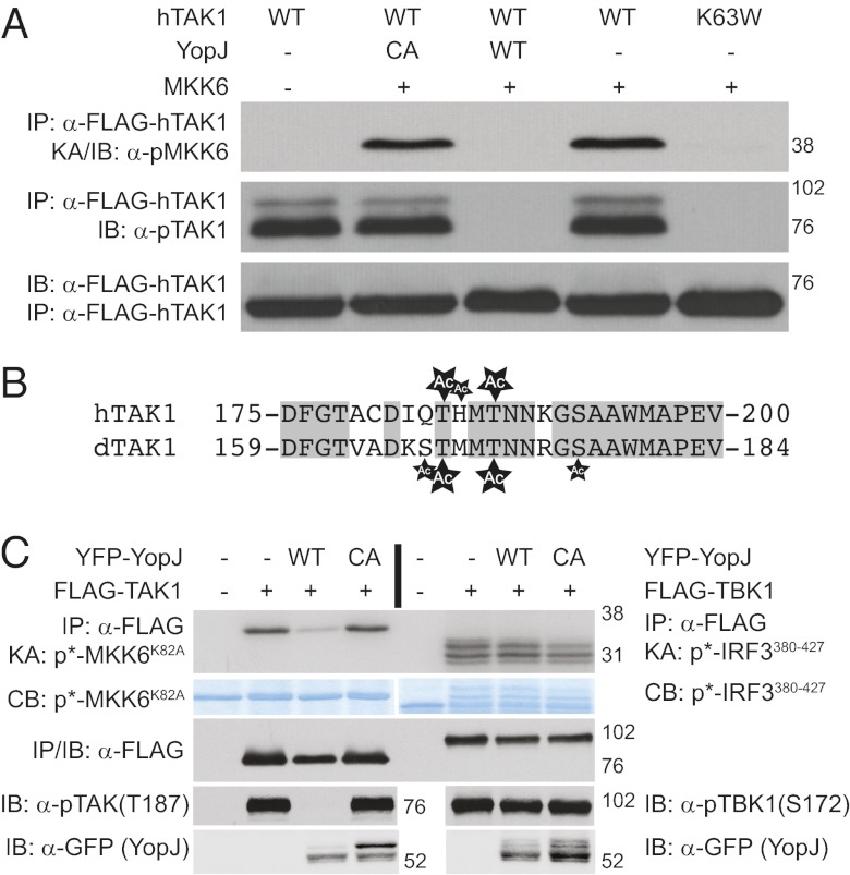
The Gram-negative bacteria Yersinia pestis, causative agent of plague, is extremely virulent. One mechanism contributing to Y. pestis virulence is the presence of a type-three secretion system, which injects effector proteins, Yops, directly into immune cells of the infected host. One of these Yop proteins, YopJ, is proapoptotic and inhibits mammalian NF-κB and MAP-kinase signal transduction pathways. Although the molecular mechanism remained elusive for some time, recent work has shown that YopJ acts as a serine/threonine acetyl-transferase targeting MAP2 kinases. Using Drosophila as a model system, we find that YopJ inhibits one innate immune NF-κB signaling pathway (IMD) but not the other (Toll). In fact, we show YopJ mediated serine/threonine acetylation and inhibition of dTAK1, the critical MAP3 kinase in the IMD pathway. Acetylation of critical serine/threonine residues in the activation loop of Drosophila TAK1 blocks phosphorylation of the protein and subsequent kinase activation. In addition, studies in mammalian cells show similar modification and inhibition of hTAK1. These data present evidence that TAK1 is a target for YopJ-mediated inhibition.


Conflict of interest statement

The authors declare no conflict of interest.

Figures

Fig. 1.
Fig. 1.
YopJ inhibits Drosophila IMD but not Toll immune signaling and functions between IMD and JNK. (A) S2* cells stably expressing YopJWT or YopJCA under control of the metallothionein promoter were pretreated with copper (to activate expression of YopJ) or not, before stimulation with Spätzle (Left) or DAP-type PGN (Right). Activation of immune signaling was monitored by Northern blotting for Diptericin and Drosomycin RNA. (B) Expression of YopJWT or YopJCA was induced with the addition of copper sulfate in S2* cells before stimulation with PGN. Ubiquitination of IMD was monitored by IMD immunoprecipitation followed by immunoblotting for ubiquitin (Upper). Anti-IMD blotting was used as a loading control and also to examine PGN-induced IMD cleavage. Anti-FLAG probing was used to verify the presence/absence of FLAG-YopJ. formula image marks unmodified full-length IMD, formula image highlights phosphorylated IMD, and formula image marks the cleaved-IMD products. (C) Phospho-JNK was monitored in whole-cell lysates. Full-length JNK blot serves as a loading control. (D) IKK activity was monitored in S2* cells expressing YopJWT or YopJCA after stimulation with PGN. IKKγ blot serves as an immunoprecipitation control.
Fig. 2.
Fig. 2.
YopJ inhibits Drosophila TAK1 kinase activity. (A) dTAK1 was overexpressed in S2* cells alone or in the presence of YopJWT or YopJCA, and activity was monitored by IP-kinase assays with catalytically inactive MKK6 serving as a substrate (row 1). As a control, immunoprecipitated FLAG-TAK1 were immunoblotted with anti-FLAG to verify kinase capture and banding pattern (row 2). To more clearly observe YopJ-mediated alteration in the dTAK1 banding pattern, FLAG-TAK1 was immunobloted directly from lysates under optimized conditions (row 3). IKK activity was similarly monitored by immunoprecipitation using an endogenous IKKγ antisera and the substrate Relish (row 4). Immuoprecipitated IKKγ samples were immunoblotted with IKKγ antisera to verify capture (row 5). The presense of FLAG-YopJ was also monitored by FLAG IP/immunoblot (row 6). (B) Anti-FLAG (TAK1) immunoblot of lysates from S2* cells expressing FLAG-TAK1 alone or in the presence of YopJWT or YopJCA were treated (or not) with λ-phosphatase. (C) Activation of endogenous dTAK1 was monitored by IP-kinase assays, with rMKK6 as substrate, from S2* cells expressing either YopJWT or YopJCA. Anti-FLAG immunoblot serves as a control for the presence of YopJ.
Fig. 3.
Fig. 3.
YopJ acetylates Drosophila TAK1. (A) Alignment of human and Drosophila TAK1 activation loops. The established phosphorylation sites on mammalian TAK1 are indicated (P). (B) Summary of tandem MS identification of Drosophila TAK1 activation loop. Phosphorylated (P) and acetylated (Ac) residues are indicated. All phospho-peptide and acetyl-peptide residues can be found in Tables S1S3. In both A and B, conserved residues are shaded in gray. (C) Activity of dTAK1 alanine substitutions in the presence or absence of YopJ assayed by IP/cold kinase assay (row 1). Immunoblots for FLAG-TAK1 and FLAG-YopJ verify the presence of the respective proteins (rows 2 and 3). (D) Activity of dTAK1 glutamic acid substitutions by IP/cold kinase assay [IP/KA-followed by immunoblot for phospho-MKK6 (S207)] (row 1). FLAG immunoblot verifies the expression of dTAK1 protein (row 2).
Fig. 4.
Fig. 4.
YopJ inhibits and acetylates mammalian kinases. (A) Wild-type or K63W hTAK1 was transiently expressed in human HEK 293T cells in the presence or absence of YFP-tagged YopJ. hTAK1 activity was monitored by cold IP-kinase assay (row 1). Phospho- and total hTAK1 were also monitored by IP/immunoblot (rows 2 and 3). (B) Alignment of hTAK1 and dTAK1 activation loops. Conserved residues are boxed (gray). Acetylated residues, as detected by MS/MS, are marked (Ac). Small Ac indicates residues that showed ambiguous acetylation. (C) hTAK1 (Left) or TBK1 (Right) were expressed in human HEK 293T cells in the presence or absence of wild-type (WT) or inactive (CA) YopJ. Kinase activities of hTAK1 and TBK1 were assayed by IP kinase assay and with phosphospecific hTAK1 or TBK1 immunoblotting. Total substrate amounts were detected by Coomassie blue staining; note the shift in IRF3 protein upon phosphorylation. As controls, total immunoprecipitated protein was detected by FLAG immunoblot, whereas YopJ levels were confirmed by GFP immunoblot.
Fig. 5.
Fig. 5.
YopJ inhibits MAP2 and MAP3 kinases. A model of YopJ-mediated inhibition, from our work and others, in both the Drosophila IMD (Left) and mammalian TNF/TLR (Right) signaling pathways. Proteins known to be acetylated by YopJ are marked with ‘Ac’ (red).

Similar articles

Cited by

References

    1. Monack DM, Mecsas J, Ghori N, Falkow S. Yersinia signals macrophages to undergo apoptosis and YopJ is necessary for this cell death. Proc Natl Acad Sci USA. 1997;94:10385–10390. - PMC - PubMed
    1. Monack DM, Mecsas J, Bouley D, Falkow S. Yersinia-induced apoptosis in vivo aids in the establishment of a systemic infection of mice. J Exp Med. 1998;188:2127–2137. - PMC - PubMed
    1. Palmer LE, Hobbie S, Galán JE, Bliska JB. YopJ of Yersinia pseudotuberculosis is required for the inhibition of macrophage TNF-alpha production and downregulation of the MAP kinases p38 and JNK. Mol Microbiol. 1998;27:953–965. - PubMed
    1. Palmer LE, Pancetti AR, Greenberg S, Bliska JB. YopJ of Yersinia spp. is sufficient to cause downregulation of multiple mitogen-activated protein kinases in eukaryotic cells. Infect Immun. 1999;67:708–716. - PMC - PubMed
    1. Schesser K, et al. The yopJ locus is required for Yersinia-mediated inhibition of NF-kappaB activation and cytokine expression: YopJ contains a eukaryotic SH2-like domain that is essential for its repressive activity. Mol Microbiol. 1998;28:1067–1079. - PubMed

Publication types

MeSH terms

LinkOut - more resources