. 2012 Jul 31;109(31):12716-21.
doi: 10.1073/pnas.1203468109. Epub 2012 Jul 16.

Cross-dressed CD8α+/CD103+ dendritic cells prime CD8+ T cells following vaccination

Affiliations

Cross-dressed CD8α+/CD103+ dendritic cells prime CD8+ T cells following vaccination

Lijin Li et al. Proc Natl Acad Sci U S A. .

Abstract

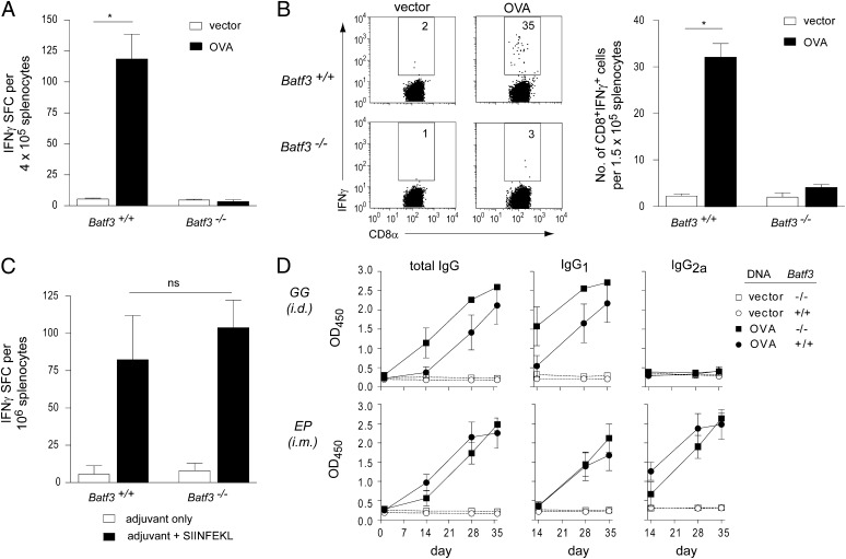
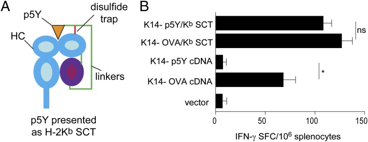
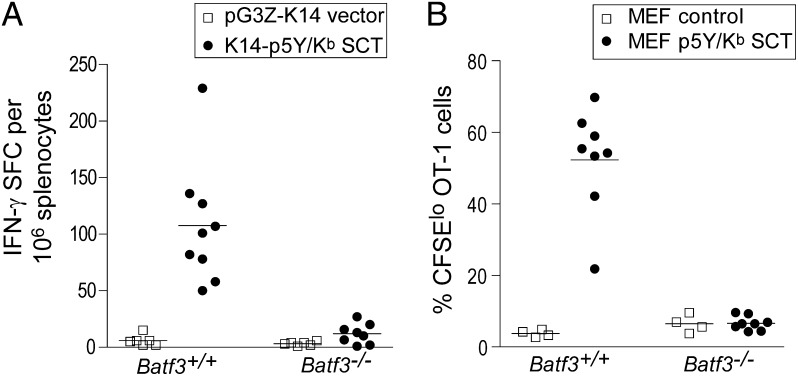
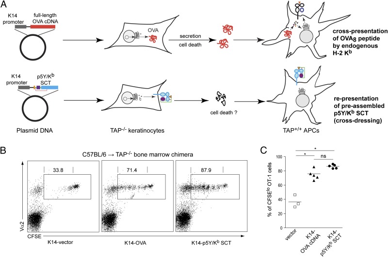
Activation of naïve cluster of differentiation (CD)8(+) cytotoxic T lymphocytes (CTLs) is a tightly regulated process, and specific dendritic cell (DC) subsets are typically required to activate naive CTLs. Potential pathways for antigen presentation leading to CD8(+) T-cell priming include direct presentation, cross-presentation, and cross-dressing. To distinguish between these pathways, we designed single-chain trimer (SCT) peptide-MHC class I complexes that can be recognized as intact molecules but cannot deliver antigen to MHC through conventional antigen processing. We demonstrate that cross-dressing is a robust pathway of antigen presentation following vaccination, capable of efficiently activating both naïve and memory CD8(+) T cells and requires CD8α(+)/CD103(+) DCs. Significantly, immune responses induced exclusively by cross-dressing were as strong as those induced exclusively through cross-presentation. Thus, cross-dressing is an important pathway of antigen presentation, with important implications for the study of CD8(+) T-cell responses to viral infection, tumors, and vaccines.


Conflict of interest statement

The authors declare no conflict of interest.

Figures

Fig. 1.
Fig. 1.
CD8+ T-cell priming is selectively ablated in Batf3−/− mice following DNA vaccination. (A and B) WT and Batf3/- mice (n = 5 per group) were immunized with OVA plasmid DNA. CD8+ T-cell responses were measured by IFN-γ enzyme-linked immunosorbent spot assay (ELISPOT) and intracellular cytokine staining. The results are representative of at least three independent experiments. (C) WT and Batf3−/− mice were vaccinated with SIINFEKL peptide and adjuvant or adjuvant alone. CD8+ T-cell responses were measured by IFN-γ ELISPOT. (D) WT and Batf3−/− mice were vaccinated with OVA plasmid DNA. Sera were collected before and after vaccination. OVA-specific total IgG, IgG1, and IgG2a were measured by ELISA, and the levels were equivalent between WT and Batf3−/− mice. *P < 0.01; ns, not significant; SFC, spot forming cells.
Fig. 2.
Fig. 2.
Molecular engineering of SCT peptide–MHC complexes precludes cross-presentation. (A) Schematic of the p5Y/Kb SCT, integrating the p5Y peptide, β2m, and H2-Kb heavy chain (HC). Of note, the disulfide bond (red line) formed between linker 1 and the α1 domain traps the low-affinity p5Y epitope in the peptide binding groove. (B) C57BL/6 mice (n = 4 per group) were vaccinated with the indicated DNA constructs. Kb/SIINFEKL-specific immune responses were measured by ELISPOT. Data are presented as means ± SEM. *P < 0.01; ns, not significant. Similar results were obtained in at least three independent experiments.
Fig. 3.
Fig. 3.
Engineered SCT vaccines prime CD8+ T-cell responses in vivo by cross-dressing. (A) WT or Batf3−/− mice were vaccinated with K14-p5Y/Kb SCT DNA or control DNA by gene gun. CD8+ T-cell responses were measured by IFN-γ ELISPOT. (B) A total of 2 × 10⁶ CFSE-labeled OT-1 cells were adoptively transferred into WT or Batf3−/− mice. Twenty-four hours later, 5 × 10⁶ necrotic fibroblasts were injected (s.c.). Ninety hours later, draining lymph nodes were harvested, and OT-1 cell proliferation was determined by flow cytometry. The percentage of OT-1 cells (identified as CD8+CD45.1+) that divided at least once (CFSElo) is shown. The results are representative of three independent experiments. Each data point represents an individual animal. (Bars indicate mean values for each experimental group.)
Fig. 4.
Fig. 4.
Cross-dressing is a robust antigen-presentation pathway in vivo. CFSE-labeled OT-1 cells were adoptively transferred into the C57BL/6→TAP−/− BM chimeras, which were subsequently vaccinated with K14-OVA or K14-p5Y/Kb SCT. (A) This experimental model system results in CD8+ T-cell priming by two distinct, nonoverlapping antigen-presentation pathways. (B) Spleen cells from vaccinated mice were collected and analyzed by flow cytometry. Representative two-color dot plots show expression of CFSE and Vα2 by viable CD8+ T cells. Gated regions represent OT-1 cells (expressing CFSE, CD8, and Vα2). Numbers indicate the percentage of CFSElo OT-1 cells that had gone through at least one cell division. (C) Summary of data from individual animals (n = 3–5 per group). *P < 0.01; ns, not significant.

References

    1. Kurts C, Robinson BW, Knolle PA. Cross-priming in health and disease. Nat Rev Immunol. 2010;10:403–414. - PubMed
    1. Dolan BP, Gibbs KD, Jr, Ostrand-Rosenberg S. Dendritic cells cross-dressed with peptide MHC class I complexes prime CD8+ T cells. J Immunol. 2006;177:6018–6024. - PubMed
    1. Dolan BP, Gibbs KD, Jr, Ostrand-Rosenberg S. Tumor-specific CD4+ T cells are activated by “cross-dressed” dendritic cells presenting peptide-MHC class II complexes acquired from cell-based cancer vaccines. J Immunol. 2006;176:1447–1455. - PubMed
    1. Wakim LM, Bevan MJ. Cross-dressed dendritic cells drive memory CD8+ T-cell activation after viral infection. Nature. 2011;471:629–632. - PMC - PubMed
    1. Hildner K, et al. Batf3 deficiency reveals a critical role for CD8alpha+ dendritic cells in cytotoxic T cell immunity. Science. 2008;322:1097–1100. - PMC - PubMed

Publication types

MeSH terms