. 2012 Jul 31;109(31):12568-73.
doi: 10.1073/pnas.1204390109. Epub 2012 Jul 16.

E-cadherin is under constitutive actomyosin-generated tension that is increased at cell-cell contacts upon externally applied stretch

Affiliations

E-cadherin is under constitutive actomyosin-generated tension that is increased at cell-cell contacts upon externally applied stretch

Nicolas Borghi et al. Proc Natl Acad Sci U S A. .

Erratum in

  • Proc Natl Acad Sci U S A. 2012 Nov 13;109(46):19034

Abstract

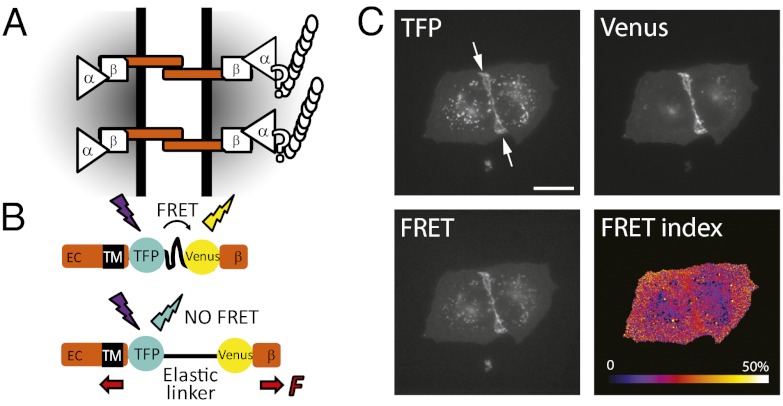
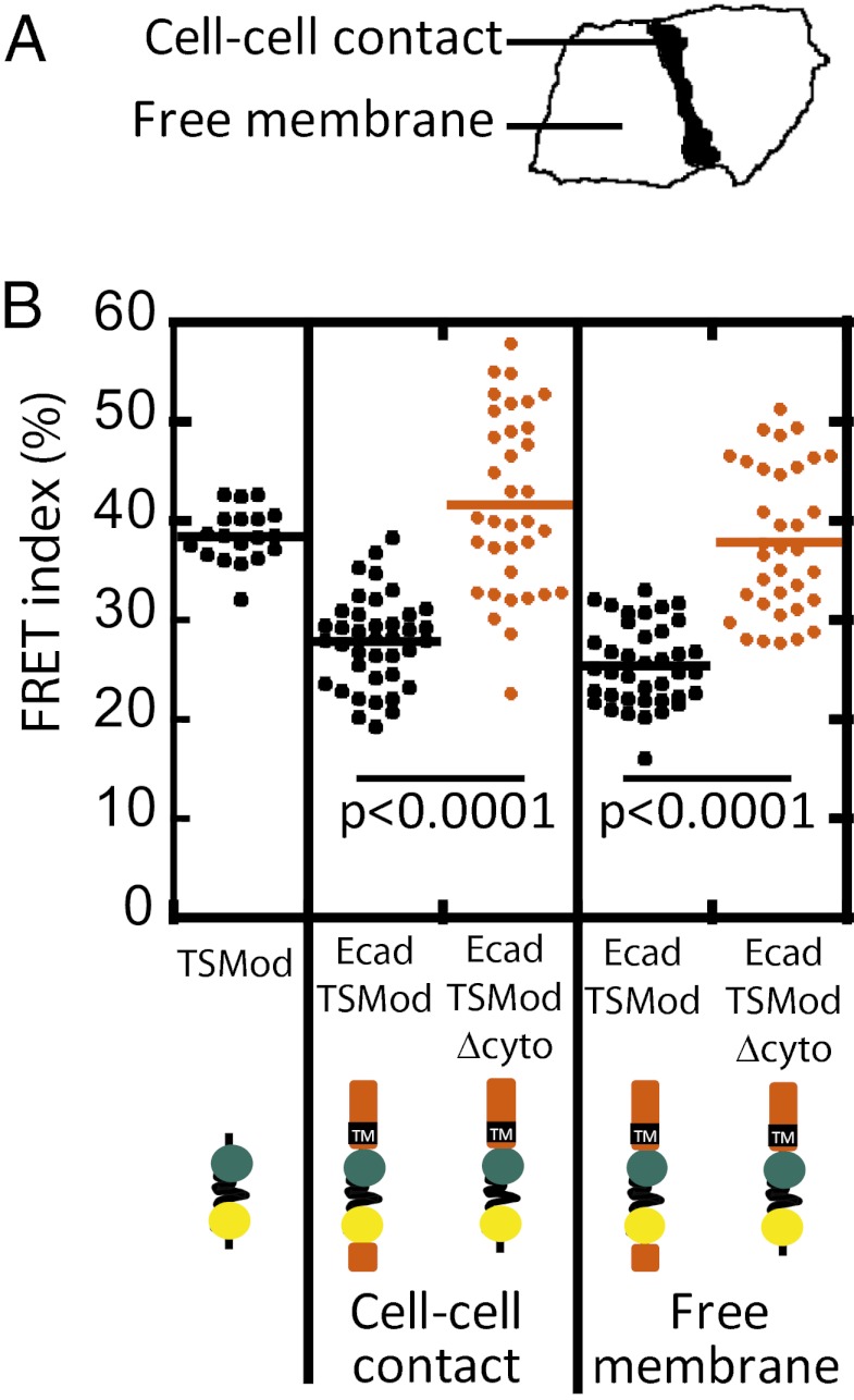
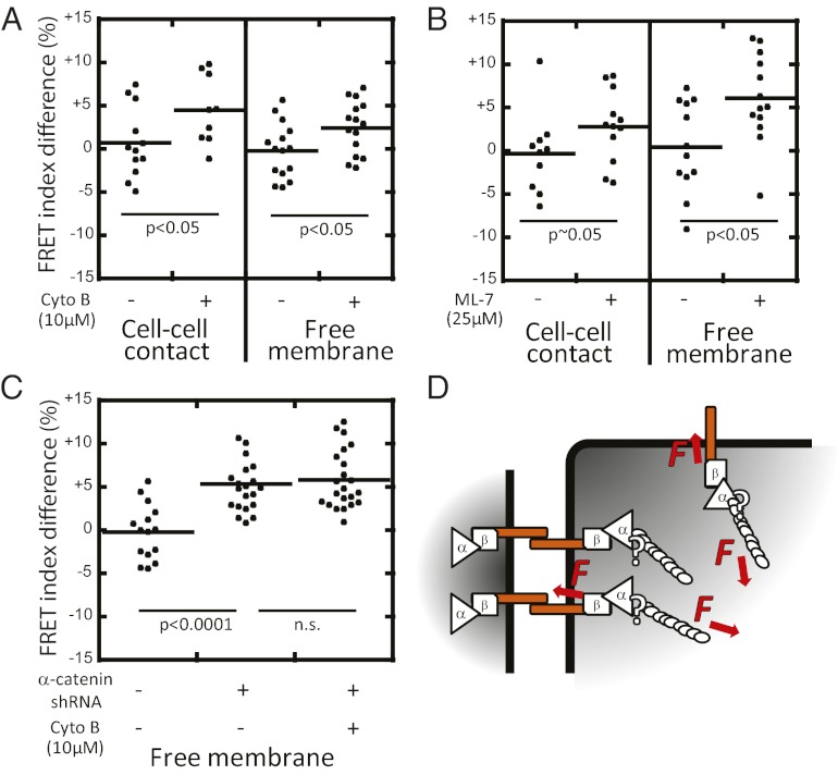
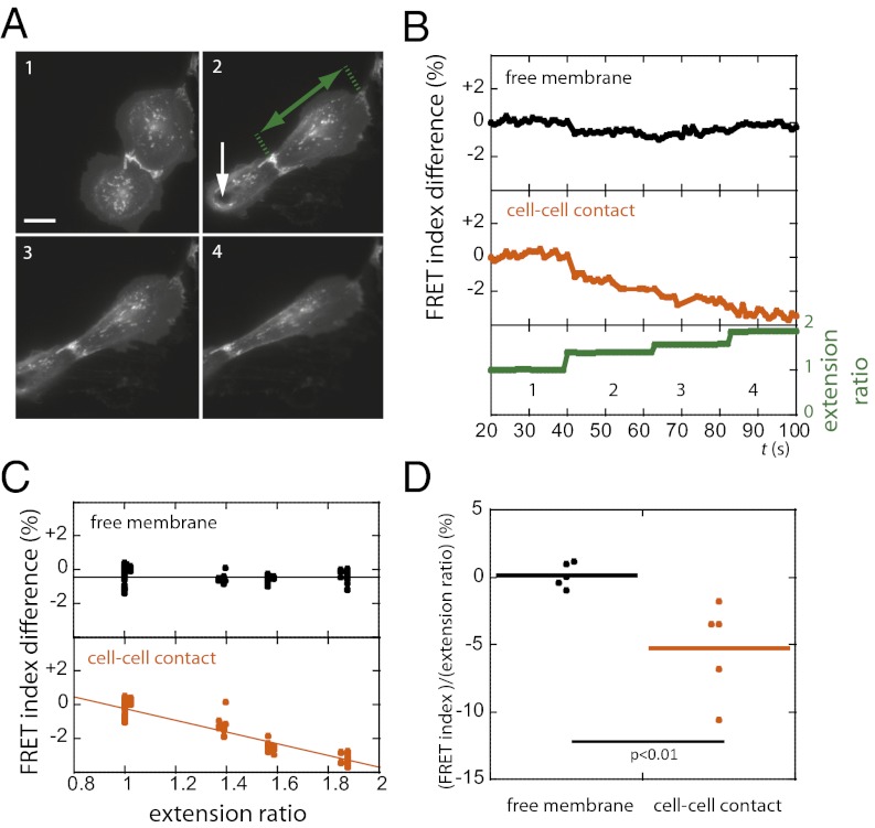
Classical cadherins are transmembrane proteins at the core of intercellular adhesion complexes in cohesive metazoan tissues. The extracellular domain of classical cadherins forms intercellular bonds with cadherins on neighboring cells, whereas the cytoplasmic domain recruits catenins, which in turn associate with additional cytoskeleton binding and regulatory proteins. Cadherin/catenin complexes are hypothesized to play a role in the transduction of mechanical forces that shape cells and tissues during development, regeneration, and disease. Whether mechanical forces are transduced directly through cadherins is unknown. To address this question, we used a Förster resonance energy transfer (FRET)-based molecular tension sensor to test the origin and magnitude of tensile forces transmitted through the cytoplasmic domain of E-cadherin in epithelial cells. We show that the actomyosin cytoskeleton exerts pN-tensile force on E-cadherin, and that this tension requires the catenin-binding domain of E-cadherin and αE-catenin. Surprisingly, the actomyosin cytoskeleton constitutively exerts tension on E-cadherin at the plasma membrane regardless of whether or not E-cadherin is recruited to cell-cell contacts, although tension is further increased at cell-cell contacts when adhering cells are stretched. Our findings thus point to a constitutive role of E-cadherin in transducing mechanical forces between the actomyosin cytoskeleton and the plasma membrane, not only at cell-cell junctions but throughout the cell surface.


Conflict of interest statement

The authors declare no conflict of interest.

Figures

Fig. 1.
Fig. 1.
(A) Working model for mechanotransduction through the E-cadherin/catenin complex. E-cadherin transmits mechanical tension between cells via transinteracting extracellular (EC) domains and to the actin cytoskeleton through β-catenin, αΕ-catenin, and possibly other proteins. (B) The tension sensitive module (TSMod) consists of the mTFP/Venus FRET pair separated by an elastic linker (GPGGA)8 derived from spider silk. TSMod was inserted into the cytoplasmic domain of E-cadherin, where it can sense forces transmitted between the transmembrane domain (TM) and the β-catenin-binding domain (β). High and low FRET indices correspond to low and high tension, respectively. (C) Fluorescence imaging of two adherent MDCK cells expressing the EcadTSMod construct in the mTFP, Venus, and FRET (mTFP excitation; Venus emission) channels, and the corresponding map of FRET index = IFRET/(IFRET + ImTFP), where I is the fluorescence intensity of the subscript channel corrected for background and spectral bleed-through. (Scale bar: 20 μm.)
Fig. 2.
Fig. 2.
(A) FRET index measured at cell–cell contacts and at the free membrane of expressing cells revealed that the EcadTSMod is under constitutive tension. (B) FRET index for TSMod in the cytoplasm, EcadTSMod and EcadTSModΔcyto at cell–cell contacts, and EcadTSMod and EcadTSModΔcyto at free membrane in MDCK cells. EcadTSMod had a lower FRET index than either cytoplasmic TSMod or EcadTSModΔcyto regardless of their subcellular localization, indicating that it was under tension. P values are calculated using a two-tailed Mann–Whitney test.
Fig. 3.
Fig. 3.
Tension on EcadTSMod requires actomyosin activity and αΕ-catenin. FRET index difference before and after perturbation with: actin polymerization inhibitor cytochalasin B (A); myosin II activity inhibitor ML-7 (B); and αΕ-catenin depletion (C). Measurements were performed at cell–cell contacts (A and B) and at contact-free plasma membrane (AC). Lack of E-cadherin recruitment to cell–cell contacts upon αE-catenin depletion prevented the measurement of a cell–cell contact FRET index in C. All three disruptions resulted in increased FRET index. Combined αE-catenin depletion and cytochalasin B addition did not increase the FRET index further, suggesting that either disruption interrupts force transmission through E-cadherin. (D) Proposed model: the cytoplasmic domain of E-cadherin is under constitutive tension generated by the actomyosin cytoskeleton and mediated by αE-catenin at cell–cell contacts and at contact-free membranes. P values are calculated using a two-tailed Mann–Whitney test.
Fig. 4.
Fig. 4.
Externally applied uniaxial stretch between pairs of cells decreased the FRET index of EcadTSMod at cell–cell junctions but not at the contact-free plasma membrane. (A) Fluorescence images from a sequence of increasing steps (1, 2, 3, 4; ∼20-s intervals) of stretch applied to a cell doublet using a microneedle. (In image 2, the white arrow shows the point of contact between the needle and the cell, and the green arrow shows the cell extension.) (B) Extension ratio (cell length in the direction of the stretch normalized by cell length at rest) and EcadTSMod FRET index at the cell–cell contact and at the free membrane for the cell that is not directly in contact with the needle in A. FRET decreased at the junction but not at the free membrane. (C) EcadTSMod FRET index vs. extension ratio from cell in A. Solid lines show linear fits. At the cell–cell contact: R2 = 0.88 and Δyx = −3.5% ± 0.1% (SEM); at contact-free membrane: R2 = 8.10−5 and Δy/Δx = 0% ± 0.1% (SEM). Slopes are significantly different with P < 0.001, t test. (D) Change in FRET index per unit change in extension ratio (n = 6 cells). FRET index decreases with increasing extension ratio at the cell–cell contact but not at the free membrane. P value is calculated using two-tailed Mann–Whitney test. (Scale bar: 20 μm.)

Comment in

References

    1. Keller R, Shook D, Skoglund P. The forces that shape embryos: Physical aspects of convergent extension by cell intercalation. Phys Biol. 2008;5:015007. - PubMed
    1. Adams DS, Keller R, Koehl MA. The mechanics of notochord elongation, straightening and stiffening in the embryo of Xenopus laevis. Development. 1990;110:115–130. - PubMed
    1. Beloussov LV, Dorfman JG, Cherdantzev VG. Mechanical stresses and morphological patterns in amphibian embryos. J Embryol Exp Morphol. 1975;34:559–574. - PubMed
    1. Farge E. Mechanotransduction in development. Curr Top Dev Biol. 2011;95:243–265. - PubMed
    1. Liu Z, et al. Mechanical tugging force regulates the size of cell-cell junctions. Proc Natl Acad Sci USA. 2010;107:9944–9949. - PMC - PubMed

Publication types