. 2012 Jul 31;109(31):12734-9.
doi: 10.1073/pnas.1204158109. Epub 2012 Jul 16.

Fighting malaria with engineered symbiotic bacteria from vector mosquitoes

Affiliations

Fighting malaria with engineered symbiotic bacteria from vector mosquitoes

Sibao Wang et al. Proc Natl Acad Sci U S A. .

Abstract

The most vulnerable stages of Plasmodium development occur in the lumen of the mosquito midgut, a compartment shared with symbiotic bacteria. Here, we describe a strategy that uses symbiotic bacteria to deliver antimalaria effector molecules to the midgut lumen, thus rendering host mosquitoes refractory to malaria infection. The Escherichia coli hemolysin A secretion system was used to promote the secretion of a variety of anti-Plasmodium effector proteins by Pantoea agglomerans, a common mosquito symbiotic bacterium. These engineered P. agglomerans strains inhibited development of the human malaria parasite Plasmodium falciparum and rodent malaria parasite Plasmodium berghei by up to 98%. Significantly, the proportion of mosquitoes carrying parasites (prevalence) decreased by up to 84% for two of the effector molecules, scorpine, a potent antiplasmodial peptide and (EPIP)(4), four copies of Plasmodium enolase-plasminogen interaction peptide that prevents plasminogen binding to the ookinete surface. We demonstrate the use of an engineered symbiotic bacterium to interfere with the development of P. falciparum in the mosquito. These findings provide the foundation for the use of genetically modified symbiotic bacteria as a powerful tool to combat malaria.


Conflict of interest statement

The authors declare no conflict of interest.

Figures

Fig. 1.
Fig. 1.
Transgenic P. agglomerans rapidly proliferate in the midgut after a blood meal. GFP-tagged P. agglomerans were administered to 2-d-old A. gambiae via a sugar meal, and, 32 h later, the insects were fed on blood. (A) Visualization of GFP-tagged bacteria in the midgut at 24 h after a blood meal. The upper midgut is from a mosquito that had been fed GFP-tagged bacteria, and the lower midgut was from a control mosquito that did not feed on recombinant bacteria. A differential interference contrast image (Right) is paired with a fluorescent image (Left). (B) GFP-fluorescent bacteria recovered from a mosquito midgut. (C) Population dynamics of GFP-tagged P. agglomerans as a function of time after a blood meal. Fluorescent bacteria were fed to mosquitoes as described above, and midguts were dissected at the indicated times after a blood meal. Fluorescent bacteria colony-forming units (CFUs) were determined by plating serially diluted homogenates of midguts on LB/kanamycin agar plates. Data were pooled from three biological replicates.
Fig. 2.
Fig. 2.
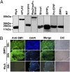
The engineered P. agglomerans strains efficiently secrete anti-Plasmodium effector proteins. (A) Western blot analysis. Culture supernatants were concentrated using Amicon Ultra-4 Centrifugal Filter Units. Aliquots originating from the same volume of supernatant were fractionated by SDS/PAGE and subjected to Western blot analysis using a rabbit anti-E-tag antibody. The supernatant from nontransformed P. agglomerans (WT) served as a negative control. Images were taken using the same exposure time. (B) In vivo binding of the secreted SM1 peptide to the mosquito midgut epithelium. A. gambiae were fed on sugar solution containing recombinant P. agglomerans strains expressing either the (SM1)2 or E-tagged HlyA alone (HlyA) and then fed on mouse blood. Midguts were dissected 18 or 24 h after blood meal, opened into a sheet, fixed, and incubated with an anti-SM1 antibody, followed by incubation with a fluorescent secondary antibody. Nuclei were stained with DAPI (blue). The differential interference contrast (DIC) images of the same field are shown to the right.
Fig. 3.
Fig. 3.
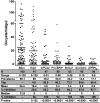
Inhibition of P. falciparum development in vector mosquitoes by recombinant P. agglomerans strains. A. gambiae mosquitoes were fed on 5% (wt/vol) sugar solution supplemented with either PBS (−Bact) or recombinant P. agglomerans strains and 32 h later fed on a P. falciparum–infected blood meal. Oocyst numbers were determined 8 d after blood meal. Each dot represents the oocyst number of an individual midgut and horizontal lines indicate mean values. Data were pooled from four biological replicates. Recombinant P. agglomerans strains were engineered to express the E-tagged HlyA leader peptide alone (HlyA) or mPLA2, Pro:EPIP, Shiva1, scorpine, and (EPIP)4 (Table S1), fused to HlyA (Fig. S3), as indicated. Inhibition, inhibition of oocyst formation relative to the −Bact control; Mean, mean oocyst number per midgut; Median, median oocyst number per midgut; N, number of mosquitoes analyzed; Prevalence, percentage of mosquitoes carrying at least one oocyst; Range, range of oocyst numbers per midgut; Tbp, transmission-blocking potential: 100 − {(prevalence of mosquitoes fed with recombinant P. agglomerans)/[prevalence of control (−Bact) mosquitoes] × 100}.
Fig. 4.
Fig. 4.
Comparison of P. falciparum development between mosquitoes that fed on an infectious blood meal at either 2 or 4 d after administration of recombinant P. agglomerans. A. gambiae females were fed on 5% (wt/vol) sugar solution supplemented with either PBS (−Bact) or recombinant P. agglomerans strains as follows: HlyA, P. agglomerans expressing the E-tagged HlyA leader peptide alone; Shiva1 + (EPIP)4 or scorpine + (EPIP)4 (Table S1), a mixture of the two recombinant bacteria in equal numbers. Two days (2 dpPa) or 4 d (4 dpPa) after administration of bacteria, mosquitoes were fed on a P. falciparum–infected blood meal. Oocyst numbers were determined 8 d after the blood meal. Each dot represents the oocyst number of an individual midgut and horizontal lines indicate mean values. Data were pooled from three biological replicates. % Inhibition, inhibition of oocyst formation relative to the −Bact control; Mean, mean oocyst number per midgut; Median, median oocyst number per midgut; N, number of mosquitoes analyzed; Prevalence, percentage of mosquitoes carrying at least one oocyst; Range, range of oocyst numbers per midgut.
Fig. 5.
Fig. 5.
Inhibition of P. berghei development in mosquitoes harboring recombinant P. agglomerans strains. A. stephensi females were fed on 5% (wt/vol) sugar solution supplemented with either PBS (−Bact) or recombinant P. agglomerans strains engineered to express E-tagged HlyA alone (HlyA), (SM1)2, pbs21scFv-Shiva1/(EPIP)4 (50–50 mixture) or scorpine/(EPIP)4 (50–50 mixture)] (Table S1 and Fig. S3) and 32 h later the five groups of mosquitoes were fed on the same P. berghei-infected mouse. Plasmodium infections were evaluated 14 d after infection. Each dot represents the number of oocysts in an individual midgut. Horizontal lines indicate mean values. Data pooled from three biological replicates. N, number of mosquitoes analyzed; Range, range of oocyst numbers per midgut; Prevalence, percentage of mosquitoes carrying at least one oocyst; Mean, mean oocyst number per midgut; Median, median oocyst number per midgut; Inhibition, inhibition of oocyst formation relative to the −Bact control.
Fig. 6.
Fig. 6.
Impact of recombinant P. agglomerans strains on lifespan of Anopheles gambiae females. A. gambiae mosquitoes were fed on 5% (wt/vol) sugar solution supplemented with either PBS (−Bact), or recombinant P. agglomerans strains as follows: HlyA, P. agglomerans expressing the E-tagged HlyA leader peptide alone; Shiva1 + (EPIP)4 or scorpine + (EPIP)4, a mixture of the two recombinant bacteria in equal numbers (Table S1 and Fig. S3). Thirty two hours later, mosquitoes were fed on a noninfected mouse, and mosquito survival was measured twice daily. Data were pooled from three independent experiments. There was no significant difference in survivorship among the groups.

Comment in

References

    1. Murray CJ, et al. Global malaria mortality between 1980 and 2010: A systematic analysis. Lancet. 2012;379:413–431. - PubMed
    1. Enayati A, Hemingway J. Malaria management: Past, present, and future. Annu Rev Entomol. 2010;55:569–591. - PubMed
    1. Trape JF, et al. Malaria morbidity and pyrethroid resistance after the introduction of insecticide-treated bednets and artemisinin-based combination therapies: A longitudinal study. Lancet Infect Dis. 2011;11:925–932. - PubMed
    1. Ghosh A, Edwards MJ, Jacobs-Lorena M. The journey of the malaria parasite in the mosquito: Hopes for the new century. Parasitol Today. 2000;16:196–201. - PubMed
    1. Whitten MMA, Shiao SH, Levashina EA. Mosquito midguts and malaria: Cell biology, compartmentalization and immunology. Parasite Immunol. 2006;28:121–130. - PubMed

Publication types

MeSH terms