

New chitosan nanobubbles for ultrasound-mediated gene delivery: preparation and in vitro characterization
- PMID: 22802689
- PMCID: PMC3396386
- DOI: 10.2147/IJN.S30912
New chitosan nanobubbles for ultrasound-mediated gene delivery: preparation and in vitro characterization
Abstract
Background: The development of nonviral gene delivery systems is one of the most intriguing topics in nanomedicine. However, despite the advances made in recent years, several key issues remain unsettled. One of the main problems relates to the difficulty in designing nanodevices for targeted delivery of genes and other drugs to specific anatomic sites. In this study, we describe the development of a novel chitosan nanobubble-based gene delivery system for ultrasound-triggered release.
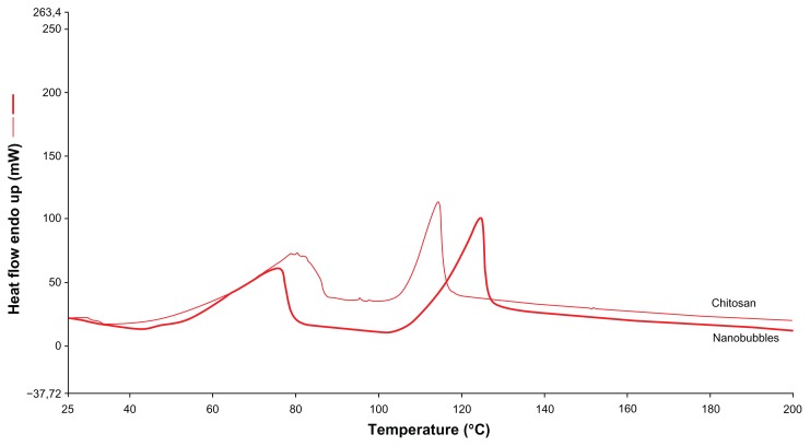
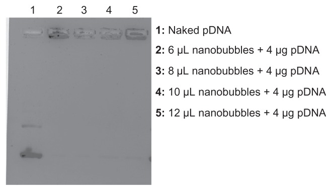
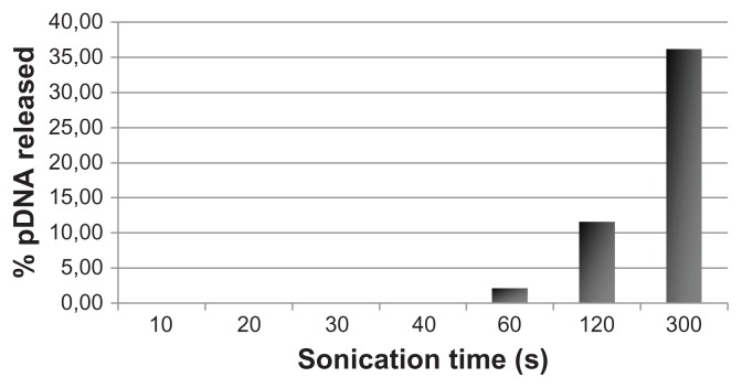
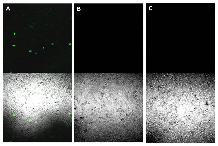
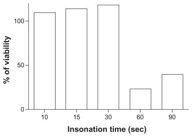
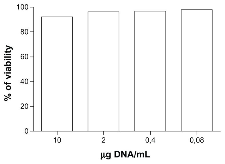
Methods and results: Chitosan was selected for the nanobubble shell because of its low toxicity, low immunogenicity, and excellent biocompatibility, while the core consisted of perfluoropentane. DNA-loaded chitosan nanobubbles were formed with a mean diameter of less than 300 nm and a positive surface charge. Transmission electron microscopic analysis confirmed composition of the core-shell structure. The ability of the chitosan nanobubbles to complex with and protect DNA was confirmed by agarose gel assay. Chitosan nanobubbles were found to be stable following insonation (2.5 MHz) for up to 3 minutes at 37°C. DNA release was evaluated in vitro in both the presence and absence of ultrasound. The release of chitosan nanobubble-bound plasmid DNA occurred after just one minute of insonation. In vitro transfection experiments were performed by exposing adherent COS7 cells to ultrasound in the presence of different concentrations of plasmid DNA-loaded nanobubbles. In the absence of ultrasound, nanobubbles failed to trigger transfection at all concentrations tested. In contrast, 30 seconds of ultrasound promoted a moderate degree of transfection. Cell viability experiments demonstrated that neither ultrasound nor the nanobubbles affected cell viability under these experimental conditions.
Conclusion: Based on these results, chitosan nanobubbles have the potential to be promising tools for ultrasound-mediated DNA delivery.
Keywords: DNA; chitosan; gene carrier; nanobubbles; transfection; ultrasound.
Figures

References
-
- Suzuki R, Oda Y, Utoguchi N, Maruyama K. Progress in the development of ultrasound-mediated gene delivery systems utilizing nano- and microbubbles. J Control Release. 2011;149:36–41. - PubMed
-
- Nomikou N, Tiwari P, Trehan T, Gulati K, McHale A. Studies on neutral, cationic and biotinylated cationic microbubbles in enhancing ultrasound-mediated gene delivery in-vitro and in-vivo. Acta Biomater. 2012;8:1273–1280. - PubMed
Publication types
MeSH terms
Substances
LinkOut - more resources
Full Text Sources
Other Literature Sources
