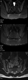
Diagnostic value of gadolinium-enhanced MR imaging of active sacroiliitis in seronegative spondyloarthropathy
- PMID: 22802778
- PMCID: PMC3389863
Diagnostic value of gadolinium-enhanced MR imaging of active sacroiliitis in seronegative spondyloarthropathy
Abstract
Background: SERONEGATIVE SPONDYLOARTHROPATHY (SPA) IS A GROUP OF DISEASES INCLUDING: ankylosing spondylitis, psoriatic spondyloarthrithis, reactive arthritis, spondyloarthrithis associated with inflammatory bowel disease, and undifferentiated spondyloarthritis. One of the diagnostic criteria of SpA is the presence of sacroiliitis. Periarticular bone marrow oedema (histologically corresponding to osteitis) is a primary symptom of the active stage of inflammation, which can be identified by MR using T2-weighted images. Its presence is essential for the therapeutic decision. The aims of this study were: 1. to compare the diagnostic value of T2-weighted images with T1 gadolinium-enhanced fat saturation (FS) images. 2. to establish if T1 gadolinium-enhanced images increase the diagnostic value of the MRI examination.
Material/methods: With the use of a 1.5T MRI scanner, 35 patients aged 19-67 years were examined. They were classified as having SpA or suspicious of SpA. The following findings were assessed: bone marrow oedema, synovitis, capsulitis/enthesistis. They were evaluated and compared on T2 and T1 gadolinium-enhanced FS images.
Results: Active sacroiliitis was identified in 21 patients, chronic in 1 patient. Two patients had signs of synovitis without any features of bone marrow oedema. One patient had fracture of the sacral bone. Ten patients had no signs of sacroiliitis. There was no significant difference in the diagnostic value between FSE T2 images and T1 gadolinium-enhanced images with FS in the evaluation of bone marrow oedema and capsulitis/enthesitis. However, T1 gadolinium-enhanced images were more sensitive than FSE T2 images in visualising synovitis.
Conclusions: MRI is a very sensitive method to identify active sacroiliitis in SpA. MRI without contrast administration is sufficient to identify bone marrow oedema as a crucial finding in active sacroiliitis. The gadolinium-enhanced images make the diagnosis easier, especially in patients with minimal bone marrow oedema because they reveal or better depict synovitis, while they do not improve visualisation of capsulitis/enthesitis.
Keywords: magnetic resonance; sacroiliitis; seronegative spondyloarthropathies.
Figures


References
-
- Elyan M, Asim Khan M. Diagnosing Ankylosing Spondylitis. J Rheumatol. 2006;33(Suppl):78. - PubMed
-
- Puhakka KB, Jurik AG, et al. MRI of sacroiliitis in early seronegative spondylorthropathy. Abnormalities correlated to clinical and laboratory findings. Rheumatology. 2004;43:234–37. - PubMed
-
- Rudwaleit M, Anne-Grethe Jurik, Hermann KA, et al. Defining active sacroiliitis on MRI for classiffication of axial spondyloarhthritis a consensual approach by the ASAS/OMERCAT MRI group. Ann Rheum Dis. published on line, 18.05 2009. - PubMed
-
- Bozygeyik Z, Ozgocmen S, Kocakoc E. Role of Diffusion-Weighted MRI in the detection of Early Active Sacroiliitis. AJR. 2008;191:980–86M. - PubMed
-
- Muche B, Bollow M, Franqois RJ, et al. Anatomic structures involved in early- and late-stage sacroiliitis in spondyloarthritis – a detailed analysis by contrast -enhanced MRI. American College of Rheumatology. 2003;48(5):1374–84. - PubMed
LinkOut - more resources
Full Text Sources
Research Materials
