

Application of the 64-slice computed tomography as a diagnostic method in acute posttraumatic ischaemia of the upper limbs - 3 case reports
- PMID: 22802783
- PMCID: PMC3389866
Application of the 64-slice computed tomography as a diagnostic method in acute posttraumatic ischaemia of the upper limbs - 3 case reports
Abstract
Background: Acute limb ischaemia' is a sudden, rapidly progressing inhibition of blood supply to a limb, characterised by appearance of new symptoms or by aggravation of the already existing ones, which may lead to amputation of the limb. Computed tomography angiography (CTA) performed with a multislice scanner belongs to methods used for arterial imaging in acute limb ischaemia. The main advantages of this method include: short examination time, low invasiveness, and possibility of a multiplanar and multivolume imaging of the vessels and adjacent tissues.
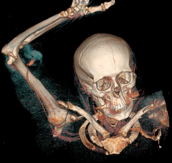
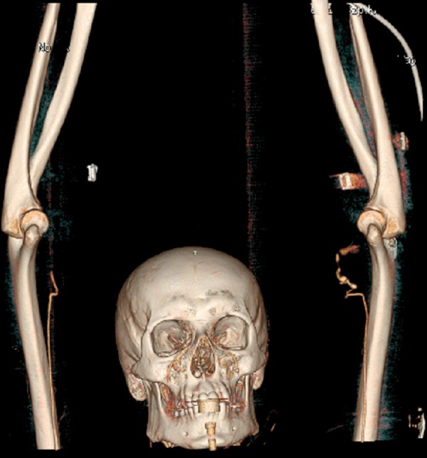
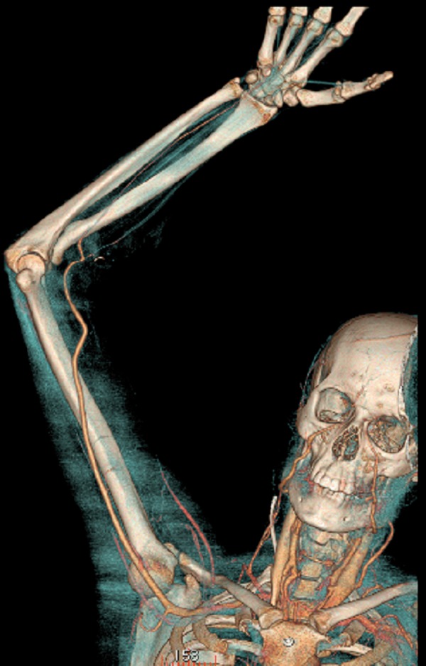
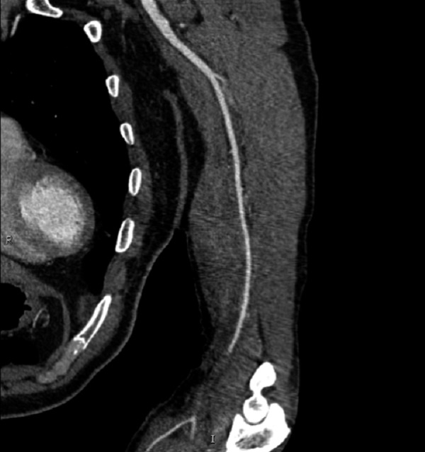
Case reports: We presented 3 cases of acute posttraumatic ischaemia of the upper limbs, diagnosed in the emergency setting with the use of a 64-slice CT scanner.
Conclusions: The CTA examination performed with the 64-slice CT unit revealed high effectiveness when used for localisation of vascular pathologies resulting in acute ischaemia of the upper limbs. Owing to this rapid radiological diagnostic method, it is now possible to perform successful limb-saving surgeries and to introduce postoperative monitoring immediately after the procedure.
Keywords: CTA; acute ischaemia of the upper limbs; computed tomography angiography.
Figures
Similar articles
-
Assessment of image quality and dose in contrast-enhanced head and neck CT angiography of New Zealand rabbit.J Xray Sci Technol. 2020;28(4):739-750. doi: 10.3233/XST-200669. J Xray Sci Technol. 2020. PMID: 32597826
-
Multi-slice CT angiography in the evaluation of patients with acute cerebrovascular disease--a promising new diagnostic tool.J Neurol. 2002 Jan;249(1):43-9. doi: 10.1007/pl00007846. J Neurol. 2002. PMID: 11954867 Clinical Trial.
-
Extravascular incidental findings in computed tomography angiography are associated with lower amputation-free survival in patients with acute lower limb ischaemia.Vascular. 2024 Feb;32(1):126-131. doi: 10.1177/17085381221135272. Epub 2022 Oct 21. Vascular. 2024. PMID: 36268567
-
[Ischaemia of the lower limbs: anaesthesia and intensive care].Ann Fr Anesth Reanim. 2004 Dec;23(12):1160-74. doi: 10.1016/j.annfar.2004.10.017. Ann Fr Anesth Reanim. 2004. PMID: 15589356 Review. French.
-
Multidetector Computed Tomography Angiography (MD-CTA) of Coronary Artery Bypass Grafts - Update 2017.Rofo. 2018 Mar;190(3):237-249. doi: 10.1055/s-0043-120528. Epub 2017 Nov 3. Rofo. 2018. PMID: 29100255 Review. English.
Cited by
-
Avoiding pitfalls of intraoperative peripheral endovascular surgery with the aid of OsiriX: expanding the use of virtual fluoroscopy.Rev Bras Cir Cardiovasc. 2014 Jul-Sep;29(3):455-8. doi: 10.5935/1678-9741.20140094. Rev Bras Cir Cardiovasc. 2014. PMID: 25372922 Free PMC article.
-
The role of interventional radiology in the treatment of lower limb vascular injuries after orthopaedic surgery.Pol J Radiol. 2019 Dec 2;84:e504-e510. doi: 10.5114/pjr.2019.91204. eCollection 2019. Pol J Radiol. 2019. PMID: 32082447 Free PMC article.
-
Dolichoectasia of the circle of Willis arteries and fusiform aneurysm of basilar artery - case report and review of the literature.Pol J Radiol. 2012 Apr;77(2):54-9. doi: 10.12659/pjr.882971. Pol J Radiol. 2012. PMID: 22844310 Free PMC article.
-
Imaging difficulties in Takayasu arteritis - case report and review of the literature.Pol J Radiol. 2012 Oct;77(4):67-71. doi: 10.12659/pjr.883633. Pol J Radiol. 2012. PMID: 23269941 Free PMC article.
References
-
- Chin AS, Rubin GD. MDCT Angiography of Peripheral Arterial Occlusive Disease. Tech Vasc Interventional Rad. 2006;9:143–49. - PubMed
-
- Norgren L, Hiatt WR, Dormandy JA, et al. Inter-Society Consensus for the Management of Peripheral Arterial Disease (TASC II) Eur J Vasc Endovasc Surg. 2007;33:1–75. - PubMed
-
- Management of peripheral arterial disease (PAD) TransAtlantic Inter-Society Consensus (TASC). Section C: acute limb ischaemia. Eur J Vasc Endovasc Surg. 2000;19:115–43. - PubMed
-
- Sontheimer DL. Peripheral vascular disease: diagnosis and treatment. Am Fam Physician. 2006;73(11):1971–76. - PubMed
Publication types
LinkOut - more resources
Full Text Sources