

Case Reports
Lesions in basal ganglia in a patient with involuntary movements as a first sign of diabetes - case report and review of the literature
Affiliations
- PMID: 22802794
- PMCID: PMC3389879
Item in Clipboard
Case Reports
Lesions in basal ganglia in a patient with involuntary movements as a first sign of diabetes - case report and review of the literature
Pol J Radiol.
2010 Jul.
Abstract
We present a case of unilateral hyperdensity of the lentiform and caudate nucleus on CT with hyperintesity on T1-weighted images on MRI in a 71-year-old woman with hemichorea-hemiballism and recently diagnosed diabetes.
Keywords: brain; computed tomography (CT); hemichorea-hemiballism; magnetic resonance imaging (MRI).
Figures
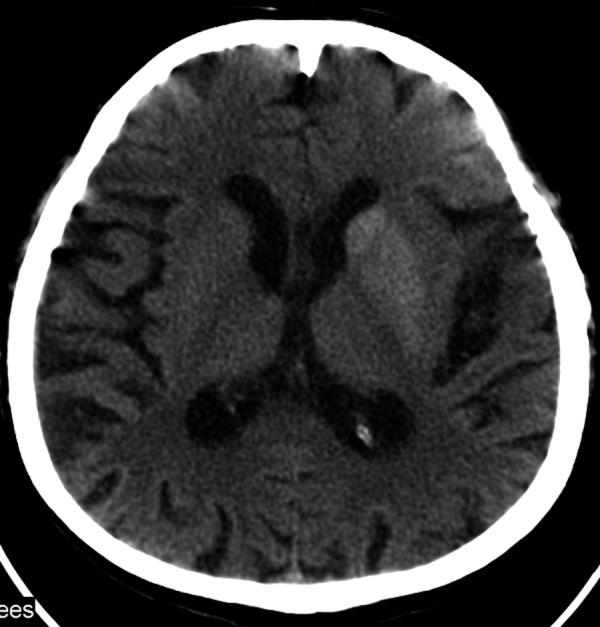
CT on the second day of symptoms. Hyperdense lentiform and caudate nucleus of the left hemisphere.
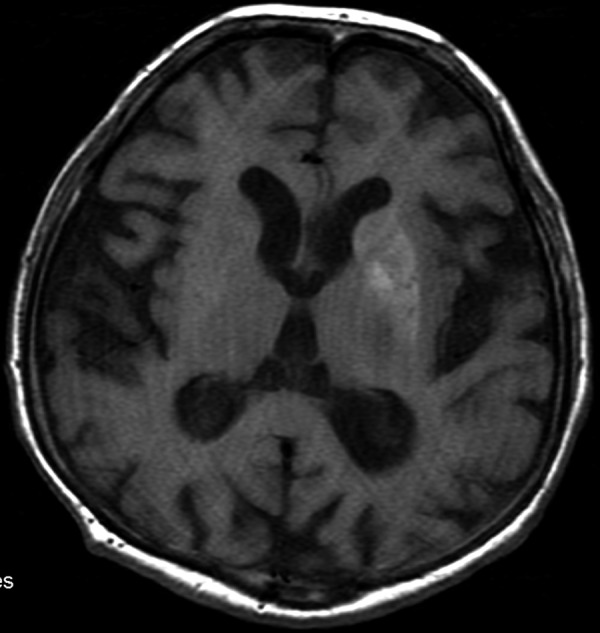
The MRI performed 4 days later. SE/T1 – Hyperintense signal from the lentiform nucleus and a less clear but also hyperintense caudate nucleus.
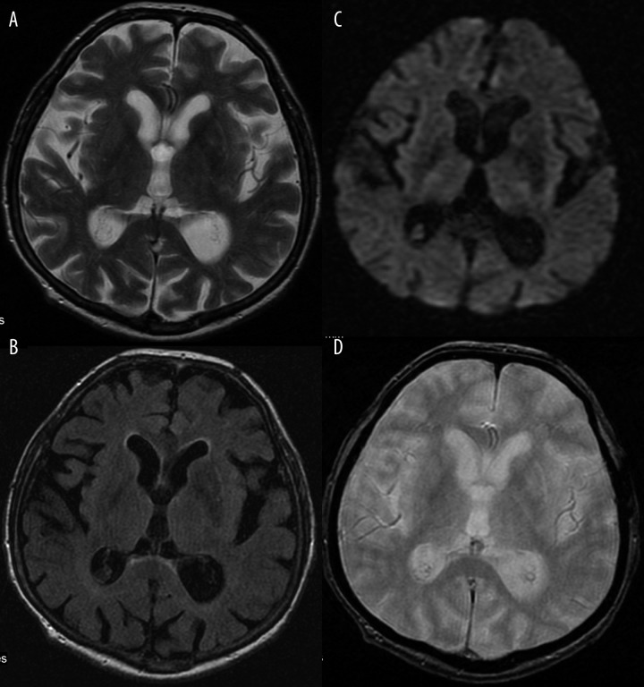
Discretely hypointense basal ganglia on the left side, within the same layer as in Figure 2. (A) FSE/T2-. (B) T2flair. (C) DWI. (D) GRE/T2*-.
References
-
- Oh SH, Lee KY, Im JH, Lee MS. Chorea associated with nonketotic hyperglycemia and hyperintensity basal ganglia lesion on T1-weighted brain MRI study: a meta-analysis of 53 cases including four present cases. J Neurol Sci. 2002;200(1–2):57–62. - PubMed
-
- Felicio AC, Chang CV, Godeiro-Junior C, et al. Hemichoreahemiballism as the first presentation of type 2 diabetes mellitus. Arq Neuropsiquiatr. 2008;66(2A):249–50. - PubMed
-
- Bekiesińska-Figatowska M, Walecki J. Zmiany hiperintensywne w mózgowiu w obrazach T1-zależnych badania rezonansu magnetycznego – przyczyny i możliwości różnicowania. Pol Przegl Radiol. 2001;66(2):15–18.
-
- Sanfield JA, Finkel J, Lewis S, et al. Alternating choreoathetosis associated with uncontrolled diabetes mellitus and basal ganglia calcification. Diabetes care. 1986;9:100–1. - PubMed
Publication types
LinkOut - more resources
Full Text Sources