

Ruptured ectopic pregnancy diagnosed with computed tomography
- PMID: 22802804
- PMCID: PMC3389893
Ruptured ectopic pregnancy diagnosed with computed tomography
Abstract
Background: The rupture of ectopic pregnancy (EP) still remains the primary and direct cause of death in the first trimester of pregnancy. Ultrasonography is known to be a modality of choice in EP diagnostics. We found a severe discrepancy between the frequency of ectopic pregnancies (EP) and the number of available computed tomography (CT) examinations.
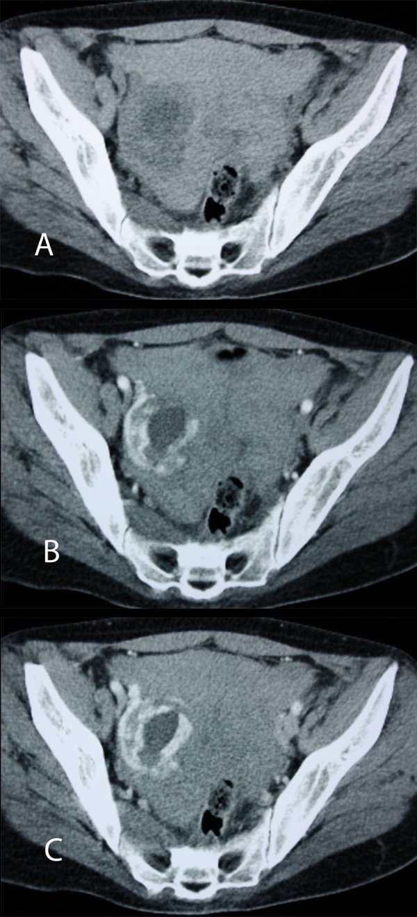
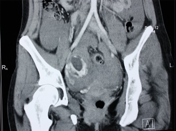
Case report: A 29-year-old woman was admitted to the emergency department with a history of abdominal pain, nausea, vomiting and collapse. Sonographic findings of a suspected EP were unclear. Moreover, not all features of intrauterine pregnancy were present. Due to the patient's life-threatening condition, an emergency multi-slice CT with MPR and VRT reconstructions was performed, revealing symptoms of a ruptured EP. In the right adnexal area, a well-vascularized, solid-cystic abnormal mass lesion was found. Intraperitoneal hemorrhage was confirmed intraoperatively, and the right fallopian tube with a tubal EP was resected. In the surgery in situ, as well as in the pathological examination of the tumor mass, a human embryo of approximately 1.5 cm in length (beginning of the 8(th) week of gestation) was found.
Conclusions: Although ultrasonography still remains the first-line imaging examination in EP diagnostics, sometimes the findings of suspected EPs are unclear and not sufficient. The rupture of EP, with serious bleeding and symptoms of shock, may require an emergent pelvic and abdominal CT inspection. A clear correlation was found between the macroscopic CT images and the intraoperatively sampled material.
Keywords: computed tomography; ectopic pregnancy; hemorrhage.
Figures
References
-
- Dialani V, Levine D. Ectopic pregnancy: a review. Ultrasound Q. 2004;20(3):105–17. - PubMed
-
- Kirsch JD, Scoutt LM. Imaging of ectopic pregnancy. Applied Radiology. 2010;39(3):10–25.
-
- Cano Alonso R, Borruel Nacenta S, Díez Martínez P, et al. Role of multidetector CT in the management of acute female pelvic disease. Emerg Radiol. 2009;16(6):453–72. - PubMed
-
- Knafel A, Basta P, Skotniczy K, et al. Pęknięcie ciąży ekotopowej – czy możemy zapobiec tej komplikacji? Ginekol Pol. 2009;80:734–39. - PubMed
-
- Levine D. Ectopic pregnancy. Radiology. 2007;245(2):385–97. - PubMed
Publication types
LinkOut - more resources
Full Text Sources