

Moyamoya disease: Diagnostic imaging
- PMID: 22802820
- PMCID: PMC3389911
Moyamoya disease: Diagnostic imaging
Abstract
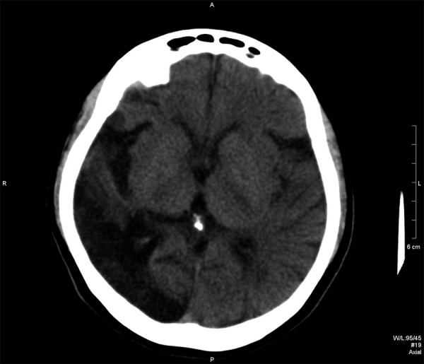
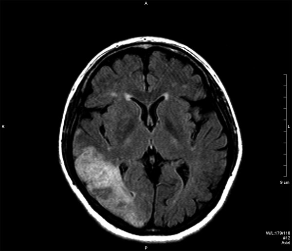
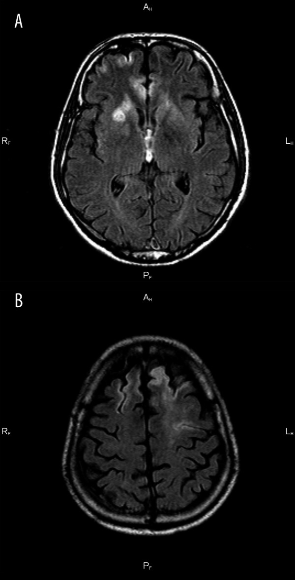
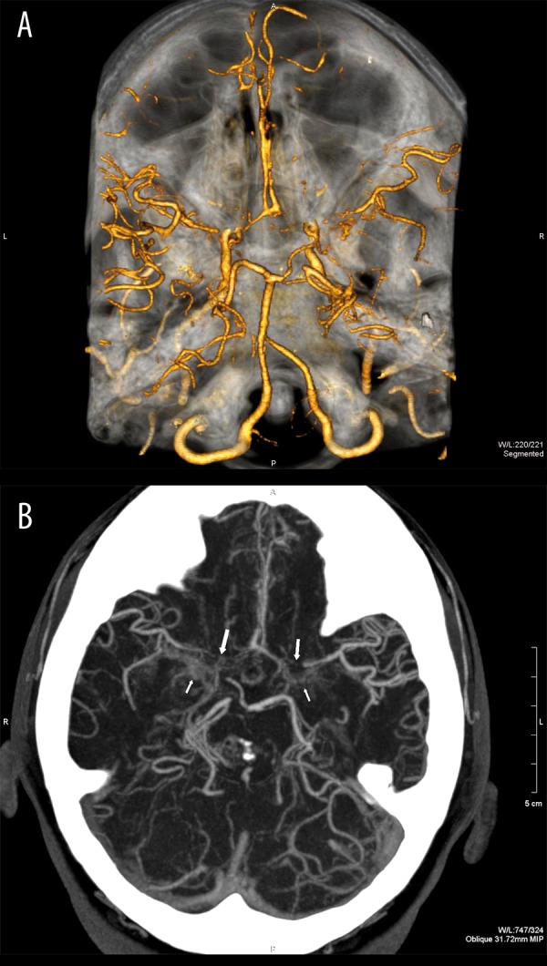
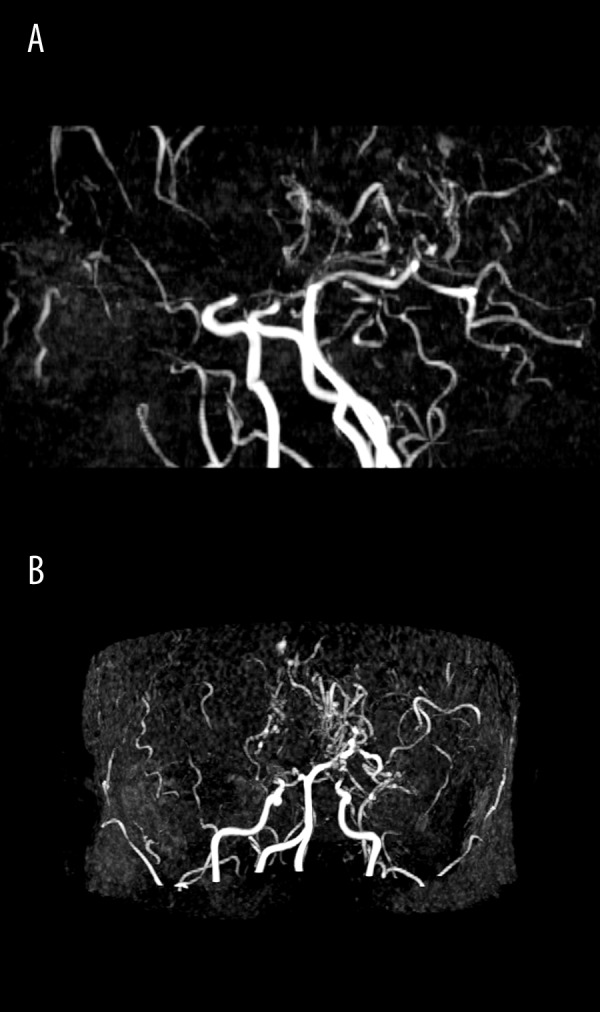
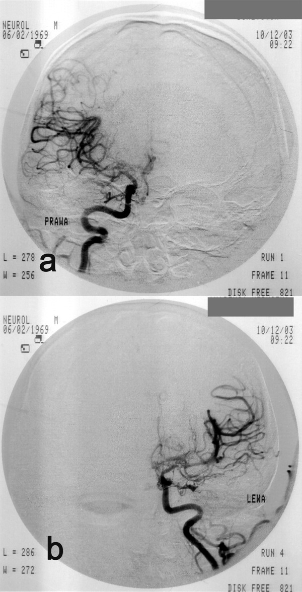
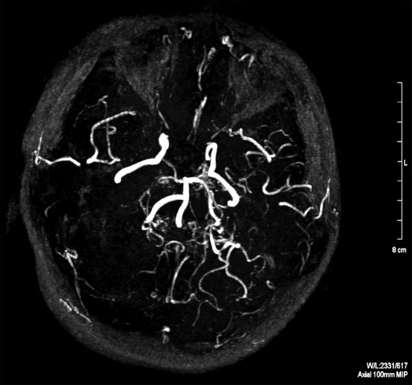
Moyamoya disease is a progressive vasculopathy leading to stenosis of the main intracranial arteries. The incidence of moyamoya disease is high in Asian countries; in Europe and North America, the prevalence of the disease is considerably lower. Clinically, the disease may be of ischaemic, haemorrhagic and epileptic type. Cognitive dysfunction and behavioral disturbance are atypical symptoms of moyamoya disease.Characteristic angiographic features of the disease include stenosis or occlusion of the arteries of the circle of Willis, as well as the development of collateral vasculature. Currently, magnetic resonance angiography and CT angiography with multi-row systems are the main imaging methods of diagnostics of the entire range of vascular changes in moyamoya disease.The most common surgical treatment combines the direct arterial anastomosis between the superficial temporal artery and middle cerebral, and the indirect synangiosis involving placement of vascularised tissue in the brain cortex, in order to promote neoangiogenesis. Due to progressive changes, correct and early diagnosis is of basic significance in selecting patients for surgery, which is the only effective treatment of the disease. An appropriate qualification to surgery should be based on a comprehensive angiographic and imaging evaluation of brain structures.Despite the rare occurrence of moyamoya disease in European population, it should be considered as one of causes of ischaemic or haemorrhagic strokes, especially in young patients.
Keywords: angiography; ischaemic stroke; moyamoya disease.
Figures
References
-
- Takeuchi K, Shimizu K. Hypoplasia of the bilateral internal carotid arteries. Brain Nerve. 1957;9:37–43.
-
- Baba T, Houkin K, Kuroda S. Novel epidemiological features of moyamoya disease. J Neurol Neurosurg Psychiatry. 2008;79(8):900–4. - PubMed
-
- Kuroda S, Houkin K. Moyamoya disease: current concepts and future perspectives. Lancet Neurol. 2008;7(11):1056–66. - PubMed
-
- Yonekawa Y, Ogata N, Kaku Y, et al. Moyamoya disease in Europe, past and present status. Clin Neurol Neurosurg. 1997;99(Suppl.2):S58–60. - PubMed
-
- Kułakowska A, Kapica-Topczewska K, Borowik H, et al. Moyamoya disease as a rare cause of ischaemic stroke-case report. Pol Merkur Lekarski. 2009;27(160):334–37. - PubMed
LinkOut - more resources
Full Text Sources