. 2012;7(7):e40192.
doi: 10.1371/journal.pone.0040192. Epub 2012 Jul 3.

The dengue vector Aedes aegypti contains a functional high mobility group box 1 (HMGB1) protein with a unique regulatory C-terminus

Affiliations

The dengue vector Aedes aegypti contains a functional high mobility group box 1 (HMGB1) protein with a unique regulatory C-terminus

Fabio Schneider Ribeiro et al. PLoS One. 2012.

Abstract

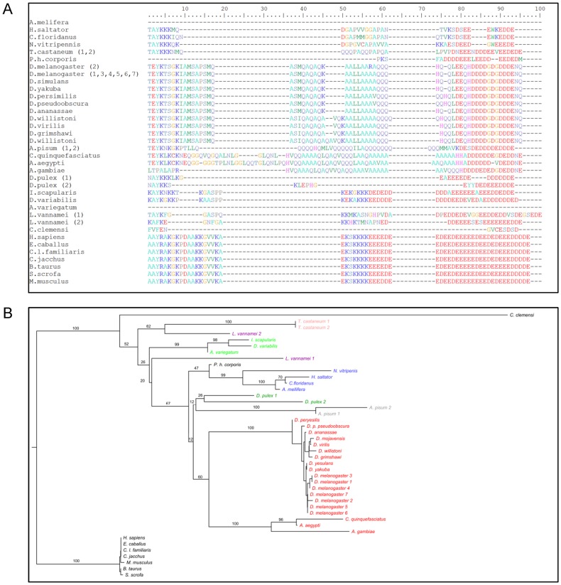
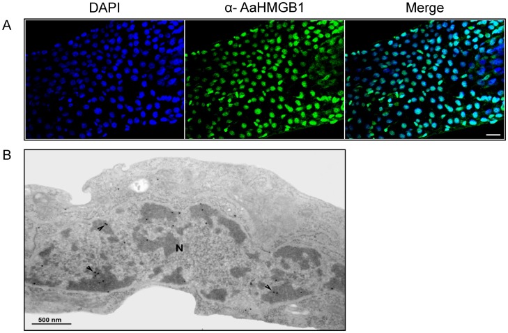
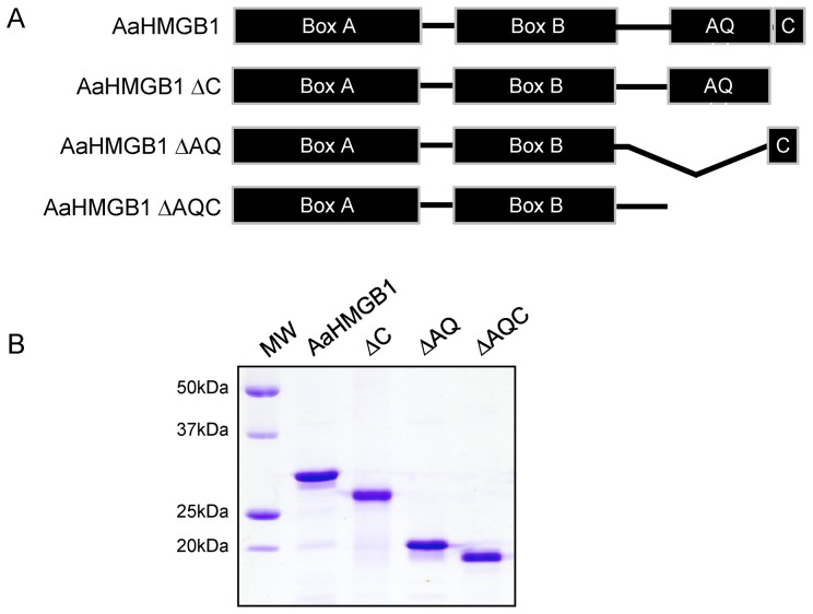
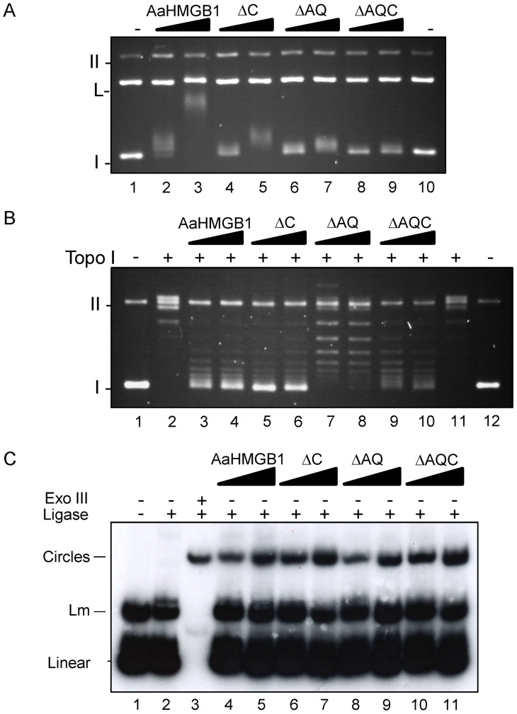
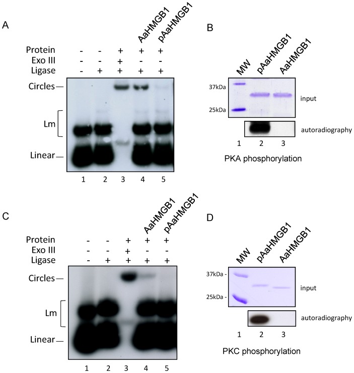
The mosquito Aedes aegypti can spread the dengue, chikungunya and yellow fever viruses. Thus, the search for key molecules involved in the mosquito survival represents today a promising vector control strategy. High Mobility Group Box (HMGB) proteins are essential nuclear factors that maintain the high-order structure of chromatin, keeping eukaryotic cells viable. Outside the nucleus, secreted HMGB proteins could alert the innate immune system to foreign antigens and trigger the initiation of host defenses. In this work, we cloned and functionally characterized the HMGB1 protein from Aedes aegypti (AaHMGB1). The AaHMGB1 protein typically consists of two HMG-box DNA binding domains and an acidic C-terminus. Interestingly, AaHMGB1 contains a unique alanine/glutamine-rich (AQ-rich) C-terminal region that seems to be exclusive of dipteran HMGB proteins. AaHMGB1 is localized to the cell nucleus, mainly associated with heterochromatin. Circular dichroism analyses of AaHMGB1 or the C-terminal truncated proteins revealed α-helical structures. We showed that AaHMGB1 can effectively bind and change the topology of DNA, and that the AQ-rich and the C-terminal acidic regions can modulate its ability to promote DNA supercoiling, as well as its preference to bind supercoiled DNA. AaHMGB1 is phosphorylated by PKA and PKC, but not by CK2. Importantly, phosphorylation of AaHMGB1 by PKA or PKC completely abolishes its DNA bending activity. Thus, our study shows that a functional HMGB1 protein occurs in Aedes aegypt and we provide the first description of a HMGB1 protein containing an AQ-rich regulatory C-terminus.


Conflict of interest statement

Competing Interests: The authors have declared that no competing interests exist.

Figures

Figure 1
Figure 1. Phylogenetic and sequence analysis of insect HMGB proteins.
(A) Phylogenetic tree of HMGB proteins from insects. The unrooted tree was built using the neighbor-joining method based on the alignment of HMGB amino acid sequences. Numbers indicate the bootstrap percentage support (10,000 replicates). (B) Alignment of the C-terminal portion of HMGB proteins from insects. The AQ-rich regions of the insect HMGB proteins run from positions 37 to 81. For comparison purposes, mammalian HMGB1 proteins were also included in the phylogeny analysis and alignment. The different Orders are represented by the different colors.
Figure 2
Figure 2. Expression of AaHMGB1 during the different stages of mosquito development.
AaHMGB1 mRNA expression was determined by quantitative Real-Time RT-PCR. Eggs (E), 1st instar larvae (L1), 2nd instar larvae (L2), 3rd instar larvae (L3), 4th instar larvae (L4), pupae (P), male (M) and female (F). Values are means of triplicate samples. Columns with different letters are significantly different from each other (a×c (P<0.01), b×c (P<0.05).
Figure 3
Figure 3. Cellular localization of native AaHMGB1 protein.
(A) Immunostaining of AaHMGB1 protein in the midguts of adult sugar-fed mosquitoes. Nuclei were stained with DAPI. AaHMGB1-polyclonal antibody (α-AaHMGB1) was used to detect the endogenous protein; Scale bar: 20 µm. (B) Transmission Electron Microscopy (TEM) of C6/36 mosquito cells using α-AaHMGB1, showing the nucleus (N). The immunogold markers show labeling of AaHMGB1 (arrows) mainly in heterochromatin regions (darker regions of the nuclei). This image is a representative of several cells observed under TEM.
Figure 4
Figure 4. Schematic diagram and SDS-PAGE of the recombinant AaHMGB1 proteins used in this study.
(A) Diagram of the recombinant 6×-his-tagged proteins: AaHMGB1, consists of two DNA-binding domains, the HMG box A and HMG box B, a alanine/glutamine-rich (AQ-rich) domain and a short acidic C-terminal domain; AaHMGB1-ΔC lacks only the short acidic C-terminal domain; AaHMGB1-ΔAQ lacks only the AQ-rich domain; AaHMGB1-ΔAQC lacks the entire C-terminus, including the AQ-rich and the short acidic C-terminal domains. (B) SDS-PAGE of the purified recombinant proteins. One microgram of each construct was loaded and analyzed on a 12% SDS-PAGE gel.
Figure 5
Figure 5. DNA transactions by recombinant AaHMGB1 proteins.
(A) Preferential binding of AaHMGB1 protein to supercoiled DNA. An equimolar mixture of supercoiled and linearized plasmid pTZ19R (∼10 nM) was pre-incubated with increasing amounts of AaHMGB1 (0.5–1 µM) and the DNA–protein complexes were resolved on a 1% agarose gel, followed by staining of the gel with ethidium bromide. Form I, supercoiled DNA; L, Linear DNA; Form II, relaxed circular DNA; (B) DNA supercoiling by AaHMGB1 and its truncated forms. Circular relaxed plasmid pTZ19R DNA was incubated in the presence of topoisomerase I (Topo I) and AaHMGB1 recombinant proteins (7–14 µM). Deproteinized DNA topoisomers were resolved on 1% agarose gels, followed by staining of the gel with ethidium bromide. Form I, supercoiled DNA; Form II, relaxed circular DNA. (C) DNA bending by AaHMGB1 and its truncated forms. A 32P-labeled 123-bp DNA fragment (∼1 nM) was pre-incubated with recombinant proteins (25–50 nM) followed by ligation with T4 DNA ligase. Exonuclease III was used to verify the identity of DNA circles. The deproteinized DNA ligation products were subjected to electrophoresis on 6% non-denaturing polyacrylamide gels and visualized by autoradiography. Lm: linear multimers. Exo III, exonuclease III. These experiments were repeated three to five times each.
Figure 6
Figure 6. Analysis of secondary and tertiary structures of AaHMGB1 proteins.
(A) CD spectra of AaHMGB1 (black line), ΔC (red line), ΔQC (green line) and ΔAQC (blue line) were performed at 25°C. Spectra were averaged from three scans at a 30 nm/min speed recorded from 190 to 260 nm, and the buffer baselines were subtracted from their respective sample spectra. (B) AaHMGB1 (black line), ΔC (red line), ΔQC (green line) and ΔAQC (blue line) were analyzed using fluorescence spectroscopy, either in the absence (native state, solid lines) or presence of 8 M urea (denatured state, dashed lines), in order to evaluate tertiary structure content. The excitation wavelength was fixed at 280 nm and the emission spectrum was recorded from 300 nm to 420 nm. Experiments were performed at 25°C.
Figure 7
Figure 7. In vitro phosphorylation of AaHMGB1.
(A) One microgram of AaHMGB1 proteins were subjected to an in vitro kinase assay with commercial kinases (CK2, PKA and PKC) and radiolabeled [γ-32P] ATP. Phosphorylations were analyzed by 12% SDS-PAGE (top panel) and autoradiography (bottom panel). Schistosoma mansoni HMGB1 (SmHMGB1) was used as a positive control for CK2 phosphorylation . (B) Immunoprecipitation of endogenous phosphorylated AaHMGB1. Total protein extract from adult mosquitoes were immune precipitated with pre-immune serum or anti-HMGB1 antibody (lane 3). Western blot analysis was carried out with anti-phospho serine monoclonal antibody (lanes 2 and 3). Endogenous AaHMGB1 (from the protein extract) was reacted against polyclonal anti-AaHMGB1 antibody (lane 1).
Figure 8
Figure 8. DNA bending assays by posphorylated AaHMGB1.
A 32P-labelled 123-bp DNA fragment (∼1 nM) was pre-incubated with 50 ng of AaHMGB1 that were phosphorylated by PKA (panels A and B, lanes 5 and 2, respectively) or not (panels A and B, lanes 4 and 3, respectively), or by PKC (panels C and D, lanes 5 and 2, respectively) or not (panels C and D, lanes 4 and 3, respectively), followed by ligation with T4 DNA ligase. Exonuclease III was used to verify the identity of DNA circles. The deproteinized DNA ligation products were subjected to electrophoresis on 6% non-denaturing polyacrylamide gels and visualized by autoradiography. Lm: linear multimers. These experiments were repeated five times.

Similar articles

Cited by

References

    1. Celona B, Weiner A, Di Felice F, Mancuso FM, Cesarini E, et al. Substantial histone reduction modulates genomewide nucleosomal occupancy and global transcriptional output. PLoS Biol. 2011;9:e1001086. - PMC - PubMed
    1. Li B, Carey M, Workman JL. The role of chromatin during transcription. Cell. 2007;128:707–719. - PubMed
    1. Groth A, Rocha W, Verreault A, Almouzni G. Chromatin challenges during DNA replication and repair. Cell. 2007;128:721–733. - PubMed
    1. Thomas JO, Travers AA. HMG1 and 2, and related ‘architectural’ DNA-binding proteins. Trends Biochem Sci. 2001;26:167–174. - PubMed
    1. Stros M. HMGB proteins: interactions with DNA and chromatin. Biochim Biophys Acta. 2010;1799:101–113. - PubMed

Publication types