. 2012 Oct;123(1):182-91.
doi: 10.1111/j.1471-4159.2012.07877.x. Epub 2012 Aug 16.

Neuroprotective effects of phenolic antioxidant tBHQ associate with inhibition of FoxO3a nuclear translocation and activity

Affiliations
Free PMC article

Neuroprotective effects of phenolic antioxidant tBHQ associate with inhibition of FoxO3a nuclear translocation and activity

Parmvir K Bahia et al. J Neurochem. 2012 Oct.
Free PMC article

Abstract

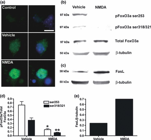
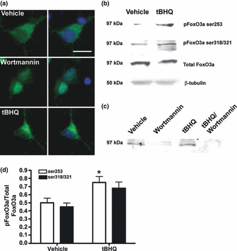
The Forkhead transcription factor, FoxO3a induces genomic death responses in neurones following translocation from the cytosol to the nucleus. Nuclear translocation of FoxO3a is triggered by trophic factor withdrawal, oxidative stress and the stimulation of extrasynaptic NMDA receptors. Receptor activation of phosphatidylinositol 3-kinase (PI3K)-Akt signalling pathways retains FoxO3a in the cytoplasm, thereby inhibiting the transcriptional activation of death-promoting genes. We hypothesized that phenolic antioxidants such as tert-Butylhydroquinone (tBHQ), which is known to stimulate PI3K-Akt signalling, would inhibit FoxO3a translocation and activity. Treatment of cultured cortical neurones with NMDA increased the nuclear localization of FoxO3a, reduced the phosphorylation of FoxO3a, increased caspase activity and up-regulated Fas ligand expression. In contrast the phenolic antioxidant, tBHQ, caused retention of FoxO3a in the cytosol coincident with enhanced PI3K- dependent phosphorylation of FoxO3a. tBHQ-induced nuclear exclusion of FoxO3a was associated with reduced FoxO-mediated transcriptional activity. Exposure of neurones to tBHQ inhibited NMDA-induced nuclear translocation of FoxO3a, prevented NMDA-induced up-regulation of FoxO-mediated transcriptional activity, blocked caspase activation and protected neurones from NMDA-induced excitotoxic death. Collectively, these data suggest that phenolic antioxidants such as tBHQ oppose stress-induced activation of FoxO3a and therefore have potential neuroprotective utility in neurodegeneration.


Figures

Fig 1
Fig 1
NMDA inhibits FoxO3a phosphorylation, stimulates FoxO3a nuclear translocation and up-regulates FasL. Cortical neurones were treated with NMDA (50 μM) for 2 h, and FoxO3a phosphorylation and FoxO3a translocation measured by immunoblotting (b, d) and immunocytochemistry, respectively, (a) and FasL expression by immunoblotting only (c, e). Reduced dephosphorylation of both pFoxO3a ser253 and pFoxO3a ser319/321 was evident following exposure to NMDA (b, d) with no change in total levels of FoxO3a or β-tubulin, but was associated with an increase in the levels of FasL (c, e). Blots shown are representative of three independent experiments. (d) Data presented as mean band intensity (mean ± SEM, n = 3) expressed as pFoxO3a relative to total FoxO3a following exposure to Vehicle control or NMDA (*p < 0.05, **p < 0.01). (e) Data presented as mean band intensity (mean ± SEM, n = 3) expressed as FasL relative to β-tubulin following exposure to Vehicle control or NMDA. To determine whether NMDA-mediated dephosphorylation of FoxO3a resulted in the nuclear accumulation of FoxO3a, cells were probed with an anti-FoxO3a antibody [(a), left and right panels] and counter-stained with Hoe 33342 (right panel only) to label nuclei. Control represents staining because of secondary antibody only. Under basal conditions, FoxO3a was distributed throughout the cytoplasm of cortical neurones with some diffuse staining evident in the nucleus (a, Vehicle). Following treatment with NMDA, FoxO3a staining was almost exclusively nuclear (a, NMDA). Scale bar = 20 μm.
Fig 2
Fig 2
tert-Butylhydroquinone (tBHQ) stimulates FoxO3a phosphorylation and promotes cytosolic accumulation of FoxO3a. Cortical neurones were treated with tBHQ (30 μM; 2 h), or wortmannin (150 nM; 2 h) and FoxO3a phosphorylation and FoxO3a translocation were measured by immunoblotting and immunocytochemistry. tBHQ increased the phosphorylation of both the S1 (pFoxO3a ser253) and S2 (pFoxO3a ser319/321) sites without changing the total levels of FoxO3a or β-tubulin (b, d). Blots shown are representative of three independent experiments. (d) Data presented as mean band intensity (mean ± SEM, n = 3) expressed as pFoxO3a relative to total FoxO3a following exposure to Vehicle control or tBHQ (*p < 0.05). Basal (vehicle) and tBHQ-induced phosphorylation of pFoxO3a ser253 was abolished by wortmannin (c). To determine whether tBHQ-induced phosphorylation of FoxO3a affected the cellular distribution of FoxO3a cells were probed with an anti-FoxO3a antibody [(a) left and right panels] and counter-stained with Hoechst 33342 (right panel only) to label nuclei. Under control conditions, FoxO3a was distributed throughout the cytoplasm of cortical neurones with some low-level diffuse staining evident in the nucleus (Fig 2, Vehicle). Following treatment with tBHQ, FoxO3a staining was strongly cytoplasmic (Fig 2, tBHQ). This was particularly evident when compared with cells treated with the PI3K inhibitor wortmannin, which resulted in pronounced nuclear accumulation of FoxO3a (Fig 2, Wortmannin). Images shown are representative of 100 individual cells. tBHQ caused strong cytoplasmic accumulation of FoxO3a in 45% of stained cells. Scale bar = 20 μm.
Fig 3
Fig 3
Inhibition of FKHD-mediated gene expression by tert-Butylhydroquinone (tBHQ). Cortical neurones (8 DIV) were transfected with pARE-luc (a, b) or FHRE-luc (c, d) and then treated with, 10 μM or 30 μM tBHQ for 24 h (a and c) or 30 μM tBHQ for different time periods as shown (b and d). Data represent mean ± SEM, analysed using one-way anova and Bonferroni post hoc test (n = 3, each concentration performed in triplicate) where *p < 0.05, **p < 0.01 ***p < 0.001 compared to vehicle control.
Fig 4
Fig 4
NMDA induction of activated caspase is accompanied by accumulation of nuclear FoxO3A and blocked by tert-Butylhydroquinone (tBHQ). (a) Representative photomicrograph showing cortical neurones (8DIV) after 6-h treatment with 0.1% ethanol vehicle (C), NMDA (50 μM) (N), tBHQ (30 μM) (T), or NMDA and tBHQ combined (N + T). Top panel shows staining for activated caspase 3 (green), and nuclear Hoe 33342 (blue). Bottom panel shows staining for foxO3A (green) and Hoe 33342 (blue), arrowheads indicate cells with strongly foxo3A-positive nuclei. Scale bar = 10 μm. (b) shows quantification of nuclei associated with activated caspase 3 at different time points after application of 0.1% ethanol vehicle (C), NMDA (50 μM) (N), tBHQ (30 μM) (T) or tBHQ and NMDA combined (T + N). (c) shows quantification of foxO3A-positive nuclei after the same treatments. In B + C, Bars show means and SEM derived from counts of six to nine fields from three experiments. Analysis was by anova and Bonferroni’s multiple comparison test: *,**,*** = p < 0.05, p < 0.01, p < 0.001 compared to vehicle control. #, ##, ### = p < 0.05, p < 0.01, p < 0.001 compared to NMDA treatment only.
Fig 5
Fig 5
tert-Butylhydroquinone (tBHQ) opposes NMDA-induced up-regulation of FKHD-mediated gene expression and protects against NMDA-evoked neuronal damage. (a) Cortical neurones (8 DIV) were transfected with FHRE-luc and were then treated with NMDA (50 μM; 6 h;formula image
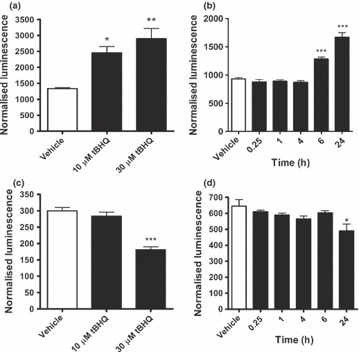
) or vehicle control (□) in the presence and absence of tBHQ (30 μM). Data represent mean ± SEM, analysed using one-way anova and Bonferroni post hoc test (n = 3, each concentration performed in triplicate). NMDA increased FHRE activity compared with vehicle control *p < 0.05 and tBHQ inhibited FKHR compared to vehicle control #p < 0.05. (b) Cultured cortical neurones (8 DIV) were incubated with increasing concentrations of NMDA in the presence of 30 μM tBHQ (formula image) or vehicle control (formula image) for 24 h. Cell viability was assessed using the MTT assay where the absorbance of reduced MTT was read at 505 nm. Neurones co-treated with tBHQ were significantly protected against 100 and 300 μM NMDA insults. Data represent mean ± SEM, analysed using one-way anova and the Bonferroni post hoc test (n = 4) where **p < 0.01 for individual NMDA concentrations versus control.

Similar articles

Cited by

References

    1. Abbott JJ, Howlett DR, Francis PT, Williams RJ. Abeta(1-42) modulation of Akt phosphorylation via alpha7 nAChR and NMDA receptors. Neurobiol. Aging. 2008;29:992–1001. - PubMed
    1. Arredondo F, Echeverry C, Abin-Carriquiry JA, Blasina F, Antúnez K, Jones DP, Go YM, Liang YL, Dajas F. After cellular internalization, quercetin causes Nrf2 nuclear translocation, increases glutathione levels, and prevents neuronal death against an oxidative insult. Free Radic. Biol. Med. 2010;49:738–747. - PubMed
    1. Bahia PK, Rattray M, Williams RJ. Dietary flavonoid (-)epicatechin stimulates phosphatidylinositol 3-kinase-dependent anti-oxidant response element activity and up-regulates glutathione in cortical astrocytes. J. Neurochem. 2008;106:2194–2204. - PubMed
    1. Barthelemy C, Henderson C, Pettmann B. Foxo3a induces motoneuron death through the Fas pathway in cooperation with JNK. BMC Neurosci. 2004;5:48. - PMC - PubMed
    1. Biggs WH, III, Cavenee WK, Arden KC. Identification and characterization of members of the FKHR (FOX O) subclass of winged-helix transcription factors in the mouse. Mamm. Genome. 2001;12:416–425. - PubMed

Publication types

MeSH terms