

Histopathological analysis of cellular localization of cathepsins in abdominal aortic aneurysm wall
- PMID: 22804761
- PMCID: PMC3444981
- DOI: 10.1111/j.1365-2613.2012.00819.x
Histopathological analysis of cellular localization of cathepsins in abdominal aortic aneurysm wall
Abstract
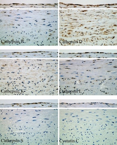
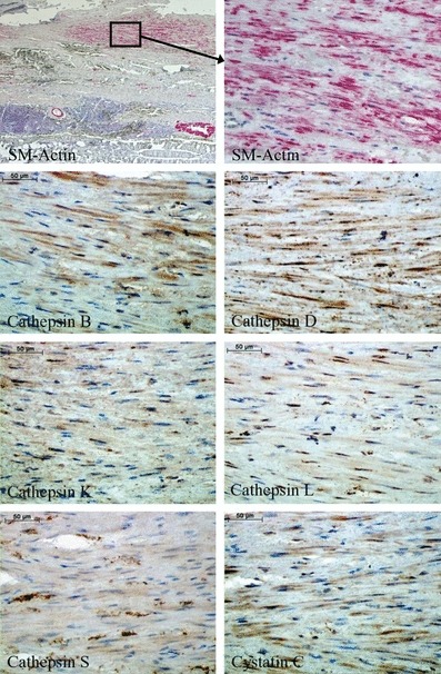
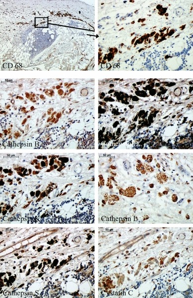
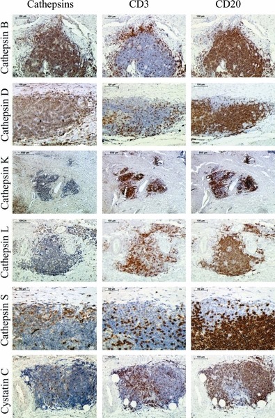
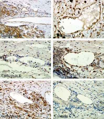
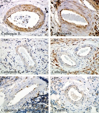
An important feature of abdominal aortic aneurysm (AAA) is the destruction of vessel wall, especially elastin and collagen. Besides matrix metalloproteinases, cathepsins are the most potent elastolytic enzymes. The expression of cathepsins with known elastolytic and collagenolytic activities in the individual cells within AAA has not yet been determined. The vessel wall of 32 AAA patients and 10 organ donors was analysed by immunohistochemistry for expression of cathepsins B, D, K, L and S, and cystatin C in all cells localized within AAA. Luminal endothelial cells (ECs) of AAA were positive for cathepsin D and partially for cathepsins B, K and S. Endothelial cells of the neovessels and smooth muscle cells in the media were positive for all cathepsins tested, especially for cathepsin B. In the inflammatory infiltrate all cathepsins were expressed in the following pattern: B > D = S > K = L. Macrophages showed the highest staining intensity for all cathepsins. Furthermore, weak overall expression of cystatin C was observed in all the cells localized in the AAA with the exception of the ECs. There is markedly increased expression of the various cathepsins within the AAA wall compared to healthy aorta. Our data are broadly consistent with a role for cathepsins in AAA; and demonstrate expression of cathepsins D, B and S in phagocytic cells in the inflammatory infiltrate; and also may reveal a role for cathepsin B in lymphocytes.
© 2012 The Authors. International Journal of Experimental Pathology © 2012 International Journal of Experimental Pathology.
Figures
Similar articles
-
Cathepsin S is associated with degradation of collagen I in abdominal aortic aneurysm.Vasa. 2018 Jun;47(4):285-293. doi: 10.1024/0301-1526/a000701. Epub 2018 Apr 6. Vasa. 2018. PMID: 29624112
-
Quantitative expression and localization of cysteine and aspartic proteases in human abdominal aortic aneurysms.Exp Mol Med. 2014 May 16;46(5):e95. doi: 10.1038/emm.2014.20. Exp Mol Med. 2014. PMID: 24833013 Free PMC article.
-
Cathepsin L expression and regulation in human abdominal aortic aneurysm, atherosclerosis, and vascular cells.Atherosclerosis. 2006 Feb;184(2):302-11. doi: 10.1016/j.atherosclerosis.2005.05.012. Epub 2005 Jun 27. Atherosclerosis. 2006. PMID: 15982660
-
B lymphocytes in abdominal aortic aneurysms.Atherosclerosis. 2015 Sep;242(1):311-7. doi: 10.1016/j.atherosclerosis.2015.07.036. Epub 2015 Jul 21. Atherosclerosis. 2015. PMID: 26233918 Review.
-
Role of Extracellular Matrix and Inflammation in Abdominal Aortic Aneurysm.Int J Mol Sci. 2022 Sep 21;23(19):11078. doi: 10.3390/ijms231911078. Int J Mol Sci. 2022. PMID: 36232377 Free PMC article. Review.
Cited by
-
Selective cathepsin S inhibition attenuates atherosclerosis in apolipoprotein E-deficient mice with chronic renal disease.Am J Pathol. 2015 Apr;185(4):1156-66. doi: 10.1016/j.ajpath.2014.11.026. Epub 2015 Feb 10. Am J Pathol. 2015. PMID: 25680278 Free PMC article.
-
Let-7f: A New Potential Circulating Biomarker Identified by miRNA Profiling of Cells Isolated from Human Abdominal Aortic Aneurysm.Int J Mol Sci. 2019 Nov 5;20(21):5499. doi: 10.3390/ijms20215499. Int J Mol Sci. 2019. PMID: 31694153 Free PMC article.
-
Assessing the targeting and fate of cathepsin k antibody-modified nanoparticles in a rat abdominal aortic aneurysm model.Acta Biomater. 2020 Aug;112:225-233. doi: 10.1016/j.actbio.2020.05.037. Epub 2020 Jun 3. Acta Biomater. 2020. PMID: 32504690 Free PMC article.
-
Cathepsins: a new culprit behind abdominal aortic aneurysm.Regen Med Res. 2013 Nov 1;1(1):5. doi: 10.1186/2050-490X-1-5. eCollection 2013 Dec. Regen Med Res. 2013. PMID: 25984324 Free PMC article. Review.
-
Endothelial cells and cathepsins: Biochemical and biomechanical regulation.Biochimie. 2016 Mar;122:314-23. doi: 10.1016/j.biochi.2015.10.010. Epub 2015 Oct 13. Biochimie. 2016. PMID: 26458976 Free PMC article. Review.
References
-
- Abisi S, Burnand KG, Waltham M, et al. Cysteine protease activity in the wall of abdominal aortic aneurysms. J. Vasc. Surg. 2007;46:1260–1266. - PubMed
-
- Carmeliet P. Mechanisms of angiogenesis and arteriogenesis. Nat. Med. 2000;6:389–395. - PubMed
-
- Chapman HA, Riese RJ, Shi GP. Emerging roles for cysteine proteases in human biology. Annu. Rev. Physiol. 1997;59:63–88. - PubMed
Publication types
MeSH terms
Substances
LinkOut - more resources
Full Text Sources
