Review
. 2012 Nov;16(11):2564-82.
doi: 10.1111/j.1582-4934.2012.01603.x.

Regenerative medicine for the treatment of spinal cord injury: more than just promises?

Affiliations
Review

Regenerative medicine for the treatment of spinal cord injury: more than just promises?

Ana Paula Pêgo et al. J Cell Mol Med. 2012 Nov.

Abstract

Spinal cord injury triggers a complex set of events that lead to tissue healing without the restoration of normal function due to the poor regenerative capacity of the spinal cord. Nevertheless, current knowledge about the intrinsic regenerative ability of central nervous system axons, when in a supportive environment, has made the prospect of treating spinal cord injury a reality. Among the range of strategies under investigation, cell-based therapies offer the most promising results, due to the multifactorial roles that these cells can fulfil. However, the best cell source is still a matter of debate, as are clinical issues that include the optimal cell dose as well as the timing and route of administration. In this context, the role of biomaterials is gaining importance. These can not only act as vehicles for the administered cells but also, in the case of chronic lesions, can be used to fill the permanent cyst, thus creating a more favourable and conducive environment for axonal regeneration in addition to serving as local delivery systems of therapeutic agents to improve the regenerative milieu. Some of the candidate molecules for the future are discussed in view of the knowledge derived from studying the mechanisms that facilitate the intrinsic regenerative capacity of central nervous system neurons. The future challenge for the multidisciplinary teams working in the field is to translate the knowledge acquired in basic research into effective combinatorial therapies to be applied in the clinic.


Figures

Fig. 1
Fig. 1
Schema of acute and chronic events following spinal cord injury.
Fig. 2
Fig. 2
Increased proliferation of endogenous stem cells in the spinal cord after SCI or after increased physical activity in comparison to control. (A) Schematic illustration of BrdU staining in thoracic spinal cord sections (Th8) of control, SCI and physical activity (running) groups. Note, the highest BrdU expression in the central canal (CC), parenchyma and around the lesion site in the SCI and running groups. Below each schematic drawing is a panel showing BrdU staining in the corresponding ventral white matter. (B) Quantitative analysis of the BrdU-positive nuclei in the ependyma of the thoracic spinal cord segments Th7-9 after SCI and (C) physical activity, revealing that the highest concentration of BrdU-labeled cells was restricted to the ependymal zone of the CC at 7–14 days after SCI and at 4–7 days after physical activity, (*P < 0.1 and **P < 0.05, Student's t-test). Modified from [16], Copyright (2009), with permission from Elsevier.
Fig. 3
Fig. 3
MAC enrichment of rat oligodendroglial progeny from spinal cord-derived neural stem cells (NSCs). In vitro differentiation to oligodendrocytes (RIP staining) at day 14 and staining for myelin basic protein (MBP) of mature oligogodendrocytes at day 21 in unsorted NSCs (A) and MAC-sorted NSCs (B). Scale bars: left hand side images = 100 μm; right hand side images = 50 μm. (C) Comparison of oligodendroglial lineage development in MAC-sorted (blue bars) and unsorted spinal NSCs (purple bars). Modified from [47], Copyright (2009), with permission from Elsevier.
Fig. 4
Fig. 4
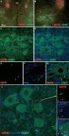
Survival of grafted human stem cells identified by hSYN immunoreactivity with a dense population of inhibitory terminals (hSYN/GAD65 immunoreactive) in the vicinity of persisting α-motoneurons. (A–G) Fluorescent microscopy images (A, B) and projected confocal images (C, D) of transverse spinal cord sections taken at 3 months after grafting and stained with human-specific SYN antibody (red), CHAT antibody (green) and SYN antibody that cross-reacts with both human and rat SYN (blue). Intense hSYN staining was found within the two bilateral grafts (A; red arrows). Numerous hSYN-stained terminals were localized in the base of the dorsal horn and extending into ventral-motoneuron pools (B, C; red). (E–G) Single optical images showing the colocalization of hSYN and SYN IR in the vicinity of CHATα-motoneurons (yellow arrows). (H-K) The majority of hSYN terminals (red) co-localized with GAD65 (blue), purple dots. Modified from [14], Copyright (2007), with permission from Elsevier.
Fig. 5
Fig. 5
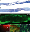
Histological evaluation of the lesion in rats with chronic SCI treated with a PHPMA-RGD hydrogel seeded with MSCs. (A, B) Longitudinal section (Luxol blue staining for myelin, Lux) of a spinal cord lesion 6 months after SCI. (A) The untreated lesion was dominated by tissue atrophy due to progressive cavitation. (B) A hydrogel seeded with MSCs (white asterisk) and implanted into a chronic spinal cord lesion (5 weeks after injury) formed a bridge across the epicenter of the chronic lesion. (C) The hydrogel was completely filled with infiltrating axons (staining for neurofilament NF160) throughout its whole volume. (D) Schwann cells (p75 staining), originating from the spinal root entry zone, crossed the spinal cord-hydrogel border and infiltrated the hydrogel (white arrow). (E) Double staining showing axons (NF 160 staining, green) growing inside the implant in close proximity with Schwann cells (p75 staining, red). (F) Regenerating axons present inside the hydrogel scaffold showed GAP-43 positivity. Scale bar: (A, B) 2 mm, (C) 500 μm, (D) 100 μm, (E) 50 μm, (F) 25 μm. Modified from [133].
Fig. 6
Fig. 6
Nanobiofunctionalized implants for spinal cord regeneration. (I) Proposed strategy: nerve implants are biofunctionalized by chitosan/siRNA nanoparticles that are taken up by cells, and enable neurite outgrowth. (II) Nanoparticles on nerve implants. (A) Particle uptake from nerve implants (filaments) into PC12 cells. Chitosan/siRNA nanoparticles (NP) were immobilized on polydioxanon (PDO) filaments (scale bar left image: 10 μm) by lyophilization. PC12 cells were seeded onto the filaments carrying NP and after 48 hrs the uptake was analyzed by microscopy. PC12 cells grew well on coated filaments (phase contrast image) and showed the efficient uptake of fluorescently labeled siRNA (red fluorescence). Scale bar right image: 20 μm. (B) Chitosan/siRNA NP functionality and RhoA mRNA reduction were determined by real time quantitative reverse transcription polymerase chain reaction (qRT PCR). Cells were lysed and mRNA was isolated and processed using the TaqMan Gene Expression Cell-to-CT Kit. RhoA mRNA levels were normalized to GAPDH mRNA levels. RhoA siRNA initiated the degradation of target mRNA compared to scr siRNA. Mean value of three independent experiments. ***P < 0.001 versus scr siRNA NP treatment. The transfection of PC12 cells with RhoA nanoparticles resulted in a 65–75% RhoA mRNA reduction compared to cells transfected with scr nanoparticles. Modified with permission from [144]. Copyright 2010 American Chemical Society.

References

    1. Fitch MT, Silver J. CNS injury, glial scars, and inflammation: Inhibitory extracellular matrices and regeneration failure. Exp Neurol. 2008;209:294–301. - PMC - PubMed
    1. Ronaghi M, Erceg S, Moreno-Manzano V, et al. Challenges of stem cell therapy for spinal cord injury: human embryonic stem cells, endogenous neural stem cells, or induced pluripotent stem cells? Stem Cells. 2010;28:93–9. - PubMed
    1. Wyndaele M, Wyndaele JJ. Incidence, prevalence and epidemiology of spinal cord injury: what learns a worldwide literature survey? Spinal Cord. 2006;44:523–9. - PubMed
    1. Willerth SM, Sakiyama-Elbert SE. Cell therapy for spinal cord regeneration. Adv Drug Deliv Rev. 2008;60:263–76. - PMC - PubMed
    1. Hulsebosch CE. Recent advances in pathophysiology and treatment of spinal cord injury. Adv Physiol Educ. 2002;26:238–55. - PubMed

Publication types