. 2012 Sep;24(9):e437-49.
doi: 10.1111/j.1365-2982.2012.01975.x. Epub 2012 Jul 16.

Relationship between enteric neurons and interstitial cells in the primate gastrointestinal tract

Affiliations

Relationship between enteric neurons and interstitial cells in the primate gastrointestinal tract

P J Blair et al. Neurogastroenterol Motil. 2012 Sep.

Abstract

Background: Morphological studies have revealed a close anatomical relationship between enteric nerve terminals and intramuscular ICC (ICC-IM) which supports a role for ICC-IM as intermediaries in enteric motor neurotransmission. Recently, a second type of interstitial cell previously described as 'fibroblast-like' but can now be identified by platelet-derived growth factor receptor-α expression, has also been implicated in enteric neurotransmission in rodents. The present study was performed to determine if enteric nerve fibers form close anatomical relationships with ICC and PDGFRα(+) cells throughout the primate GI tract.

Methods: Immunohistochemical experiments and confocal microscopy were performed to examine the relationship between excitatory and inhibitory motor neurons, ICC and PDGFRα(+) cells throughout the monkey GI tract.

Key results: The pan neuronal marker. Protein gene product 9.5 (PGP9.5) was used to label all enteric neurons and substance-P (sub-P) and neuronal nitric oxide synthase (nNOS) to label excitatory and inhibitory neurons, respectively. Double labeling with Kit revealed that both classes of nerve fibers were closely apposed with ICC-IM in the stomach, small intestine and colon (taenia and inter-taenia regions), but not with ICC at the level of the myenteric plexus (ICC-MY). Varicose enteric nerve fibers were closely associated with ICC-IM for distances up to 250 μm. Both excitatory and inhibitory nerve fibers were also closely apposed to PDGFRα(+) cells throughout the primate GI tract.

Conclusions & inferences: The close anatomical relationship between enteric nerve fibers and ICC-IM and PDGFRα(+) cells throughout the GI tract of the Cynomolgus monkey provides morphological evidence that these two classes of interstitial cells may provide a similar physiological function in primates as has been attributed in rodent animal models.


Conflict of interest statement

CONFLICT OF INTEREST

The authors have no competing interests to declare.

Figures
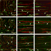
Figure 1
Figure 1
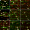
Relationship between enteric neurons and ICC in the gastric fundus. (A,D,G) Cryostat sections through the fundus tunica muscularis revealing CM and LM (cm & lm, respectively). (A) Double label of PGP9.5+ neurons (green) and Kit+ ICC (red) closely apposed to each other in both muscle layers (arrows). (D) Sub-P+ nerve fibers (green) and Kit+ ICC (red) in close apposition (arrows). (G) nNOS+ neurons (green) and Kit+ ICC (red) in close morphological association (arrows in CM). (B,C,E,F,H,I) Double labeled images of flat mounts through the CM of the fundus with Kit and PGP9.5 (B,C) sub-P (E,F) and nNOS (H,I) showing the close anatomical relationship between enteric neurons and ICC (arrows). (B,E,H) Multiple stacks through 10–30 μm. (C,F,I) single sections of 0.4 μm displaying morphological relationship between enteric neurons and ICC in the same optical plane. Single sections in C,F, and I were taken from the confocal stacks in B,E,H. Numerous small rounded Kit+ cells (*) with a morphology resembling mast cells were observed within the LM/myenteric plexus region. N.B. ICC were observed transversing from the CM into the LM (#) in cryostat sections (A, D). Scale bars are indicated on each panel.
Figure 2
Figure 2
Close morphological relationship between enteric neurons and ICC in the gastric antrum. (A–D) Cryostat sections (A,B) and flat mount sections (C,D) of PGP9.5+ neurons (green) and Kit+ ICC (red). PGP9.5+ nerve fibers were observed in close association with ICC (arrows) in cryostat sections cut parallel (A) or transverse (B) to the CM and in flat mounts (C,D) within the CM (cm) but not the LM (lm). (E–H) Double labeling with sub-P and Kit in the antrum revealed a close anatomical relationship between excitatory nerve fibers and ICC. Cryostat sections cut parallel (E) and transverse (F) to the CM and flat mounts (G,H) cut parallel to CM fibers showed close apposition between sub-P containing neurons and ICC-IM (arrows). (I–L) nNOS neurons and ICC were also in close apposition to one another. (I, J) cryostat sections (K,L) flat mounts of nNOS (green) and Kit+ ICC (red) closely apposed to one another in the CM (cm; arrows). Both nerve fibers and ICC were sparse in the LM. (D,H,L) Single sections (0.4 μm) taken from the reconstructed stacks in C,G, and K, respectively. ICC-MY also surrounded myenteric ganglia (mg) but did not penetrate ganglionic sheaths. Numerous small rounded mast cells (*) were observed in the LM and occasionally within the CM. Scale bars are as indicated in each panel.
Figure 3
Figure 3
Morphological relationship between enteric neurons and ICC in the small intestine. (A) Cryostat cross-section through the intestine wall labeled with PGP9.5 and Kit. PGP9.5+ nerve fibers (green) were evenly distributed throughout the CM but Kit+ ICC (red) were observed in close apposition to PGP9.5+ nerve fibers predominantly at the level of the deep muscular plexus (DMP, arrows). (B,C) Flat mounts showing the DMP. PGP9.5+ nerves fibers were in close apposition to ICC (arrows). (D) Flat mount of the myenteric plexus region (also seen in A, arrowheads) showing little or no morphological relationship between PGP9.5+ nerves fibers and ICC-MY in this region of the intestinal wall. (E–G) Relationship between sub-P containing nerve fibers and Kit+ ICC in the intestine. (I–K) Relationship between nNOS containing nerve fibers and Kit+ ICC. Both sub-P and nNOS containing neurons were readily observed throughout the CM but few fibers were observed in the LM. (F,G) and (J,K) show flat mounts of sub-P and nNOS nerve fibers and Kit+ ICC at the level of the DMP, respectively. Excitatory and inhibitory neurons were apposed to ICC in this region of the intestine (arrows). (C,G,K) Single sections of reconstructed stacks in (B,F,J), respectively. (H,L) Cryostat sections labeled with an anti-histamine antibody to identify mast cells in the intestine. Mast cells were located in the mucosa, lamina propria, tunica muscularis and serosa. Scale bars are as indicated on each panel.
Figure 4
Figure 4
Relationship between enteric neurons and ICC within the taenia (t) and inter-taenia (it) regions of the proximal colon. (A,F,K) Cryostat cross-sections of PGP9.5+, sub-P and nNOS containing neurons (green) with Kit+ ICC (red), respectively. Close appositions are seen in the CM and taenia region of the colon (open arrows). (B,C) CM (cm in A,F,K) double labeled with PGP9.5 (green) and Kit (red). (G,H) CM double labeled with sub-P and Kit. (L,M) CM double labeled with nNOS and Kit. (C,H,M) Single sections of reconstructed stacks taken from (B,G,L). ICC ran parallel to and in close apposition with excitatory and inhibitory neurons. (D,I,N) & (E,J,O) Taenia LM and serosal surface of taenia double labeled with PGP9.5 (D,E), sub-P (I,J) and nNOS (N,O) and Kit, respectively. Enteric nerve fibers formed close anatomical associations with ICC throughout the taenia. ICC were observed to form an anastomosing network (*) along and between nerve fibers adjacent to the serosa of the taenia. Scale bars are as indicated in each panel.
Figure 5
Figure 5
Relationship between enteric neurons and PDGFRα+ cells in the gastric fundus. (A,B) Double labeling with PGP9.5 and PDGFRα antibodies reveal close apposition between enteric neurons (red) and PDGFRα+ cells (green) in CM and LM. PGP9.5+ fibers ran parallel to PDGFRα+ cells for at least 130 μm. Double labeling with sub-P (C,D) or nNOS (E,F) reveal that both excitatory and inhibitory nerve fibers run parallel to PDGFRα+ cells for at least 150 μm. Scale bars are as indicated in each panel.
Figure 6
Figure 6
Anatomical relationships between enteric neurons and PDGFRα+ cells in the gastric antrum. (A,D,G) Cryostat sections through the antral wall labeled with PGP9.5 (A, red), sub-P (D, red) and nNOS (G, red) and PDGFRα (green) antibodies. (B,C) Flat mounts of antrum labeled with PGP9.5 and PDGFRα antibodies showing close apposition of PGP9.5+ nerve fibers and PDGFRα+ cells (arrows). Flat mounts labeled with sub-P (E,F) and nNOS (H,I) along with PDGFRα. Sub-P and nNOS nerve fibers (arrows) were closely apposed to PDGFRα+ cells for at least 100 μm and 230 μm, respectively. (C,F,I) Single sections of reconstructed stacks taken from (B,E,H). Scale bars are as indicated in each panel.
Figure 7
Figure 7
Enteric neurons and PDGFRα+ cells in the small intestine. (A–C) PGP9.5+ neurons and PDGFRα+ cells in cryostat sections (A) and flat mounts (B,C). (D–F) Sub-P+ neurons and PDGFRα+ cells in cryostat sections (D) and flat mounts (E,F). (G–I) nNOS+ neurons and PDGFRα+ cells in sections (G) and flat mounts (H,I). Both classes of enteric neurons (red) were closely apposed to PDGFRα+ cells (green, arrows). Note the even distribution of PDGFRα+ cells and close association with nerve fibers throughout the CM compared to the dense distribution of ICC at the DMP and scant distribution within the CM (see Fig. 4). PDGFRα+ cells also surrounded myenteric ganglia (arrowheads) but did not infiltrate ganglionic sheaths. (C,F,I) Single sections of reconstructed stacks taken from (B,E,H) show both cell types are closely apposed in one optical section. Scale bars are as indicated in each panel.
Figure 8
Figure 8
Relationship between enteric neurons and PDGFRα+ cells in the colon. (A–D) PGP9.5+ neurons (red) and PDGFRα+ cells (green) in the taenia (t) and inter-taenia (it) regions of the proximal colon. (A) Cryostat cross-section in the region of the taenia and non-taenia revealing LM (lm) and CM (cm). Close relationships between enteric nerve fibers and PDGFRα+ cells exist (open arrows). (B,C) Confocal reconstructions of PGP9.5+ fibers and PDGFRα+ cells in the CM showing the close apposition (closed arrows). (D) Myenteric ganglia revealing PDGFRα+ cells lie on the surface and between myenteric ganglia. (E–H) Sub-P+ neurons and PDGFRα+ cells and (I–L) nNOS+ neurons and PDGFRα+ cells in the proximal colon. Both neuron types (red) were closely apposed to PDGFRα+ cells (green; arrows) in the CM (F,G for sub-P and J,K for nNOS). (H) A reconstruction through the myenteric region showing a single sub-P+ nerve fiber not encased within a ganglionic sheath and with no apparent structural relationship with PDGFRα+ cells. (L) nNOS nerve fibers also observed in close apposition to PDGFRα+ cells in the taenia muscle layer.

Similar articles

Cited by

References

    1. Maeda H, Yamagata A, Nishikawa S, Yoshinaga K, Kobayashi S, Nishi K. Requirement of c-kit for development of intestinal pacemaker system. Development. 1992;116:369–75. - PubMed
    1. Ward SM, Burns AJ, Torihashi S, Sanders KM. Mutation of the proto-oncogene c-kit blocks development of interstitial cells and electrical rhythmicity in murine intestine. J Physiol. 1994;480(Pt 1):91–7. - PMC - PubMed
    1. Ward SM, Beckett EA, Wang X, Baker F, Khoyi M, Sanders KM. Interstitial cells of Cajal mediate cholinergic neurotransmission from enteric motor neurons. J Neurosci. 2000;20:1393–403. - PMC - PubMed
    1. Huizinga JD, Thuneberg L, Kluppel M, Malysz J, Mikkelsen HB, Bern-stein A. W/kit gene required for interstitial cells of Cajal and for intestinal pacemaker activity. Nature. 1995;373:347–9. - PubMed
    1. Ordög T, Ward SM, Sanders KM. Interstitial cells of cajal generate electrical slow waves in the murine stomach. J Physiol. 1999;518(Pt 1):257–69. - PMC - PubMed

Publication types

MeSH terms

Substances