. 2012 Aug 15;31(16):3480-93.
doi: 10.1038/emboj.2012.185. Epub 2012 Jul 17.

The Reb1-homologue Ydr026c/Nsi1 is required for efficient RNA polymerase I termination in yeast

Affiliations

The Reb1-homologue Ydr026c/Nsi1 is required for efficient RNA polymerase I termination in yeast

Alarich Reiter et al. EMBO J. .

Abstract

Several DNA cis-elements and trans-acting factors were described to be involved in transcription termination and to release the elongating RNA polymerases from their templates. Different models for the molecular mechanism of transcription termination have been suggested for eukaryotic RNA polymerase I (Pol I) from results of in vitro and in vivo experiments. To analyse the molecular requirements for yeast RNA Pol I termination, an in vivo approach was used in which efficient termination resulted in growth inhibition. This led to the identification of a Myb-like protein, Ydr026c, as bona fide termination factor, now designated Nsi1 (NTS1 silencing protein 1), since it was very recently described as silencing factor of ribosomal DNA. Possible Nsi1 functions in regard to the mechanism of transcription termination are discussed.


Conflict of interest statement

The authors declare that they have no conflict of interest.

Figures

Figure 1
Figure 1
Establishment of an in vivo system to study Pol I termination. (A) Schematic representation of one repeat unit of the yeast rDNA locus and of the genetic modifications performed by insertion of DNA elements into ITS1. The direction of transcription at the 35S rRNA gene and 5S rRNA gene is marked with arrows. The coding sequences for mature 18S, 5.8S, 25S and 5S rRNAs (rectangles), external transcribed spacers (ETS1, ETS2), internal transcribed spacers (ITS1, ITS2), as well as a T-rich element (filled square), two predicted Reb1-binding sites (Reb1-BS; filled circle), a sequence coding for a Rnt1-cleavage site (Rnt1, arrowhead) and a RFB (filled rounded rectangle) are indicated. Additional DNA elements were inserted in the ITS1 region of every rDNA repeat at the AflII restriction site: WT refers to a WT-ITS1 without insertion. ITS1-TTF-I-BS refers to an insertion of a single DNA-binding site of the mouse Pol I transcription termination factor TTF-I (filled oval). ITS1-LexA-BS refers to an insertion of a DNA-binding cluster for the bacterial LexA protein (filled hexagons). ITS1-T-rich-TERM refers to the DNA element(s) at the 3′-end of the yeast 35S rRNA gene, which has been implicated in Pol I termination, starting with a T-rich element and ending after the RFB (for description of the symbols see above). ITS1-TERM lacks the T-rich element and is referred to as the terminator element in this study. ITS1-TERM-mut1 and ITS1-TERM-mut2, respectively, are derivatives that carry mutations in the putative Reb1 DNA-binding site, which either abolish or enhance Reb1 binding in vitro (Lang and Reeder, 1993). The respective sequences of the WT Reb1-binding site as well as the two mutated sequences are indicated, with the altered bases labelled in grey. (B) Integration of the yeast terminator element into ITS1 leads to growth reduction, whereas the integration of DNA-binding sites of heterologous proteins shows no growth phenotype. Yeast strains (y1598, y1599, y2038, y2042, y2273, y2274, y2339) carrying rDNA loci with a ITS1-WT, or ITS1 DNA element insertions as described in Figure 1A (ITS1-T-rich-TERM, ITS1-TTF-I-BS, ITS1-LexA-BS, ITS1-TERM, ITS1-TERM-mut1, ITS1-TERM-mut2) were grown to an OD600 of 0.2 at 30°C. The same amount of cells was spotted on YPD plates in a 1:10 serial dilution series, as indicated above the panels and incubated for 2 days at 30°C.
Figure 2
Figure 2
The Reb1-homologue Ydr026c associates with the yeast terminator element in vivo. (A) Schematic representation and amino-acid sequence alignment of Ydr026c and Reb1. Regions sharing homology with the Myb-DNA-binding domain are depicted as blue boxes in the schematic representation or as blue lines on top of the alignment. Yellow or green rectangles denote similar or identical amino-acid residues, respectively. (B) ChEC analyses reveal Ydr026c binding at the terminator element integrated at ITS1 in vivo. Yeast strains (y2050, y2054, y2090, y2093, y2241) carrying rDNA loci with a ITS1-WT, or ITS1 DNA element insertions as described in Figure 1A (ITS1-TERM, ITS1-TERM-mut1), and expressing either Reb1–MNase, or Ydr026c–MNase fusion proteins from the respective chromosomal gene locus, were grown to exponential phase. After formaldehyde crosslinking, cells were harvested and crude nuclei were isolated. The nuclei suspension was incubated in the absence (0) or presence of calcium activating DNA cleavage by MNase fusion proteins for the times indicated on top of the panels (min ChEC). DNA was isolated, linearized with the indicated restriction enzyme (KpnI), separated in an agarose gel and analysed in a Southern blot by indirect endlabelling using the indicated probe (KpnI (ITS2)) (Supplementary Table 4). The cartoon on the right shows a map of the corresponding rDNA-fragment of around 10 kb. The positions of the 18S, 5.8S and 25S rRNA coding sequences, of the integrated terminator element or derivative (ITS1-TERM, ITS1-TERM-mut1) and of the target sequence of the radioactive probe are depicted. An arrow on the left marks the position of the full-length KpnI fragment. An asterisk on the left labels an Reb1–MNase mediated cleavage event at the promoter-proximal Reb1-binding site. (C) ChEC analysis reveals specific Ydr026c–MNase mediated cleavage at the terminator element located at the 3′-end of the rRNA gene. (y2050, y2090) carrying rDNA loci with a ITS1-WT and expressing either Reb1–MNase, or Ydr026c–MNase fusion proteins from the respective chromosomal gene locus, were grown to exponential phase. ChEC analysis were performed as in (B); however different restriction enzymes and probes (Supplementary Table 4) were used to analyse the 35S rRNA gene 5′-end and upstream region (panels labelled ‘Promoter’), and the 35S rRNA gene 3′-end and downstream region (panels labelled ‘Terminator’), respectively. The cartoon on the right shows a map of the corresponding rDNA-fragments of around 5.6 and 2.1 kb, respectively. The positions of the 5S, 5.8S, 18S and 25S rRNA coding sequences, Reb1-binding sites, the RFB and the transcription start sites (arrows) and of the target sequence of the radioactive probe are depicted. The L-element refers to the left flanking region of the yeast rDNA locus, which is part of the IGS in yeast strains carrying the reconstituted rDNA loci. An arrow on the left marks the position of the full-length XcmI and AvaII fragments. Asterisks on the left label DNA-fragments of unknown origin, potentially cross-hybridizing with probes rDNp or 5S. We note an additional band between RFB and 5S, which appears after Calcium addition to nuclei from cells expressing an Reb1–MNase fusion protein. This fragment might be a product of non-specific MNase cleavage at a hypersensitive site. (D) ChIP reveals a reciprocal relationship of Reb1 and Ydr026c binding at the promoter and at the 3′-end of the 35S rRNA gene. Yeast strains (y2050, y2090) expressing Ydr026c–, and Reb1–MNase fusion proteins with a C-terminal triple HA-tag were grown to exponential phase. Cells were treated with formaldehyde and ChIP experiments were performed as described in Materials and methods. The amounts of specific DNA-fragments present in the input and retained on the beads were determined by quantitative PCR with primer pairs amplifying the promoter region (969/970, see region 1 in the schematic representation in Figure 5), the ITS2 region (2864/2865, see region 4 in the schematic representation in Figure 5), or the terminator element (2884/2885, see region 7 in the schematic representation in Figure 5). The bar graphs depict percent of total input DNA retained after ChIP of the respective triple HA-tagged protein (see legend of the graph). Error bars represent the standard deviation of three independent ChIP experiments, each of which was analysed in triplicate quantitative PCR reactions.
Figure 3
Figure 3
Growth defects observed in yeast strains carrying the terminator element integrated at the rDNA ITS1 region depend on expression of Ydr026c. Yeast strains (y1599, y2042, y2276, y2281) carrying rDNA loci with a ITS1-WT, or an ITS1 DNA element insertion as described in Figure 1A (ITS1-TERM), and being WT in YDR026C or being deleted in the gene (ydr026cΔ) were transformed with plasmid 1803 (pGAL-YDR026C) for galactose inducible expression of the protein. Cells were grown in selective complete media lacking leucine (SC-LEU) and containing raffinose to an OD600 of 0.2 at 30°C. The same amount of cells was spotted on SC-Leu plates containing either glucose (GLC) or galactose (GAL) as carbon source in 1:10 serial dilution series, as indicated above the panels and incubated for 2–3 days at 30°C.
Figure 4
Figure 4
Binding of mouse TTF-I, but not of bacterial LexA to their respective binding sites integrated at ITS1 of yeast rDNA leads to growth defects. (A) Yeast strains carrying TTF-I-binding sites integrated at ITS1 of the rDNA are impaired in growth upon heterologous inducible expression of mouse TTF-I protein. Yeast strains (y1598, y1599, y2038) carrying rDNA loci with a ITS1-WT, or ITS1 DNA element insertions as described in Figure 1A (ITS1-TTF-I-BS, ITS1-LexA-BS), were transformed with plasmids 230, 1806, 1808 (pGAL, pGAL-LexA–MNase, pGAL-TTF-I–MNase) as a control, or for galactose inducible expression of the respective DNA-binding protein. Cells were grown in selective complete media lacking leucine (SC-LEU) and containing raffinose to an OD600 of 0.2 at 30°C. The same amount of cells was spotted on SC-Leu plates containing either glucose (GLC) or galactose (GAL) as carbon source in 1:10 serial dilution series, as indicated above the panels and incubated for 2 and 3 days at 30°C, respectively. (B) Heterologous expression of TTF-I–MNase and LexA–MNase leads to specific cleavage events at their binding sites integrated at ITS1 of the rDNA. Yeast strains (y1598, y1599, y2038) carrying rDNA loci with a ITS1-WT, or ITS1 DNA element insertions as described in Figure 1A (ITS1-TTF-I-BS, ITS1-LexA-BS), were transformed with plasmids 1806, 1808 (pGAL-LexA–MNase, pGAL-TTF-I–MNase) for galactose inducible expression of the respective DNA-binding protein. Cells were grown in selective complete media lacking leucine (SC-LEU) and containing raffinose to an OD600 0.3, before galactose was added to a final concentration of 2% and incubation was continued for 4 h at 30°C. Cells were subjected to ChEC analysis as described in the legend to Figure 2B. An arrow on the left marks the position of the full-length KpnI fragment. Asterisks on the left label DNA-fragments of unknown origin, potentially cross-hybridizing with probe ITS2.
Figure 5
Figure 5
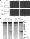
Pol I association with rRNA gene sequences with or without DNA-binding sites for Pol I termination factors inserted into ITS1 and correlation with steady-state rRNA levels. (A) Effects of YDR026C deletion on Pol I occupancy in proximity of the rDNA terminator element as analysed by ChIP. Yeast strains (y1620, y2094, y2229, y2234) carrying rDNA loci with a ITS1-WT, or an ITS1 DNA element insertion as described in Figure 1A (ITS1-TERM), being WT in YDR026C or being deleted in the gene (ydr026cΔ), and expressing the Pol I subunit Rpa190 as a MNase fusion protein with a C-terminal triple HA-tag were grown to exponential phase. ChIP experiments were performed as described in the legend to Figure 2D. The amounts of specific DNA-fragments present in the input and retained on the beads were determined by quantitative PCR with primer pairs amplifying the regions 1–9 of the rDNA depicted in the schematic representation on the bottom of the figure. Primer pairs used were 3249/3250 (1), 3022/3023 (2), 969/970 (3), 712/713 (4), 1049/2863 (5) 2864/2865 (6), 710/711 (7), 2882/2883 (8), 2884/2885 (9), 2886/2887 (10), 2421/2422 (11), 2419/2420 (12), 920/921 (13), 613/614 (14). The bar graphs depict percent of total input DNA retained after ChIP of the triple HA-tagged Rpa190. Error bars represent the standard deviation of three independent ChIP experiments, each of which was analysed in triplicate quantitative PCR reactions. Note: the slight enrichment of fragments upstream of the Pol I promoter and the 5S rRNA gene co-purifying with Pol I, compared with the PDC1 fragment could be due to some residual Pol I association with the ‘non-transcribed’ rDNA regions. Alternatively, the 5S rRNA gene and upstream sequences could be enriched by being part of larger DNA-fragments produced during the sonication process for ChIP sample preparation, which contain the 35S rRNA coding sequence and can thus be co-precipitated with Pol I. (B) Deletion of YDR026c leads to an increased production of radiolabelled RNAs hybridizing to DNA sequences downstream of the terminator region in transcription run on experiments. After transcription run-on conditions, radiolabelled RNA from isolated nuclei of either WT cells (y1599) or the YDR026c deletion strain (y2276) were hybridized to single-stranded DNA immobilized on a Nylon membrane. The single-stranded DNA probes were derived by PCR from genomic DNA using the same primer pairs as for the ChIP experiment described in Figure 5A. After hybridization with RNAs from YDR026C, WT and YDR026c deletion strains membranes were exposed to a imaging plate (Fujifilm) and radioactive signals were quantified using the FLA-3000 imaging system (Fujifilm). The hybridization signal obtained with DNA probe 3 spanning the Pol I promoter was used for normalization. Mean values and standard deviations of the fold enrichment over amplicon 3 of three hybridization experiments (one biological replicate and one technical replicate) are depicted in form of a bar graph. Note: the quantitative data were not corrected for content of incorporated 32P-UMP. (C) Insertion of the terminator element into ITS1 leads to a decreased density of Pol I molecules downstream of the Reb1-binding site as visualized in EM analysis. Miller chromatin spreading analyses were performed with yeast strains (y1599, y2042) carrying rDNA loci with a ITS1-WT, or an ITS1 DNA element insertion as described in Figure 1A (ITS1-TERM). Two representative electron micrographs from typical ‘Christmas-tree’-like structures likely corresponding to actively transcribed rRNA genes in the two different strains are shown on the left. A bar on the bottom indicates the scale of the images. The length of the regions with high polymerase density (electron-dense stem of the ‘Christmas trees’) with extending nascent rRNA transcripts (branches of the ‘Christmas trees’) was determined for a total number of 20 and 26 molecules for strains y2042 and y1599, respectively (see Supplementary Figure S2 for details). The average length and standard deviation are depicted in the graph on the right. (D) Insertion of the terminator element into ITS1 leads to an imbalance in the steady-state level of 18S and 25S rRNAs. Yeast strains (y1599, y2042, y2273, y2274) carrying rDNA loci with a ITS1-WT, or ITS1 DNA element insertions as described in Figure 1A (ITS1-T-rich-TERM, ITS1-TERM, ITS1-TERM-mut1) were grown to exponential phase. RNA was extracted, separated by gel electrophoresis and Northern blot analysis was performed as described in Materials and methods. Membranes were hybridized with radioactively labelled oligonucleotides 205 and 212 specific for 18S, and 25S rRNA, respectively. Radioactive signals on the blot were quantified by phospho imager analysis as described in Materials and methods. The graph depicts the 25S/18S rRNA ratio, which was normalized to the value obtained with strain y1599 (ITS1-WT), which was arbitrarily set to 1.

Similar articles

Cited by

References

    1. Bartsch I, Schoneberg C, Grummt I (1988) Purification and characterization of TTFI, a factor that mediates termination of mouse ribosomal DNA transcription. Mol Cell Biol 8: 3891–3897 - PMC - PubMed
    1. Braglia P, Heindl K, Schleiffer A, Martinez J, Proudfoot NJ (2010a) Role of the RNA/DNA kinase Grc3 in transcription termination by RNA polymerase I. EMBO Rep 11: 758–764 - PMC - PubMed
    1. Braglia P, Kawauchi J, Proudfoot NJ (2010b) Co-transcriptional RNA cleavage provides a failsafe termination mechanism for yeast RNA polymerase I. Nucleic Acids Res 39: 1439–1448 - PMC - PubMed
    1. Bryk M, Banerjee M, Murphy M, Knudsen KE, Garfinkel DJ, Curcio MJ (1997) Transcriptional silencing of Ty1 elements in the RDN1 locus of yeast. Genes Dev 11: 255–269 - PubMed
    1. Chasman DI, Lue NF, Buchman AR, LaPointe JW, Lorch Y, Kornberg RD (1990) A yeast protein that influences the chromatin structure of UASG and functions as a powerful auxiliary gene activator. Genes Dev 4: 503–514issn: 0890-9369 - PubMed

Publication types

MeSH terms

Substances