. 2013 Jan;6(1):156-66.
doi: 10.1038/mi.2012.59. Epub 2012 Jul 18.

Airway CD8(+) T cells induced by pulmonary DNA immunization mediate protective anti-viral immunity

Affiliations
Free PMC article

Airway CD8(+) T cells induced by pulmonary DNA immunization mediate protective anti-viral immunity

M Bivas-Benita et al. Mucosal Immunol. 2013 Jan.
Free PMC article

Abstract

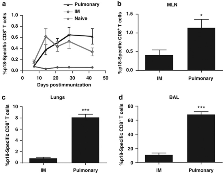
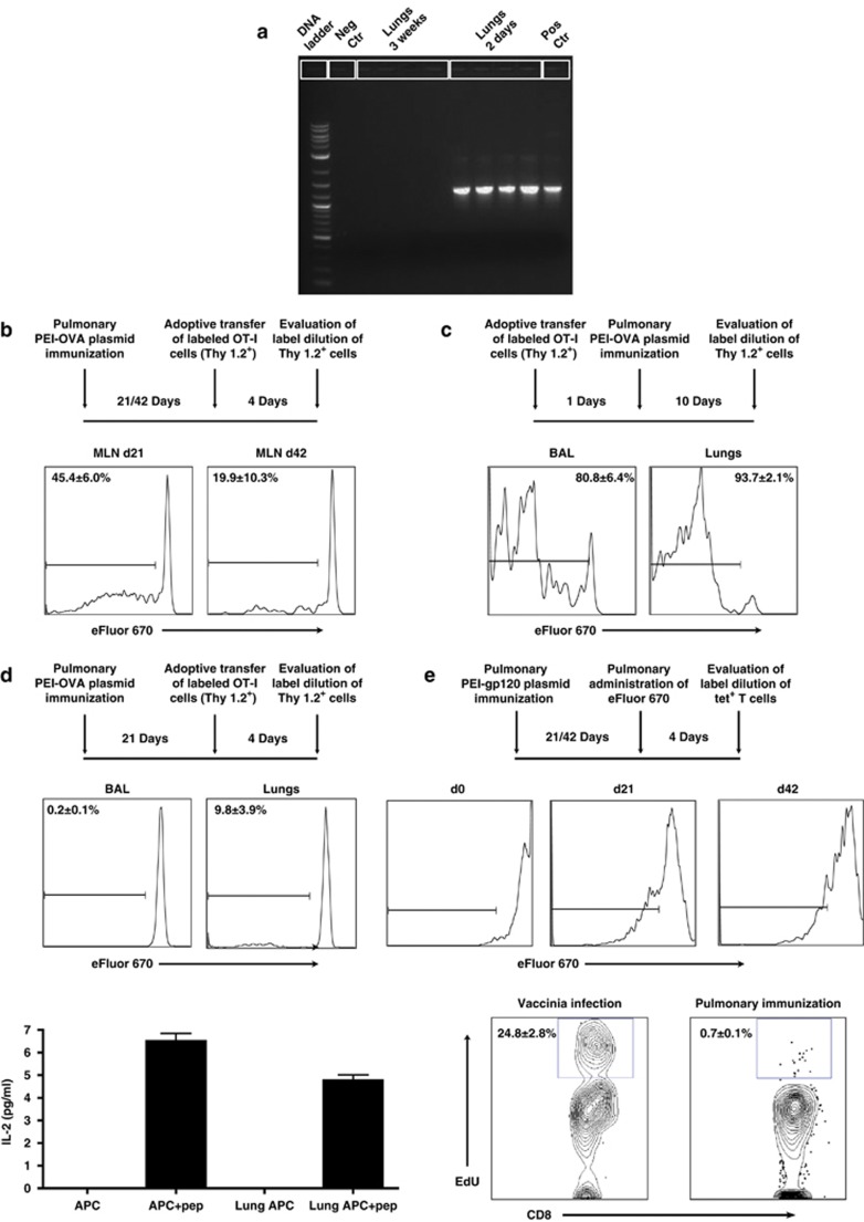
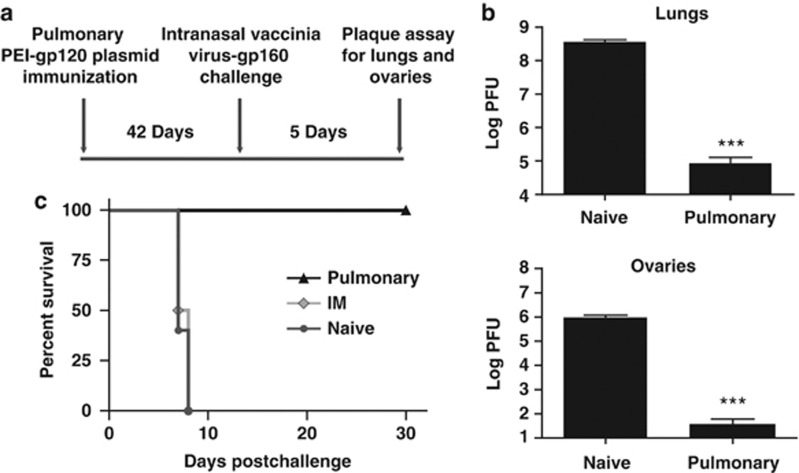
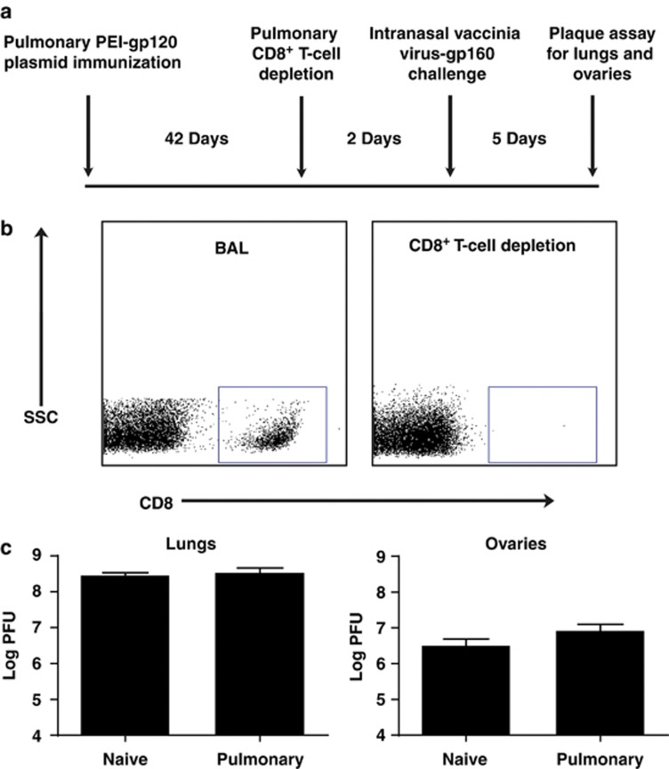
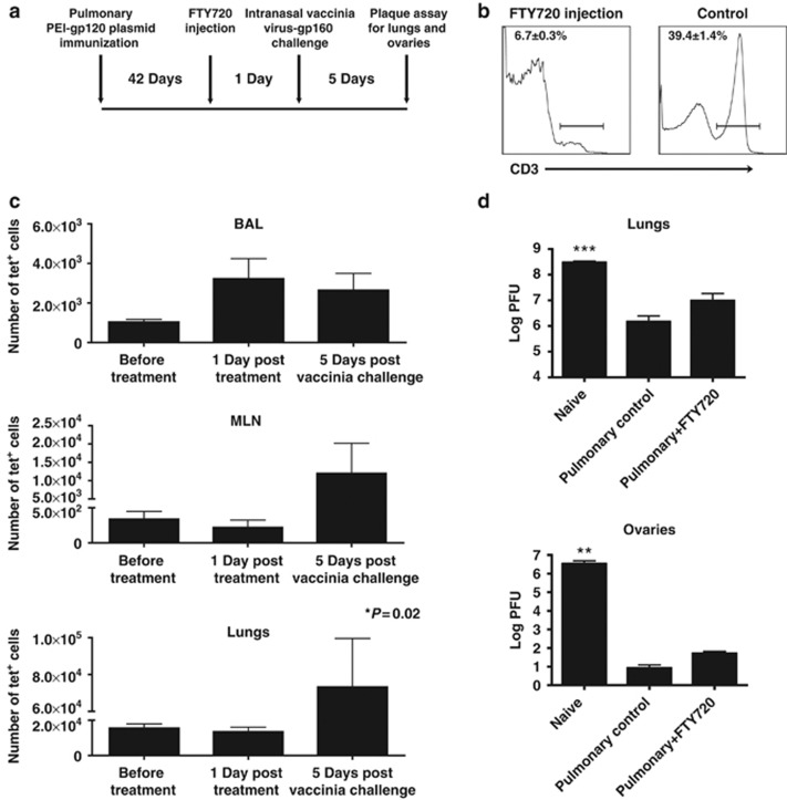
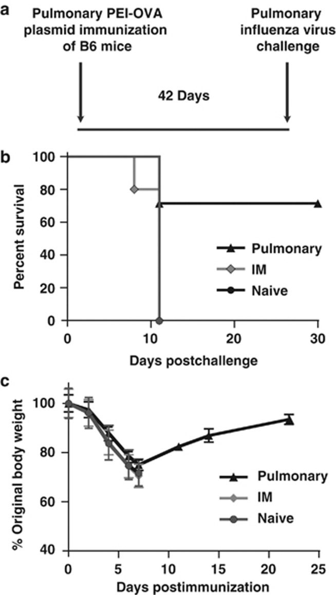
Vaccination strategies for protection against a number of respiratory pathogens must induce T-cell populations in both the pulmonary airways and peripheral lymphoid organs. In this study, we show that pulmonary immunization using plasmid DNA formulated with the polymer polyethyleneimine (PEI-DNA) induced antigen-specific CD8(+) T cells in the airways that persisted long after antigen local clearance. The persistence of the cells was not mediated by local lymphocyte proliferation or persistent antigen presentation within the lung or airways. These vaccine-induced CD8(+) T cells effectively mediated protective immunity against respiratory challenges with vaccinia virus and influenza virus. Moreover, this protection was not dependent upon the recruitment of T cells from peripheral sites. These findings demonstrate that pulmonary immunization with PEI-DNA is an efficient approach for inducing robust pulmonary CD8(+) T-cell populations that are effective at protecting against respiratory pathogens.


Figures

Figure 1
Figure 1
Effect of the route of immunization on systemic and mucosal p18-specific CD8+ T-cell responses. Mice were immunized by the intramuscular (IM) route with 40 μg of plasmid DNA expressing HIV-1 HXB2 gp120 protein or by the pulmonary route with 40 μg of this DNA formulated with polyethyleneimine (PEI). (a) Kinetics of the p18-specific CD8+ T-cell response in the peripheral blood of immunized mice. p18-specific CD8+ T-cell responses in (b) mediastinal lymph nodes (MLN), (c) pulmonary lymphocytes and (d) broncho-alveolar lavage (BAL) lymphocytes of mice on day 42 following IM immunization with DNA-gp120 or by the pulmonary route with PEI-DNA-gp120. Data represent means of 4–8 mice per group±s.e.
Figure 2
Figure 2
Persistence of pulmonary CD8+ T cells is independent of the duration of antigen presentation in the airways and lungs or local T-cell proliferation. (a) Lungs were harvested from B6 mice 2 days and 3 weeks post pulmonary PEI-DNA-OVA immunization and plasmid DNA isolated from the tissue. The OVA sequence was amplified from plasmid DNA by PCR. The plasmid DNA-OVA used for immunization served as positive control (pos ctr) while plasmid DNA without the OVA insert served as the negative control (neg ctr). (b) B6.PL (Thy 1.1+) mice were immunized by the pulmonary route with PEI-DNA-OVA and 21 and 42 days later, received adoptively transferred labeled OT-I cells (Thy 1.2+) by intravenous injection. The dilution of eFluor 670 in donor Thy1.2+ tetramer+ CD8+ T cells was evaluated in mediastinal lymph nodes (MLN) of the immunized mice 4 days post transfer. Representative histograms show the average percentages±s.e. of proliferating Thy1.2+ tetramer+ CD8+ T cells (3–4 mice per group). (c) Labeled OT-I cells (Thy 1.2+) were adoptively transferred to the airway lumen of B6.PL mice (Thy 1.1+). One day later, mice were immunized by the pulmonary route with PEI-DNA-OVA, and 10 days later, donor tetramer-binding CD8+ T cells were isolated from the BAL and lungs, and evaluated for the dilution of eFluor 670 staining. (d) B6-PL (Thy 1.1+) mice were immunized by the pulmonary route with PEI-DNA-OVA and 21 days later received adoptively transferred labeled OT-I cells (Thy 1.2+) in their airway lumens. The dilution of eFluor 670 in donor Thy1.2+ tetramer+ CD8+ T cells was evaluated 4 days later in BAL and lungs. Representative histograms show the average percentages±s.e. of proliferating Thy1.2+ tetramer+ CD8+ T cells (6–8 mice per group). Local antigen presentation was also evaluated in lungs of B6 mice immunized by the pulmonary route with PEI-DNA-OVA. Lungs were harvested 6 weeks following immunization and co-cultured with the RF.33.70 hybridoma overnight. The levels of IL-2 secretion from the RF33.70 hybridoma in response to SIINFEKL presentation were determined using a mouse IL-2 enzyme-linked immunosorbent assay. Bars represent the average±s.e. of secreted IL-2 (3 mice per group). (e) Mice were inoculated by the pulmonary route with PEI-DNA-gp120, and 3 and 6 weeks later eFluor 670 was applied to the airways to stain local resident T cells. Four days later, the dilution of eFluor 670 staining of tetramer+ CD8+ T cells in the BAL was assessed. Histograms representative of 3–8 mice per time point are shown. Proliferation of antigen-specific CD8+ T cells was also evaluated in the BAL of immunized Balb/c mice 6 weeks following pulmonary PEI-DNA-gp120 immunization. Mice were injected with 250 μg 5-ethynyl-2′-deoxyuridine (EdU) intraperitoneally 12 h before sacrifice. Cells isolated from the BAL were stained with monoclonal antibodies to CD4 and CD8, fixed, permeabilized, and EdU detected with the Alexa Fluor 647 Click-iT EdU flow cytometry assay kit. Representative plot show the average percentages±s.e. of EdU incorporation by BAL CD8+ T cells (3 mice per group). BAL CD8+ T cells from mice infected intranasally with rVac-gp160 were used as a positive control for local proliferation. APC, antigen-presenting cells; BAL, broncho-alveolar lavage; ctr, control; IL-2, interleukin 2; pep, peptide; PEI-DNA-OVA, polyethyleneimine-DNA-Ovalbumin complexes.
Figure 3
Figure 3
Pulmonary PEI-DNA immunization protects against intranasal vaccinia virus challenge. (a) Mice were primed by intramuscular (IM) inoculation with 40 μg DNA-gp120 or pulmonary inoculation of PEI-DNA-gp120 and challenged 6 weeks later by intranasal instillation of a lethal dose (2 × 10⁶ PFU) of vaccinia virus-gp160. Protective immunity was monitored by evaluating (b) viral titers in the lungs and ovaries, and (c) mouse survival. Statistically significant differences between groups were determined using the Student's t-test (***P<0.001). Data represent means of 4–9 mice per group±s.e. PEI, polyethyleneimine; PFU, plaque-forming units.
Figure 4
Figure 4
Protective immunity following pulmonary PEI-DNA-gp120 immunization is mediated by CD8+ T cells. (a) Mice were inoculated by the pulmonary route with PEI-DNA-gp120 and 6 weeks later CD8+ T cells were depleted by pulmonary instillation of depleting antibodies. Mice were challenged 2 days later by intranasal instillation of a lethal dose (2 × 10⁶ PFU) of vaccinia virus-gp160. Tissue viral titers were evaluated 5 days post-challenge. (b) Depletion of CD8+ T cells in BAL of immunized mice. (c) Vaccinia virus-gp160 titers in lungs of CD8+ T-cell–depleted mice. Data represent means of 3–5 mice per group±s.e. BAL, broncho-alveolar lavage; PEI-DNA-gp120, polyethyleneimine-DNA-gp120 complexes; PFU, plaque-forming units; SSC, side scatter.
Figure 5
Figure 5
Protection following pulmonary DNA immunization is independent of the peripheral T-cell supply. (a) Mice were immunized by the pulmonary route with PEI-DNA-gp120, and 6 weeks later the mice were treated with FTY720. The following day the mice were challenged by intranasal instillation of a lethal dose (2 × 10⁶ PFU) of vaccinia virus-gp160. Five days later, lungs and ovaries were harvested and assessed for viral titers. (b) The average percentages ± s.e. of circulating T cells in the blood of 5–7 mice per group with or without FTY720 treatment. (c) The total numbers of tetramer-positive CD8+ T cells (mean±s.e.) before FTY720 treatment, 1 day post treatment and 5 days following intranasal vaccinia virus-gp160 challenge. (d) Viral titers in the lungs and ovaries of unimmunized, pulmonary immunized, and pulmonary immunized+FTY720-treated mice that were challenged intranasally with vaccinia virus-gp160. Data represent means of 3–5 mice per group±s.e. BAL, broncho-alveolar lavage; MLN, mediastinal lymph nodes; PEI-DNA-gp120, polyethyleneimine-DNA-gp120 complexes; PFU, plaque-foming units.
Figure 6
Figure 6
CD8+ T-cell immunity induced by pulmonary DNA immunization protects against influenza virus challenge. (a) C57BL/6 mice were immunized by the pulmonary route with PEI-DNA-OVA, and 6 weeks later they were infected intranasally with 105 EID50 PR8-SIINFEKL influenza virus. (b) Protective immunity was assessed by the survival rates of mice post infection and (c) mouse weight loss following challenge. Data represent means of 5–7 mice per group±s.e. IM, intramuscular; PEI-DNA-gp120, polyethyleneimine-DNA-gp120 complexes.

References

    1. Kohlmeier J.E., Woodland D.L. Immunity to respiratory viruses. Annu. Rev. Immunol. 2009;27,:61–82. - PubMed
    1. Hogan R.J., et al. Activated antigen-specific CD8+ T cells persist in the lungs following recovery from respiratory virus infections. J. Immunol. 2001;166,:1813–1822. - PubMed
    1. de Bree G.J., van Leeuwen E.M., Out T.A., Jansen H.M., Jonkers R.E., van Lier R.A. Selective accumulation of differentiated CD8+ T cells specific for respiratory viruses in the human lung. J. Exp. Med. 2005;202,:1433–1442. - PMC - PubMed
    1. Woodland D.L., Scott I. T cell memory in the lung airways. Proc. Am. Thorac. Soc. 2005;2,:126–131. - PMC - PubMed
    1. Bivas-Benita M., et al. Pulmonary delivery of chitosan-DNA nanoparticles enhances the immunogenicity of a DNA vaccine encoding HLA-A*0201-restricted T-cell epitopes of Mycobacterium tuberculosis. Vaccine. 2004;22,:1609–1615. - PubMed

Publication types

MeSH terms