. 2012 Dec;56(6):2316-27.
doi: 10.1002/hep.25938.

Nicotinamide adenine dinucleotide phosphate oxidase in experimental liver fibrosis: GKT137831 as a novel potential therapeutic agent

Affiliations

Nicotinamide adenine dinucleotide phosphate oxidase in experimental liver fibrosis: GKT137831 as a novel potential therapeutic agent

Tomonori Aoyama et al. Hepatology. 2012 Dec.

Abstract

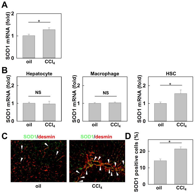
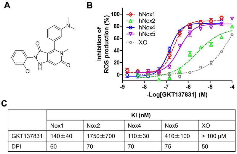
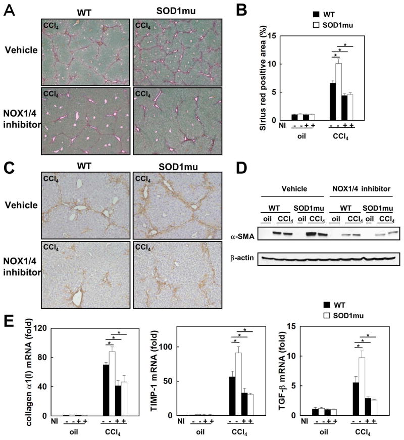
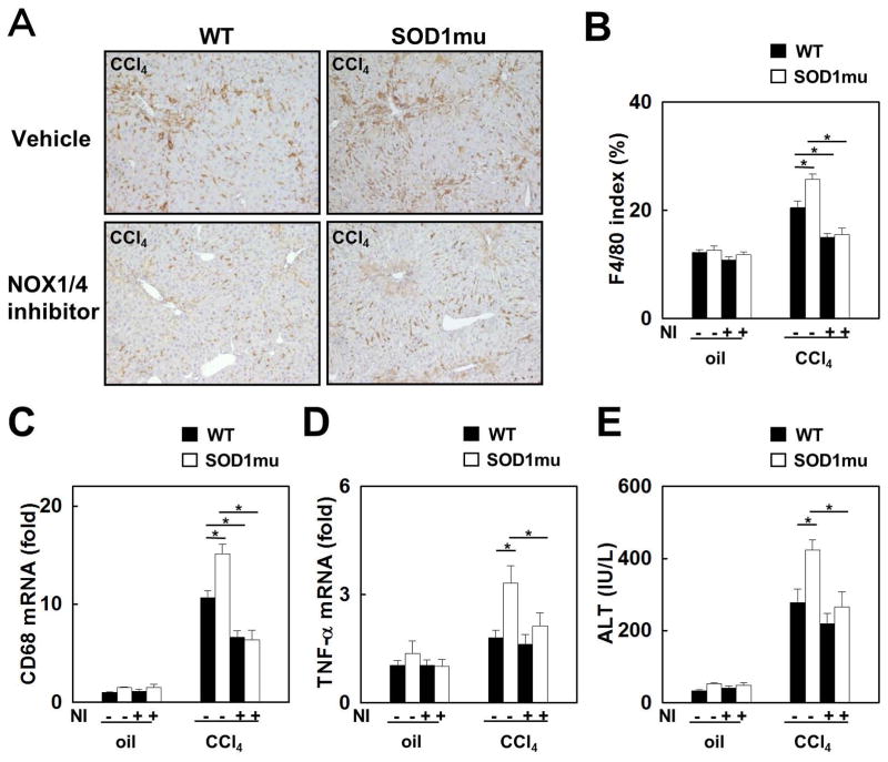
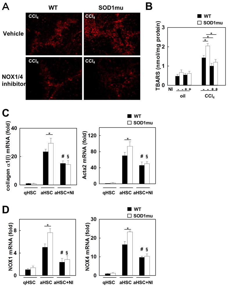
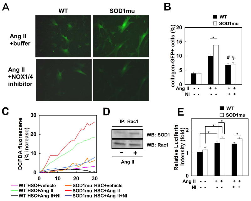
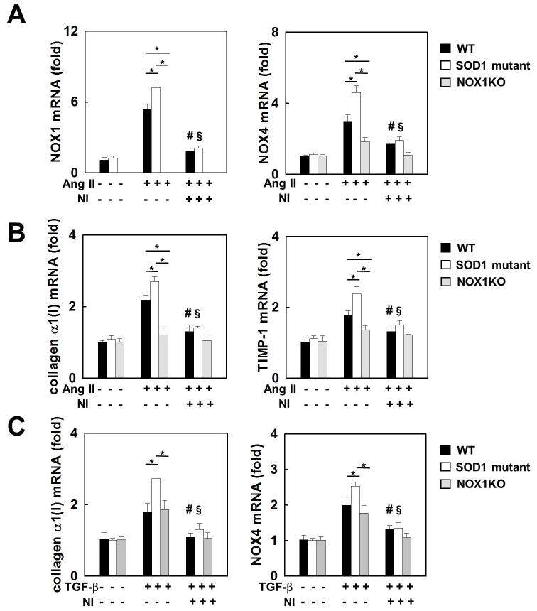
Nicotinamide adenine dinucleotide phosphate (NADPH) oxidase (NOX) generates reactive oxygen species (ROS) in hepatic stellate cells (HSCs) during liver fibrosis. In response to fibrogenic agonists, such as angiotensin II (Ang II), the NOX1 components form an active complex, including Ras-related botulinum toxin substrate 1 (Rac1). Superoxide dismutase 1 (SOD1) interacts with the NOX-Rac1 complex to stimulate NOX activity. NOX4 is also induced in activated HSCs/myofibroblast by increased gene expression. Here, we investigate the role of an enhanced activity SOD1 G37R mutation (SODmu) and the effects of GKT137831, a dual NOX1/4 inhibitor, on HSCs and liver fibrosis. To induce liver fibrosis, wild-type (WT) and SOD1mu mice were treated with CCl(4) or bile duct ligation (BDL). Then, to address the role of NOX-SOD1-mediated ROS production in HSC activation and liver fibrosis, mice were treated with a NOX1/4 inhibitor. Fibrosis and ROS generation was assessed by histology and measurement of thiobarbituric acid reactive substances and NOX-related genes. Primary cultured HSCs isolated from WT, SODmu, and NOX1 knockout (KO) mice were assessed for ROS production, Rac1 activity, and NOX gene expression. Liver fibrosis was increased in SOD1mu mice, and ROS production and Rac1 activity were increased in SOD1mu HSCs. The NOX1/4 inhibitor, GKT137831, attenuated liver fibrosis and ROS production in both SOD1mu and WT mice as well as messenger RNA expression of fibrotic and NOX genes. Treatment with GKT137831 suppressed ROS production and NOX and fibrotic gene expression, but not Rac1 activity, in SOD1mut and WT HSCs. Both Ang II and tumor growth factor beta up-regulated NOX4, but Ang II required NOX1.

Conclusions: SOD1mu induces excessive NOX1 activation through Rac1 in HSCs, causing enhanced NOX4 up-regulation, ROS generation, and liver fibrosis. Treatment targeting NOX1/4 may be a new therapy for liver fibrosis.


Figures

Fig. 1
Fig. 1. SOD1 expression is enhanced in hepatic stellate cells (HSCs) in the fibrotic liver
Livers from WT mice were analyzed after 12 injections of CCl4 or vehicle (n=5) (A) Hepatic mRNA expression of SOD1 was measured by quantitative real-time PCR. (B) mRNA levels of SOD1 in hepatocytes, macrophages, and HSCs isolated from vehicle- or CCl4 treated WT mice detected by quantitative real-time PCR (n=3). (C) SOD1 expression (green) was detected by fluorescence microscopy via containing with desmin (red). (D) Percentage of SOD1 positive cells in desmin positive cells.
Fig. 2
Fig. 2. Pharmacological profile of GKT137831, a dual Nox1/Nox4 inhibitor
(A) Chemical structure of the dual Nox1/Nox4 inhibitor GKT137831. (B) Inhibition of Nox-dependent ROS production by GKT137831: concentration-response curves of GKT137831 on membranes prepared from cells specifically overexpressing hNox1 (◇), hNox2 (△), hNox4 (○), hNox5 (▽) and on Xanthine Oxidase (XO) (○). Km of NADPH for hNox1, hNox2, hNox4 and hNox5 and was 70±10mM, 16±3mM, 120±20mM and 70 ±10mM respectively and Km of Xanthine for XO was 6±1mM. Results are from one experiment performed in triplicate, representative of at least three performed. Values are means±SEM. (C) Inhibition constants (Ki) of GKT137831 and DPI on hNox1, hNox2, hNox4, hNox5 and XO.
Fig. 3
Fig. 3. Enhanced liver fibrosis in SOD1mu mice is suppressed by inhibition of NOX1/4 with GKT137831
Livers from WT or SOD1mu mice were analyzed after 12 injections of CCl4 or vehicle (n=5). In last half period of injections, some mice in each strain were treated by NOX1/4 inhibitor daily. (A) Fibrillar collagen deposition was evaluated by sirius red staining (original magnification ×40), and (B) its quantification is shown. The expression of a-SMA in the liver was detected by (C) immunohistochemistry staining and (D) Western blotting (original magnification ×100). (E) Hepatic expression of collagen α1(I), TIMP-1 and TGF-β1 mRNA was measured by quantitative real-time PCR. NI: NOX1/4 inhibitor. *P<0.05
Fig. 4
Fig. 4. Enhanced liver inflammation in SOD1mu mice is suppressed by NOX1/4 inhibition
Livers from WT or SOD1mu mice were analyzed after 12 injections of CCl4 or vehicle (n=5). In last half period of injections, some mice in each strain were treated by NOX1/4 inhibitor daily. Immunohistochemistry for (A) F4/80 and (B) its quantification are shown (original magnification ×100). (C, D) Hepatic mRNA expression of CD68 and TNF-α was measured by quantitative real-time PCR. (E) Serum ALT levels were measured. NI: NOX1/4 inhibitor. *P<0.05
Fig. 5
Fig. 5. Enhanced lipid peroxidation and expression of fibrogenic genes in SOD1mu mice was reduced by NOX1/4 inhibition
(A) Livers from WT or SOD1mu mice were analyzed after 12 injections of CCl4 or vehicle (n=5). In last half period of injections, some mice in each strain were treated by NOX1/4 inhibitor daily. Representative images of 4-hydroxynonenal (4-HNE) immunofluorescent staining (original magnification ×100). (B) Hepatic malondialdehyde levels were measured in livers from (A) using thiobarbituric acid reactive substances (TBARS) assay. NI: NOX1/4 inhibitor. *P<0.05. HSCs isolated from WT or SOD1mu mice were incubated with NOX1/4 inhibitor (20 μM) or vehicle (n=3). mRNA levels of (C) collagen α1(I) and Acta2 and (D) NOX1 and NOX4 were measured by quantitative real-time PCR. qHSC: quiescent HSC. aHSC: activated HSC. NI: NOX1/4 inhibitor. *P<0.05. #P<0.05 compared to WT aHSC. §P<0.05 compared with SOD1mu aHSC.
Fig. 6
Fig. 6. Increased fibrogenic response and ROS production stimulated by Ang II in SOD1mu HSCs are suppressed by NOX1/4 inhibition
HSCs isolated from WT and SOD1mu colI-GFP transgenic mice were stimulated by Ang II (10−6 M), with NOX1/4 inhibitor (20 μM), or vehicle for 24 hours (n=3). (A) Representative photomicrographs of GFP-positive HSCs and (B) their quantification are shown (original magnification ×200). *P<0.05, #P<0.05 compared to WT HSC stimulated by Ang II. §P<0.05 compared to SOD1mu HSC stimulated by Ang II. (C) HSCs from WT and SOD1mu mice were loaded with redox-sensitive dye CM-H2DCFDA (10 μM) for 20 minutes. Cells were then washed twice and subsequently stimulated by Ang II (10−6 M) with NOX1/4 inhibitor (20 μM) or vehicle. Fluorescence signals were quantified continuously for 30 minutes using fluorometer (n=3). (D) Rac1 immunoprecipitation (IP) from cell extracts from human HSC LX-2 cell line followed by Western blot (WB) for SOD1 or Rac1. (E) WT or SOD1mu HSCs were stimulated by Ang II with NOX1/4 inhibitor (20 μM) or vehicle for 24 hours. Rac1 activity was measured by ELISA (n=3). Ang II: angiotensin II, NI: NOX1/4 inhibitor. *P<0.05.
Fig. 7
Fig. 7. Upregulation of NOX4 by Ang II and TGF-β
HSCs isolated from WT, SOD1mu, and NOX1KO mice were stimulated by Ang II (10−6 M), TGF-β (10 ng), with NOX1/4 inhibitor (20 μM), or vehicle for 24 hours (n=3). mRNA levels of (A) NOX1 and NOX4, and (B) collagenα1(I) and TIMP-1 in HSCs after Ang II stimulation. (C) mRNA levels of collagen α1(I) and NOX4 after TGF-β stimulation. Ang II: angiotensin II, NI: NOX1/4 inhibitor. *P<0.05. #P<0.05 compared to WT HSC stimulated by Ang II or TGF-β. §P<0.05 compared to SOD1mu HSC stimulated by Ang II.

References

    1. Bataller R, Brenner DA. Liver fibrosis. J Clin Invest. 2005;115:209–218. - PMC - PubMed
    1. Friedman SL. Mechanisms of hepatic fibrogenesis. Gastroenterology. 2008;134:1655–1669. - PMC - PubMed
    1. Fink SA, Jacobson IM. Managing patients with hepatitisB-related or hepatitisC-related decompensated cirrhosis. Nat Rev Gastroenterol Hepatol. 2011;8:285–295. - PubMed
    1. Klein S, Mittendorfer B, Eagon JC, Patterson B, Grant L, Feirt N, Seki E, et al. Gastric bypass surgery improves metabolic and hepatic abnormalities associated with nonalcoholic fatty liver disease. Gastroenterology. 2006;130:1564–1572. - PubMed
    1. Popov Y, Schuppan D. Targeting liver fibrosis: strategies for development and validation of antifibrotic therapies. Hepatology. 2009;50:1294–1306. - PubMed

Publication types

MeSH terms

Substances