. 2012 Sep 27;120(13):2669-78.
doi: 10.1182/blood-2011-08-375873. Epub 2012 Jul 17.

Role of SHP2 phosphatase in KIT-induced transformation: identification of SHP2 as a druggable target in diseases involving oncogenic KIT

Affiliations

Role of SHP2 phosphatase in KIT-induced transformation: identification of SHP2 as a druggable target in diseases involving oncogenic KIT

Raghuveer Singh Mali et al. Blood. .

Abstract

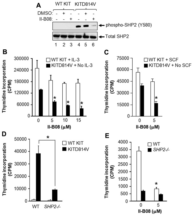
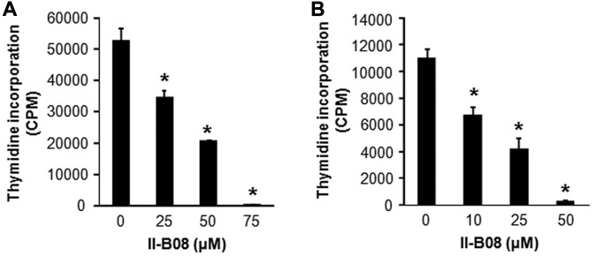
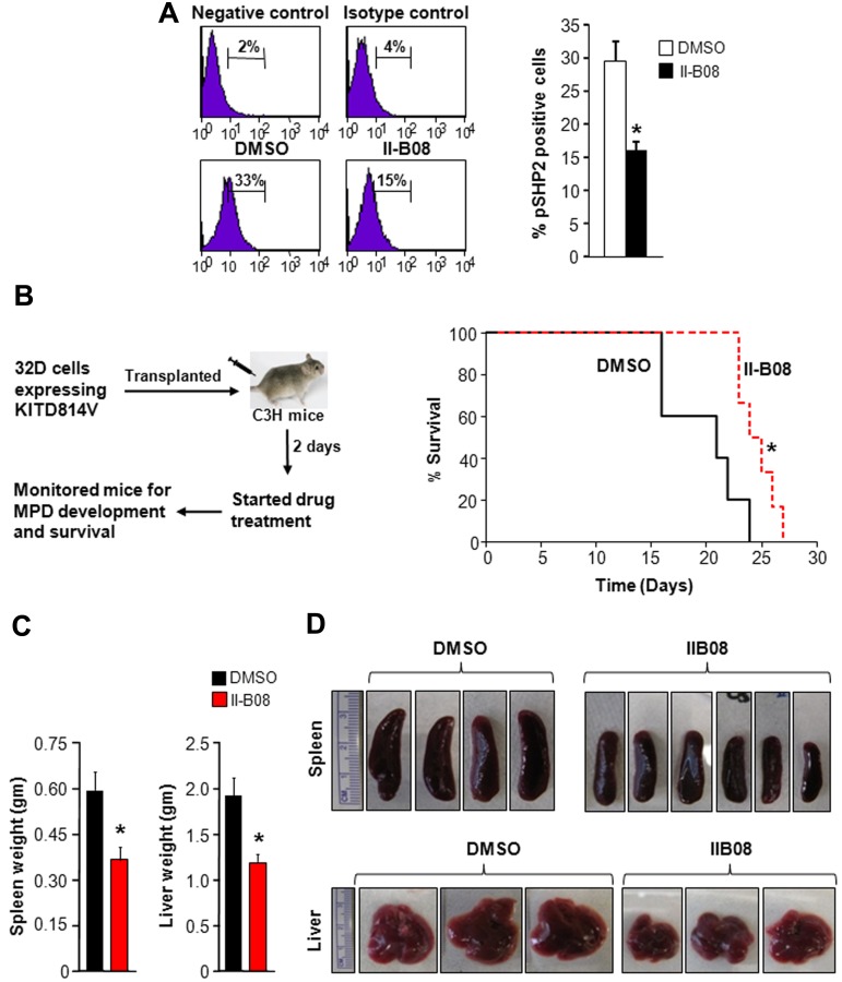
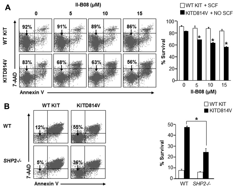
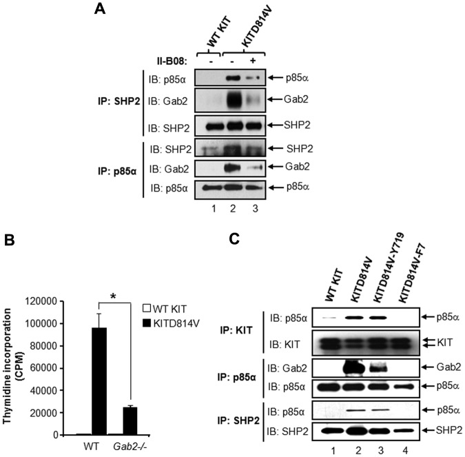
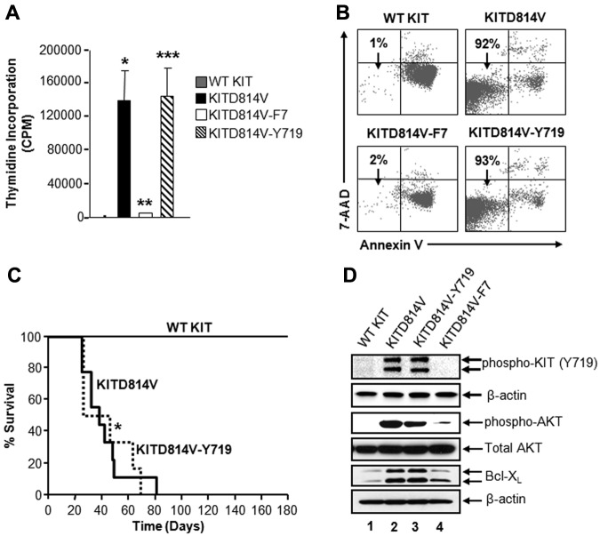
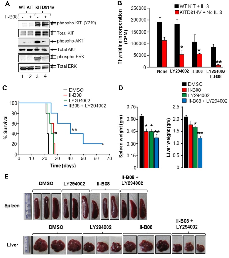
Intracellular mechanism(s) that contribute to promiscuous signaling via oncogenic KIT in systemic mastocytosis and acute myelogenous leukemia are poorly understood. We show that SHP2 phosphatase is essential for oncogenic KIT-induced growth and survival in vitro and myeloproliferative disease (MPD) in vivo. Genetic disruption of SHP2 or treatment of oncogene-bearing cells with a novel SHP2 inhibitor alone or in combination with the PI3K inhibitor corrects MPD by disrupting a protein complex involving p85α, SHP2, and Gab2. Importantly, a single tyrosine at position 719 in oncogenic KIT is sufficient to develop MPD by recruiting p85α, SHP2, and Gab2 complex to oncogenic KIT. Our results demonstrate that SHP2 phosphatase is a druggable target that cooperates with lipid kinases in inducing MPD.


Figures

Figure 1
Figure 1
SHP2 is essential for constitutive growth of cells bearing oncogenic KITD814V. (A) The 32D cells were transduced with retrovirus bearing WT KIT or KITD814V as described in “Expression of WT and mutant KIT receptors in 32D cells and primary HSC/Ps.” Cells bearing WT KIT or KITD814V were starved of serum and growth factors for 6 hours followed by treatment with SHP2 inhibitor, II-B08 (20μM), for 1 hour, after which cells were lysed and equal amount of protein lysates were subjected to Western blot analysis using an anti–phospho-SHP2 (Y580) or total SHP2 antibody. Similar results were observed in 3 independent experiments. (B) The 32D cells bearing WT KIT or KITD814V were starved of serum and growth factors for 6 hours and subjected to proliferation assay in the presence or absence of indicated concentration of II-B08. Assay was performed in the presence of IL-3 (10 ng/mL) for cells bearing WT KIT and in the absence of growth factors for cells bearing oncogenic KITD814V. Bars represent the mean thymidine incorporation (CPM ± SD) from 1 of the 3 independent experiments performed in quadruplicate. *P < .005. (C) Primary BM-derived WT KIT or KITD814V-expressing cells from WT mice were starved for 6 hours and subjected to thymidine incorporation assay in the presence or absence of indicated concentrations of II-B08. Assays were performed in the presence of murine SCF (50 ng/mL) for cells bearing WT KIT and in the absence of growth factors for cells bearing oncogenic KITD814V. Bars represent the mean thymidine incorporation (CPM ± SD) from 1 of 3 independent experiments performed in quadruplicate. *P < .001. (D) Primary BM-derived cells expressing WT KIT or KITD814V from WT or SHP2−/− mice were starved and subjected to proliferation assay in the absence of growth factors by thymidine incorporation. Bars represent the mean thymidine incorporation (mean ± SD) from 1 of 3 independent experiments performed in quadruplicate. *P < .01, WT-KITD814V vs SHP2−/−-KITD814V. (E) Primary BM-derived cells expressing KITD814V from WT or SHP2−/− mice were starved and subjected to proliferation assay in the presence or absence of II-B08 (5μM) by thymidine incorporation. Bars represent the mean thymidine incorporation (mean ± SD) from 1 of 2 independent experiments performed in triplicate. *P < .05, WT-KITD814V-0μM vs WT-KITD814V-5μM.
Figure 2
Figure 2
SHP2 inhibitor II-B08 inhibits the constitutive growth of human leukemic mast cell line HMC1.2 and human CD34+ cells bearing KITD816V as well as primary BM-derived AML blasts. (A) Human leukemic mast cell line HMC 1.2 or (B) human CD34+ cells transduced with KITD816V were starved for 6 hours in serum- and cytokine-free media and treated with indicated amounts of SHP2 inhibitor II-B08. After 48 hours, proliferation was evaluated by [3H] thymidine incorporation. Bars represent the mean thymidine incorporation (CPM ± SD) from 1 independent experiment performed in quadruplicate. *P < .001.
Figure 3
Figure 3
In vivo SHP2 inhibitor treatment of KITD814V-bearing mice enhances their survival and modulates MPD. (A) Mice received 100 mg/kg II-B08 intraperitoneally each day for 7 days. Mice were harvested after 24 hours of final injection, and peripheral blood, BM, spleen, and thymus were analyzed. Cells from spleen were stained with anti–phospho-SHP2 antibody by intracellular staining and analyzed by flow cytometry. Left panel: representative phospho-SHP2 flow micrographs. Right panel: average percentage of phospho-SHP2-positive cells from 4 mice. A 50% reduction in the phosphorylation of SHP2 was observed in mice treated with II-B08 compared with DMSO. (B) Kaplan-Meier survival analysis of leukemic mice treated with DMSO or II-B08. The 32D cells bearing oncogenic KITD814V were injected into syngeneic C3H/HeJ mice through tail vein. Mice were treated with either vehicle DMSO (n = 5) or II-B08 (50 mg/kg body weight; n = 6) at 12-hour intervals for 14 days. Left panel: schematic of drug treatment model. Right panel: survival curve. Significantly prolonged survival of mice treated with II-B08 was observed compared with mice treated with DMSO. *P < .05. (C-D) Reduced splenomegaly and hepatomegaly in KITD814V-bearing mice treated with II-B08. Average weights (C) and pictures (D) of spleen and liver of mice transplanted with cells bearing oncogenic KITD814V and treated with DMSO or II-B08. Significant reduction in spleen and liver weights was observed in mice treated with II-B08 compared with mice treated with DMSO. n = 5 or 6. *P < .05.
Figure 4
Figure 4
SHP2 is essential for oncogenic KITD814V-induced ligand-independent survival. (A) Primary BM-derived WT KIT or KITD814V-expressing cells were starved and treated with indicated concentrations of II-B08 for 48 hours. Assays were performed in the presence of SCF (50 ng/mL) for cells bearing WT KIT and in the absence of growth factors for cells bearing KITD814V. Cells were harvested and stained with PE-conjugated anti–annexin V antibody and 7-amino-actinomycin D followed by flow cytometric analysis. Left panel: representative flow micrographs. Right panel: bars represent the mean percent of surviving cells from 2 independent experiments. *P < .05. (B) Primary BM cells bearing WT KIT or KITD814V from WT or SHP2−/− mice were starved of serum and growth factors for 6 hours and cultured in media containing serum for 48 hours in the absence of growth factors. After 48 hours, cells were harvested and stained with PE-conjugated anti–annexin V antibody and 7-amino-actinomycin D followed by flow cytometric analysis. Left panel: representative flow micrographs. Right panel: bars represent the mean percent of surviving cells from 1 of 3 independent experiments. *P < .01.
Figure 5
Figure 5
Tyrosine 719 in KIT receptor is sufficient for recruiting protein complex involving p85α, SHP2, and Gab2 to KITD814V. (A) The 32D cells bearing WT KIT or KITD814V were starved for 8 hours in serum- and growth factor-free medium followed by incubation with or without II-B08 (20μM) for 1 hour. After incubation, equal amounts of protein lysates were subjected to immunoprecipitation with anti-SHP2 or anti-p85α antibodies followed by Western blot analysis using anti-p85α, anti-SHP2, or anti-Gab2 antibodies. Similar results were observed in 2 independent experiments. (B) Primary BM-derived cells expressing WT KIT or KITD814V from WT or Gab2−/− mice were starved and subjected to proliferation assay in the absence of growth factors by thymidine incorporation. Bars represent the mean (± SD) thymidine incorporation from 1 of 3 independent ex-eriments performed in quadruplicate. *P < .01, WT-KITD814V vs Gab2−/−-KITD814V. (C) The 32D cells bearing WT KIT, KITD814V, KITD814V-Y719, or KITD814V-F7 were starved of serum and growth factors for 8 hours, and equal amounts of protein lysates were subjected to immunoprecipitation with an anti-KIT antibody, anti-p85α antibody, or anti-SHP2 antibody followed by Western blot analysis using anti-KIT, anti-p85α, anti-SHP2, or anti-Gab2 antibodies as indicated. Similar results were observed in 2 or 3 independent experiments.
Figure 6
Figure 6
Tyrosine 719 associated p85α, SHP2, and Gab2 complex is sufficient for KITD814V-induced ligand-independent growth and survival in vitro and MPD in vivo. (A) Primary HSC/Ps derived from Wsh/sh mice, which lack endogenous expression of KIT receptor, were transduced with the indicated chimeric KIT receptors and sorted to homogeneity based on EGFP expression. Cells were starved in serum- and growth factor-free media for 6 hours and subjected to proliferation assay in the absence of growth factors by thymidine incorporation. Bars represent the mean (CPM ± SD) thymidine incorporation from 1 of the 3 independent experiments performed in quadruplicate. *P < .05, WT KIT vs KITD814V. **P < .05, KITD814V vs KITD814V-F7. ***P < .05, KITD814V-F7 vs KITD814V-Y719. (B) The 32D cells bearing the indicated chimeric receptors were starved of serum and growth factors and cultured for 48 hours in the absence of growth factors. Cells were harvested and stained with anti–annexin V antibody and 7-amino-actinomycin D followed by flow cytometric analysis. Double-negative cells in the lower left quadrant are indicated as surviving cells. Representative dot blots are shown. (C) Kaplan-Meier survival analysis of mice transplanted with the primary HSC/Ps bearing indicated KIT receptors (n = 8-18 per group). Results show that restoration of Y719 alone is sufficient to induce transformation in vivo (median survival, 55 days; n = 15). (D) The 32D cells bearing the indicated chimeric KIT receptors were starved in serum- and growth factor-free medium for 8 hours. Starved cells were lysed, and equal amounts of protein lysates were subjected to Western blot analysis using an anti–phospho-KIT (Y719), anti–phospho-AKT, total AKT, Bcl-xL, and β-actin antibodies as indicated. Similar results were observed in 3 independent experiments.
Figure 7
Figure 7
SHP2 inhibitor II-B08 enhances the efficacy of PI3K inhibitor in repressing oncogenic KITD814V-induced growth. (A) The 32D cells bearing WT KIT or KITD814V were starved for 6 hours followed by treatment with or without II-B08 (20μM) for 1 hour. After incubation, cells were lysed and equal amounts of protein lysates were subjected to Western blot analysis using an anti–phospho-KIT (Y719), total KIT, anti–phospho-AKT, total AKT, anti–phospho-ERK, or total ERK antibody as indicated. Similar results were observed in 3 independent experiments. (B) The 32D cells bearing WT KIT or KITD814V were starved of serum and growth factors for 6 hours and subjected to proliferation assay in the presence or absence of PI-3K inhibitor (LY294002, 2μM) and SHP2 inhibitor (II-B08, 5μM) alone or in combination. Bars represent the mean thymidine incorporation (CPM ± SD) from 1 of 3 independent experiments performed in quadruplicate. *P < .05, no GF vs LY294002 or II-B08. **P < .05, LY294002 or II-B08 vs LY294002 and II-B08. (C) Kaplan-Meier survival analysis of MPD mice treated with II-B08 or LY294002 alone or in combination. The 32D cells bearing oncogenic KITD814V were injected into syngeneic C3H/HeJ mice through tail vein. After 48 hours, mice were treated with either vehicle DMSO or II-B08 (50 mg/kg body weight) or LY294002 (10 mg/kg body weight) or a combination of II-B08 and LY294002 (50 + 10 mg/kg body weight, respectively) at 24-hour intervals for 21 days (n = 5). Significantly prolonged survival of mice treated with II-B08 or LY294002 alone or in combination was observed compared with mice treated with DMSO. *P < .05, DMSO vs II-B08 or LY294002. **P < .05, II-B08 or LY294002 vs II-B08 and LY294002. (D-E) Reduced splenomegaly and hepatomegaly in mice treated with II-B08 or LY294002 or combination. Average weights (D) and pictures (E) of spleens and livers from mice transplanted with cells bearing oncogenic KITD814V and treated with DMSO or II-B08 or LY294002 or combination of II-B08 and LY294002. Significant reduction in spleen and liver weights was observed in mice treated with II-B08 or LY294002 or combination of II-B08 and LY294002 compared with mice treated with DMSO. n = 4. *P < .05, DMSO vs II-B08 or LY294002. **P < .05, II-B08 or LY294002 vs II-B08 and LY294002.

Comment in

  • KIT's ship comes in.
    Braun BS. Braun BS. Blood. 2012 Sep 27;120(13):2541-2. doi: 10.1182/blood-2012-08-445247. Blood. 2012. PMID: 23019203 No abstract available.

Similar articles

Cited by

References

    1. Hirota S, Isozaki K, Moriyama Y, et al. Gain-of-function mutations of c-kit in human gastrointestinal stromal tumors. Science. 1998;279(5350):577–580. - PubMed
    1. Longley BJ, Reguera MJ, Ma Y. Classes of c-KIT activating mutations: proposed mechanisms of action and implications for disease classification and therapy. Leuk Res. 2001;25(7):571–576. - PubMed
    1. Beghini A, Peterlongo P, Ripamonti CB, et al. C-kit mutations in core binding factor leukemias. Blood. 2000;95(2):726–727. - PubMed
    1. Nagata H, Worobec AS, Oh CK, et al. Identification of a point mutation in the catalytic domain of the protooncogene c-kit in peripheral blood mononuclear cells of patients who have mastocytosis with an associated hematologic disorder. Proc Natl Acad Sci U S A. 1995;92(23):10560–10564. - PMC - PubMed
    1. Kitayama H, Kanakura Y, Furitsu T, et al. Constitutively activating mutations of c-kit receptor tyrosine kinase confer factor-independent growth and tumorigenicity of factor-dependent hematopoietic cell lines. Blood. 1995;85(3):790–798. - PubMed

Publication types

MeSH terms