. 2012 Dec;27(12):2452-63.
doi: 10.1002/jbmr.1701.

Protein kinase C-delta deficiency perturbs bone homeostasis by selective uncoupling of cathepsin K secretion and ruffled border formation in osteoclasts

Affiliations

Protein kinase C-delta deficiency perturbs bone homeostasis by selective uncoupling of cathepsin K secretion and ruffled border formation in osteoclasts

Viviana Cremasco et al. J Bone Miner Res. 2012 Dec.

Abstract

Bone homeostasis requires stringent regulation of osteoclasts, which secrete proteolytic enzymes to degrade the bone matrix. Despite recent progress in understanding how bone resorption occurs, the mechanisms regulating osteoclast secretion, and in particular the trafficking route of cathepsin K vesicles, remain elusive. Using a genetic approach, we describe the requirement for protein kinase C-delta (PKCδ) in regulating bone resorption by affecting cathepsin K exocytosis. Importantly, PKCδ deficiency does not perturb formation of the ruffled border or trafficking of lysosomal vesicles containing the vacuolar-ATPase (v-ATPase). Mechanistically, we find that cathepsin K exocytosis is controlled by PKCδ through modulation of the actin bundling protein myristoylated alanine-rich C-kinase substrate (MARCKS). The relevance of our finding is emphasized in vivo because PKCδ-/- mice exhibit increased bone mass and are protected from pathological bone loss in a model of experimental postmenopausal osteoporosis. Collectively, our data provide novel mechanistic insights into the pathways that selectively promote secretion of cathepsin K lysosomes independently of ruffled border formation, providing evidence of the presence of multiple mechanisms that regulate lysosomal exocytosis in osteoclasts.


Conflict of interest statement

Disclosure

All the authors state that they have no conflicts of interest

Figures

Figure 1
Figure 1. PKCδ is required for cathepsin K secretion
(A) WT and PLCγ2−/− pre-osteoclasts were stimulated by adhesion to vitronectin and phosphorylation of PKCδ was assessed in total cell lysates, using ERK as a loading control. (B) WT and PKCδ−/− osteoclasts were grown on bone slices, fixed and stained with FITC-phalloidin (in green) and with a monoclonal antibody against cathepsin K (in red). Magnification 63X. (C) Quantification of the percentage of actin rings with cathepsin K localized inside. (D) Cells as in (B) were analyzed by confocal microscopy, and Z stack images were reconstructed using LSM software. (E–F) WT and PKCδ−/− osteoclasts were stimulated with either bone particles (E) or PMA (F). Culture supernatant (sup) was collected and subjected to western blot analysis for the presence of cathepsin K. Cells in each well were lysed and served as total protein control (TCL). (G) WT and PKCδ−/− osteoclasts were grown on bone slices for 10 days, cells were removed and resorption pits visualized by staining with peroxidase-conjugated wheat-germ agglutinin. Black lines delineate the resorbed areas. (H) Area of bone resorption was determined using Image J software (n=5). (I) Supernatant from cultures as in (G) was collected and analyzed for the presence of collagen type I fragment using a colorimetric Elisa method. Data are expressed as average +/− standard deviation. An asterisk (*) depicts differences with a p value <0.05.
Figure 2
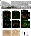
Figure 2. PKCδ-deficiency does not affect ruffled border formation or trafficking lysosomes containing the vATPase
(A) Transmission electron-microscopy was performed on PKCδ−/− osteoclasts plated on bone slices. Figures show actin rings (AR) and ruffled borders (RB). Scale bar 1 μm. (B) WT and PKCδ−/− macrophages were grown on bone slices in osteoclastogenic medium for 10 days. Mature osteoclasts were fixed and stained with FITC-phalloidin (in green) and with antibodies against the subunit E of the v-ATPase, Lamp1 or Lamp2 (in red). Cells were analyzed by confocal microscopy, and Z stack images were reconstructed using LSM software. Magnification 63X. (C–D) WT and PKCδ−/− macrophages were grown in the presence of M-CSF and RANKL on osteologic hydroxyapatite–coated slides for 4 days. Resorptive pits were visualized using a microscope without phase, magnification 20X, and quantified as percentage of resorbed area. Data are expressed as average +/− standard deviation.
Figure 3
Figure 3. PKCδ regulates bone homeostasis in physiological and pathological conditions
(A) Femurs of 8 week-old WT and PKCδ−/− mice were subjected to μCT analysis to detect trabecular bone. Images show representative 3D reconstructions. (B) Quantitative analysis of bone parameters were obtained from μCT data. Data are expressed as average +/− standard deviation. An asterisk (*) depicts differences with a p <0.05. (C) Histomorphometric analysis of the number of osteoclasts per bone surface from sections of 8 week-old mice (n=4/group). (D) WT and PKCδ−/− female mice were subjected to ovariectomy and trabecular bone volume versus total bone volume (BV/TV) was determined using vital μCT analysis before (pre OVX) and 1 month after ovariectomy (post OVX). Images show representative 3D reconstructions. (E) Quantitative analysis of percent trabecular bone volume versus total bone volume (n=4/group). An asterisk (*) depicts differences with a p value <0.05.
Figure 4
Figure 4. MARCKS regulates cathepsin K secretion downstream of PKCδ
(A) WT and PKCδ−/− osteoclasts were grown on bone slices, fixed and stained with FITC-phalloidin (in green) and with a monoclonal antibody against MARCKS (in red). Cells were analyzed by confocal microscopy and Z stack images were reconstructed using LSM software (AR=actin ring; RB=ruffled border). (B) WT and PKCδ−/− pre-osteoclasts were stimulated by adhesion to vitronectin. Cells were lysed and equal amounts of total cell lysates were western blotted for the indicated proteins. Actin served as a loading control. (C) WT and MARCKS−/− osteoclasts were stimulated for the indicated times with bone particles, and culture medium supernatant (sup) was analyzed for the presence of cathepsin K. Cells in each well were lysed and served as total protein control (TCL). (D) WT and MARCKS−/− osteoclasts were grown on bone slices for 10 days, cells were removed, and resorption pits visualized by staining with peroxidase-conjugated wheat-germ agglutinin. Black lines delineate the resorbed areas. (E) Area of bone resorption per field was determined using Image J software. (F) Supernatant medium from culture as in (D) was analyzed for the presence of collagen type I fragments using a colorimetric Elisa method. Data are expressed as average +/− standard deviation. An asterisk (*) depicts differences with a p value <0.05. (G) WT osteoclasts were treated with Latrunculin A and medium supernatant (sup) was collected and subjected to western blot for cathepsin K. Cells in the well were lysed as a total protein control (TCL).
Figure 5
Figure 5. MARCKS silencing in PKCδ−/− osteoclasts rescues cathepsin K lysosome exocytosis
(A) WT and PKCδ−/− macrophages were lentivirally transduced with two different MARCKS silencing constructs or with shRNA directed towards Luciferase as a negative control. Efficiency of MARCKS knock-down was determined using semi quantitative RT-PCR. GAPDH served as a loading control. (B) Cells as in (A) were cultured in osteoclastogenic medium for 7 days and stimulated for different time points with bone particles. Medium supernatant (sup) was collected and subjected to western blot analysis for the presence of cathepsin K. Cells in the well were lysed as a total protein control (TCL). (C) Cells as in (A) were grown on bone slices in osteoclastogenic medium for 10 days. Cells were then removed and resorption pits visualized by staining with peroxidase-conjugated wheat-germ agglutinin. Black lines delineate the resorbed areas. (D) Area of bone resorption per field was determined using Image J software and graphed for each sample. Data are expressed as average +/− standard deviation. An asterisk (*) depicts differences with a p <0.05.
Figure 6
Figure 6. Specific regulation of cathepsin K lysosomal secretion by PKCδ and MARCKS
(A) MARCKS (M, in yellow) binds to actin and tethers filaments to the plasma membrane of the ruffled border. PKCδ-mediated phosphorylation (P) of MARCKS displaces MARCKS from the membrane and impairs its actin-binding capacity. MARCKS displacement produces a local release of actin filaments, thus allowing cathepsin K vesicle secretion. (B) In absence of PKCδ, MARCKS is stabilized at the membrane. While failure to disrupt the actin barrier does not affect ruffled border formation or translocation of vesicles containing the v-ATPase, it impairs cathepsin K release, thus inhibiting the cell resorptive ability.

Similar articles

Cited by

References

    1. Blott EJ, Griffiths GM. Secretory lysosomes. Nat Rev Mol Cell Biol. 2002;3(2):122–31. - PubMed
    1. Novack DV, Teitelbaum SL. The osteoclast: friend or foe? Annu Rev Pathol. 2008;3:457–84. - PubMed
    1. Zaidi M. Skeletal remodeling in health and disease. Nat Med. 2007;13(7):791–801. - PubMed
    1. Teitelbaum SL, Ross FP. Genetic regulation of osteoclast development and function. Nature Reviews Genetics. 2003;4:638–649. - PubMed
    1. Collin O, Tracqui P, Stephanou A, Usson Y, Clement-Lacroix J, Planus E. Spatiotemporal dynamics of actin-rich adhesion microdomains: influence of substrate flexibility. J Cell Sci. 2006;119(Pt 9):1914–25. - PubMed

Publication types

MeSH terms

Substances