

doi: 10.1002/chem.201201805.
Epub 2012 Jul 17.
Cell-penetrating peptides as delivery vehicles for a protein-targeted terbium complex
Affiliations
- PMID: 22807190
- PMCID: PMC3729426
- DOI: 10.1002/chem.201201805
Item in Clipboard
Cell-penetrating peptides as delivery vehicles for a protein-targeted terbium complex
Chemistry.
.
Abstract
Release after transmission: Arginine-rich, cell-penetrating peptides (CPPs) mediate cytoplasmic delivery of trimethoprim (TMP)-terbium complex conjugates and selective, intracellular labeling of E. coli dihydrofolate reductase (eDHFR) fusion proteins. A disulfide bond linking CPP and cargo is reduced following uptake. CPP conjugation can be used to deliver otherwise cell-impermeable, ligand-fluorophore conjugates.
Copyright © 2012 WILEY-VCH Verlag GmbH & Co. KGaA, Weinheim.
Figures
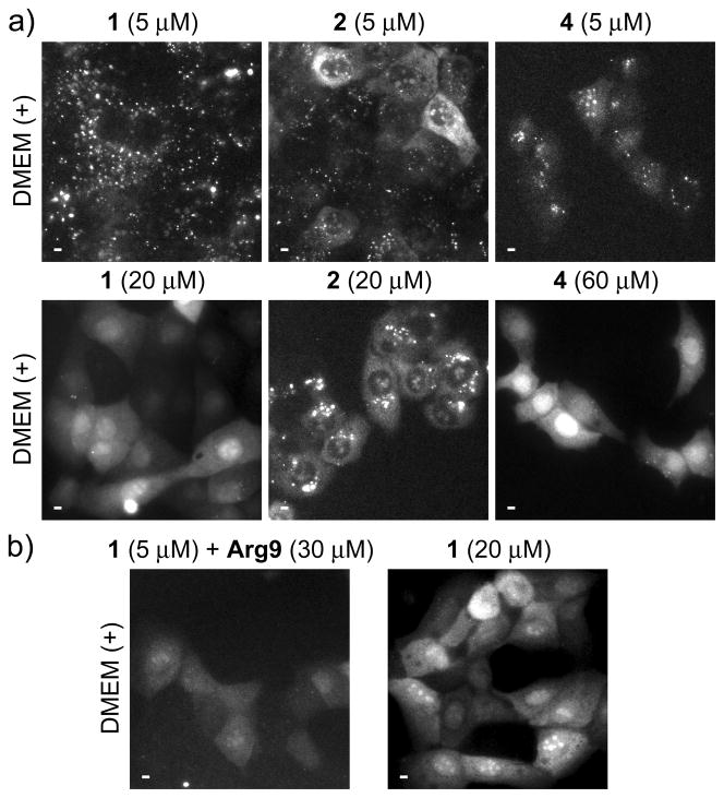
Effects of extracellular peptide concentration on uptake and distribution. Micrographs, (a–b): time-gated luminescence (delay = 10 μs, λex = 365 nm, λem = 540 ± 10 nm) Scale bars, 10 μm. MDCKII cells were incubated for 30 min. at 37 °C in Dulbecco’s modified eagle medium with fetal bovine serum (DMEM (+)) that contained indicated concentrations of peptides 1, 2, 4 or Arg9. a) Incubation in DMEM (+) containing low (5 μM) concentrations of indicated peptides results in punctate Tb3+ luminescence (top) while incubation in DMEM (+) above a threshold concentration (20 μM, 1, 2; 60 μM, 4) results in diffuse distribution of Tb3+ luminescence throughout cytoplasm and nucleus (bottom). b) Cells incubated in DMEM (+) containing 1 (5 μM) plus Arg9 (30 μM) show diffuse Tb3+ luminescence (left) similar to that seen in cells incubated in DMEM (+) containing higher concentrations of 1 (20 μM, right).
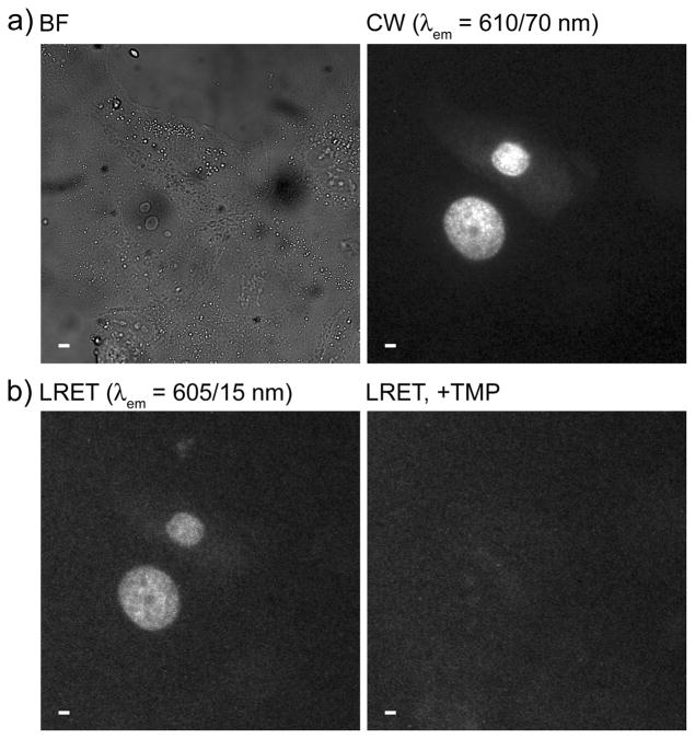
Arg9 mediates cytoplasmic delivery of 4 and specific labeling of H2B-TagRFPT-eDHFR as evidenced by time-gated LRET imaging of Tb3+-to-TagRFP-T sensitized emission. MDCKII cells transiently expressing H2B-TagRFPT-eDHFR were incubated for 30 min. at 37 °C in DMEM (−) containing 4 (10 μM), washed 2X in PBS and reimmersed in DMEM (+) containing 1 mM Patent Blue™ prior to imaging. a) Bright field (BF) and continuous wave (CW) fluorescence (λex = 545 ± 15 nm, λem = 610 ± 35 nm) images reveal nucleus-localized TagRFP-T fluorescence in expressing cells. b) Time-gated LRET (delay = 10 μs, λex = 365 nm, λem = 605 ± 7 nm) image shows long-lived, Tb3+-sensitized TagRFP-T emission (left) that disappears when TMP (final conc. = 100 μM) was added to medium (right). Scale bars, 10 μm.
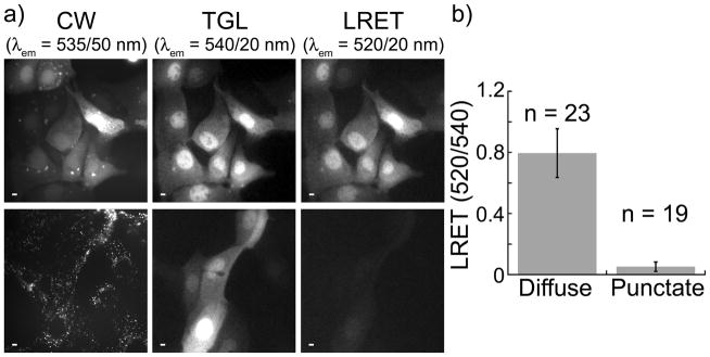
Intacellular disulfide reduction releases cargo from carrier peptide. MDCKII cells were incubated for 10 min. at 37 °C in DMEM (−) containing 17 (10 μM), washed 2X in PBS and reimmersed in DMEM (+) containing 1 mM Patent Blue™ prior to imaging. Micrographs: CW, continuous wave fluorescence (λex = 480 ± 40 nm, λem = 535 ± 50 nm); TGL, time-gated luminescence (delay = 10 μs, λex = 365 nm, λem = 540 ± 10 nm); LRET, luminescence resonance energy transfer (delay = 10 μs, λex = 365 nm, λem = 520 ± 10 nm). Scale bars, 10 μm. a) (top) Representative images of cells acquired immediately following wash step. Fluorescein fluorescence, Tb3+ luminescence, and Tb3+-to-fluorescein LRET signals are diffuse throughout cytoplasm and nucleus. (bottom), Representative images of cells acquired ~2 h post-wash. Fluorescein signal is punctate while Tb3+ luminescence and diminished LRET signals remain diffuse. b) Mean, normalized, time-gated LRET signal (520 nm/540 nm) measured in the nuclear region of cells exhibiting diffuse (immediate) and punctate (2 h post-wash) patterns of continuous wave, fluorescein fluorescence. Error bars represent standard deviation of the mean values (n = no. of cells) obtained from 4 separate experiments.
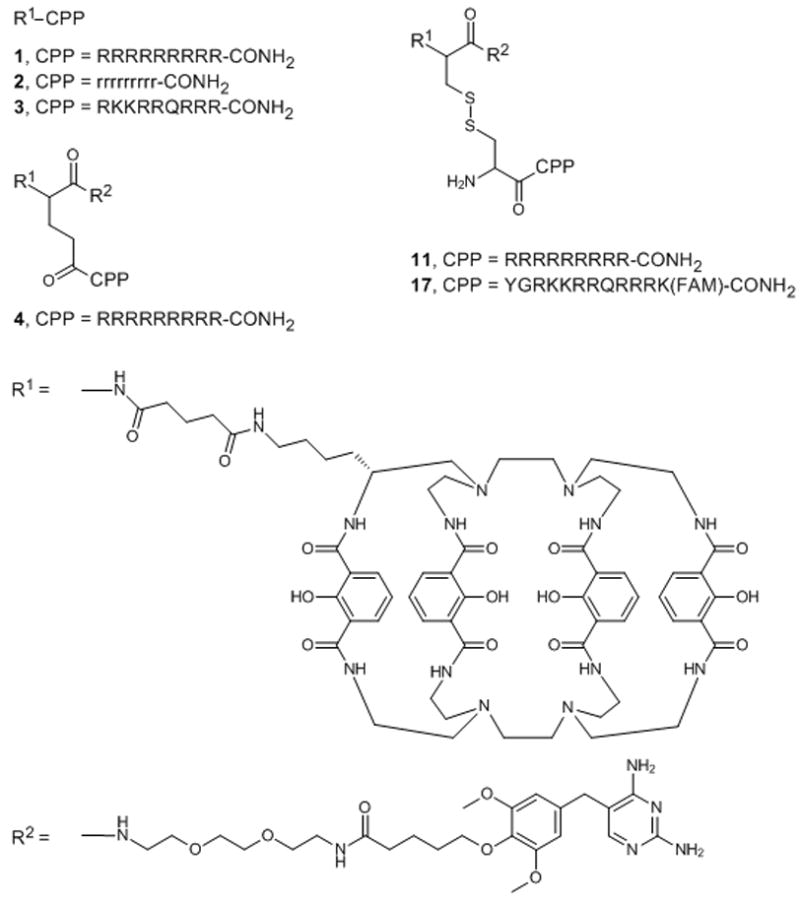
Chemical structures of the peptide conjugates used in this study. Abbreviations: R1, linker-functionalized derivative of Lumi4; R2, triethyleneglycolamino derivative of trimethoprim (TEGTMP); capital letters, L-amino acids; small letters, D-amino acids; FAM, 5,6-carboxyfluorescein.
References
-
- Carreon JR, Stewart KM, Mahon KP, Jr, Shin S, Kelley SO. Bioorg Med Chem Lett. 2007;17:5182–5185. - PubMed
-
- Rajapakse HE, Reddy DR, Mohandessi S, Butlin NG, Miller LW. Angew Chem Int Ed Engl. 2009;48:4990–4992. - PMC - PubMed
- Rajapakse HE, Gahlaut N, Mohandessi S, Yu D, Turner JR, Miller LW. Proc Natl Acad Sci U S A. 2010;107:13582–13587. - PMC - PubMed
- Reddy DR, Pedro Rosa LE, Miller LW. Bioconjug Chem. 2011;22:1402–1409. - PMC - PubMed
Publication types
MeSH terms
Substances
Grants and funding
LinkOut - more resources
Full Text Sources
Other Literature Sources
