. 2012 Jul;72(1):53-64.
doi: 10.1002/ana.23651. Epub 2012 Jul 17.

Aquaporin 4-specific T cells in neuromyelitis optica exhibit a Th17 bias and recognize Clostridium ABC transporter

Affiliations
Free PMC article

Aquaporin 4-specific T cells in neuromyelitis optica exhibit a Th17 bias and recognize Clostridium ABC transporter

Michel Varrin-Doyer et al. Ann Neurol. 2012 Jul.
Free PMC article

Abstract

Objective: Aquaporin 4 (AQP4)-specific autoantibodies in neuromyelitis optica (NMO) are immunoglobulin (Ig)G1, a T cell-dependent Ig subclass, indicating that AQP4-specific T cells participate in NMO pathogenesis. Our goal was to identify and characterize AQP4-specific T cells in NMO patients and healthy controls (HC).

Methods: Peripheral blood T cells from NMO patients and HC were examined for recognition of AQP4 and production of proinflammatory cytokines. Monocytes were evaluated for production of T cell-polarizing cytokines and expression of costimulatory molecules.

Results: T cells from NMO patients and HC proliferated to intact AQP4 or AQP4 peptides (p11-30, p21-40, p61-80, p131-150, p156-170, p211-230, and p261-280). T cells from NMO patients demonstrated greater proliferation to AQP4 than those from HC, and responded most vigorously to p61-80, a naturally processed immunodominant determinant of intact AQP4. T cells were CD4(+), and corresponding to association of NMO with human leukocyte antigen (HLA)-DRB1*0301 and DRB3, AQP4 p61-80-specific T cells were HLA-DR restricted. The T-cell epitope within AQP4 p61-80 was mapped to 63-76, which contains 10 residues with 90% homology to a sequence within Clostridium perfringens adenosine triphosphate-binding cassette (ABC) transporter permease. T cells from NMO patients proliferated to this homologous bacterial sequence, and cross-reactivity between it and self-AQP4 was observed, supporting molecular mimicry. In NMO, AQP4 p61-80-specific T cells exhibited Th17 polarization, and furthermore, monocytes produced more interleukin 6, a Th17-polarizing cytokine, and expressed elevated CD40 and CD80 costimulatory molecules, suggesting innate immunologic dysfunction.

Interpretation: AQP4-specific T-cell responses are amplified in NMO, exhibit a Th17 bias, and display cross-reactivity to a protein of an indigenous intestinal bacterium, providing new perspectives for investigating NMO pathogenesis.


Figures

FIGURE 3
FIGURE 3
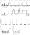
Cross-reactivity between aquaporin 4 (AQP4) p63–76 and Clostridium perfringens adenosine triphosphate-binding cassette (ABC) transporter permease (TP) p204–217. (A) The T-cell epitope within AQP4 p61–80 was mapped by testing recall proliferation of AQP4 p61–80-reactive T cells from neuromyelitis optica (NMO) patients to truncated AQP4 peptides (10μg/ml) in the presence of irradiated autologous antigen-presenting cells (APC). (B) AQP4 p63–76 appeared to contain p61–80 core determinant. Proliferation was measured by [3H]thymidine incorporation after 3 days. Data are representative of 3 independent experiments. (C) Sequence homology between AQP4 p63–76 and C. perfringens ABC-TP p204–217 was identified using the protein–protein Basic Local Alignment Search Tool from National Center for Biotechnology Information. Top bracket represents the predicted core binding motif for human leukocyte antigen (HLA)-DRB1*0301 and HLA-DRB3*0202 within AQP4 p63–76 (netMHCII-1.1 and netMHCII-2.2 programs). (D) 5,6-Carboxylfluorescein diacetate succinimidyl ester-labeled peripheral blood mononuclear cells (PBMC) from 3 NMO patients were stimulated with antigens (10μg/ml) and cultured for 10 days before evaluating proliferation by flow-activated cell sorting. (E) PBMC from 4 NMO patients were initially stimulated for 10 days with AQP4 p63–76 or ABC-TP p204–217 at 10μg/ml. Recall responses to peptides in the presence of irradiated autologous APC were evaluated by [3H]thymidine incorporation after 3 days. Paired t tests were performed to compare counts per minute (cpm) values of each antigen to cpm values of no-antigen controls, *p < 0.05, **p < 0.01. In A, B, and E, data are presented as means of duplicate or triplicate wells; error bars throughout indicate standard error of the mean. CDI = cell division index.
FIGURE 1
FIGURE 1
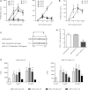
T cells from neuromyelitis optica (NMO) patients recognize discrete determinants of aquaporin 4 (AQP4). Peripheral blood mononuclear cells (PBMC) were tested for proliferation to (A) pools of AQP4 peptides (n = 8 NMO and n = 3 healthy controls [HC]) and to (B) individual AQP4 peptides identified from those pools. In A and B, PBMC were cultured for 6 days in the presence of AQP4 pools (10μg/ml) or AQP4 peptides (10μg/ml), respectively, then pulsed with [3H]thymidine and harvested 18 hours later. In A, positive wells were defined as values > control counts per minute average values + 3 standard deviations. (C) AQP4 determinants are represented within a human AQP4 topological diagram using TOPO2 transmembrane protein display software (http://www.sacs.ucsf.edu/TOPO2/). (D, E) PBMC were examined by 5,6-carboxylfluorescein diacetate succinimidyl ester (CFSE) dilution for proliferation to individual AQP4 peptides (10μg/ml), recombinant human (rh) AQP4 (5μg/ml), or in E, tetanus toxoid (TT; 1μg/ml) after 10 days of culture. CFSE was measured in CD3+, CD4+, and CD8+ T cells by flow-activated cell sorting and quantified by cell division index (CDI). CDI > 2 (broken lines) was considered positive. (F) Recall T-cell proliferation ([3H]thymidine incorporation) to individual AQP4 peptides (10μg/ml) or rhAQP4 (5μg/ml) was detected after initial stimulation with rhAQP4 (5μg/ml) for 10 days. In A and E, error bars indicate standard error of the mean; in B, D, and F, horizontal lines indicate mean values. *p < 0.05 Mann–Whitney U test. Ag = antigens.
FIGURE 2
FIGURE 2
Human leukocyte antigen (HLA)-DR serves as a restriction element for aquaporin 4 (AQP4)-specific T cells. (A, B) 5,6-carboxylfluorescein diacetate succinimidyl ester (CFSE)-labeled peripheral blood mononuclear cells (PBMC) from neuromyelitis optica (NMO) patients were cultured for 10 days with antigens (Ag) alone or in combination with antibodies against HLA-DR, HLA-DQ, or HLA-DP or isotype control antibodies. T-cell proliferation was evaluated by flow-activated cell sorting analysis of CFSE dilution. Inhibitory effects of blocking antibodies were examined on proliferating CD4+ T cells (n = 7 NMO in A and n = 4 NMO in B). T-cell proliferation is expressed as cell division index (CDI). (C) PBMC from healthy controls (HC) were similarly examined after stimulation with AQP4 p61–80 (n = 2). Error bars represent standard error of the mean. *p < 0.05, Mann–Whitney U test.
FIGURE 4
FIGURE 4
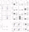
Aquaporin 4 (AQP4) p61–80-specific T cells exhibit a proinflammatory bias. Peripheral blood mononuclear cells (PBMC) were stained with 5,6-Carboxylfluorescein diacetate succinimidyl ester (CFSE) and cultured for 10 days with AQP4 peptides (10μg/ml) or recombinant human (rh) AQP4 (5μg/ml). (A) CD4+CFSElow proliferating T cells were analyzed for interleukin (IL)-17 and interferon (IFN)-γ production by intracellular staining after stimulation with phorbol 12-myristate 13-acetate/Ionomycin for 5 hours. (B) Frequencies of IL17+IFN-γ, IL17+IFN-γ+, and IL17IFN-γ+ were examined among proliferating p61–80-specific CD4+ T cells (n = 8 NMO and n = 5 healthy controls [HC]), p156–170-specific CD4+ T cells (n = 6 NMO and n = 3 HC), and rhAQP4-specific CD4+ T cells (n = 6 NMO and n = 5 HC). Frequencies of IL-17 and IFN-γ single positive T cells were used to calculate Th17/Th1 ratio. (C) PBMC were examined by fluorescence-activated cell sorting (FACS) for expression of regulatory T cells (Treg)markers including CD4, CD127, and CD25. (D) CFSE-labeled PBMC were cultured for 10 days with AQP4 p61–80 (10μg/ml) or rhAQP4 (5μg/ml). Proliferating CD4+ T cells (cell division index > 2) were examined by FACS for expression of CD25high, defined as the top half of CD25+ cells, and Foxp3 (n = 8 NMO p61–80, n = 6 HC p61–80, n = 7 NMO rhAQP4, and n = 5 HC rhAQP4). Box and whisker plots include the median, distribution, and range. **p < 0.01 Mann–Whitney U test.
FIGURE 5
FIGURE 5
CD14+ monocytes from neuromyelitis optica (NMO) patients exhibit increased expression of certain costimulatory molecules and production of interleukin (IL)-6. (A) Peripheral blood mononuclear cells (PBMC) were rested for 4 hours at 37°C. Expression of costimulatory (CD80, CD86, and CD40) and major histocompatibility complex class II molecules was analyzed by flow-activated cell sorting gating on the CD14+ population (n = 8 NMO and n = 8 healthy controls [HC]). (B, C) PBMC were stimulated with LPS lipopolysaccharide (LPS; 1μg/ml) for 4 hours. Expression of IL-6 in CD14+ monocytes was analyzed by intracellular cytokine staining, before and after LPS stimulation. In C, horizontal lines indicate mean values; in A and B, error bars represent standard error of the mean. *p < 0.05, **p < 0.01 Mann–Whitney U test. MFI = mean fluorescent intensity.

References

    1. Graber DJ, Levy M, Kerr D, Wade WF. Neuromyelitis optica pathogenesis and aquaporin 4. J Neuroinflammation. 2008;5:22. - PMC - PubMed
    1. Lennon VA, Kryzer TJ, Pittock SJ, et al. IgG marker of optic-spinal multiple sclerosis binds to the aquaporin-4 water channel. J Exp Med. 2005;202:473–477. - PMC - PubMed
    1. Nurieva RI, Chung Y. Understanding the development and function of T follicular helper cells. Cell Mol Immunol. 2010;7:190–197. - PMC - PubMed
    1. Bennett JL, Lam C, Kalluri SR, et al. Intrathecal pathogenic anti-aquaporin-4 antibodies in early neuromyelitis optica. Ann Neurol. 2009;66:617–629. - PMC - PubMed
    1. Bradl M, Misu T, Takahashi T, et al. Neuromyelitis optica: pathogenicity of patient immunoglobulin in vivo. Ann Neurol. 2009;66:630–643. - PubMed

Publication types

MeSH terms