. 2012 Aug 31;287(36):30358-67.
doi: 10.1074/jbc.M112.384339. Epub 2012 Jul 17.

Characterization of a proteasome and TAP-independent presentation of intracellular epitopes by HLA-B27 molecules

Affiliations

Characterization of a proteasome and TAP-independent presentation of intracellular epitopes by HLA-B27 molecules

Adriana Magnacca et al. J Biol Chem. .

Abstract

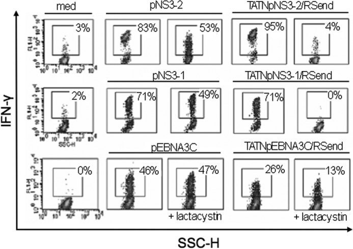
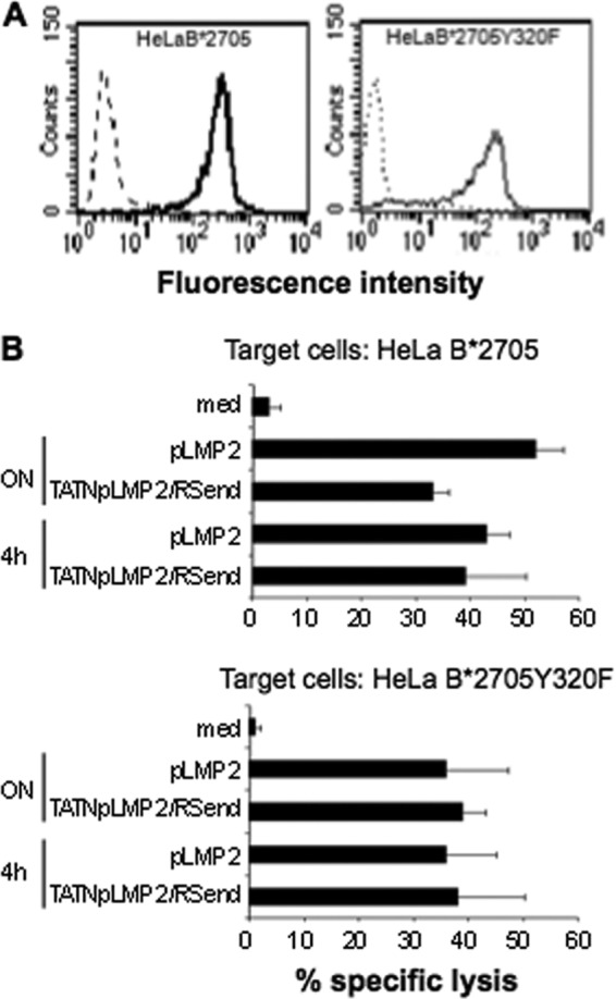
Nascent HLA-class I molecules are stabilized by proteasome-derived peptides in the ER and the new complexes proceed to the cell surface through the post-ER vesicles. It has been shown, however, that less stable complexes can exchange peptides in the Trans Golgi Network (TGN). HLA-B27 are the most studied HLA-class I molecules due to their association with Ankylosing Spondylitis (AS). Chimeric proteins driven by TAT of HIV have been exploited by us to deliver viral epitopes, whose cross-presentation by the HLA-B27 molecules was proteasome and TAP-independent and not restricted to Antigen-Presenting Cells (APC). Here, using these chimeric proteins as epitope suppliers, we compared with each other and with the HLA-A2 molecules, the two HLA-B*2705 and B*2709 alleles differing at residue 116 (D116H) and differentially associated with AS. We found that the antigen presentation by the two HLA-B27 molecules was proteasome-, TAP-, and APC-independent whereas the presentation by the HLA-A2 molecules required proteasome, TAP and professional APC. Assuming that such difference could be due to the unpaired, highly reactive Cys-67 distinguishing the HLA-B27 molecules, C67S mutants in HLA-B*2705 and B*2709 and V67C mutant in HLA-A*0201 were also analyzed. The results showed that this mutation did not influence the HLA-A2-restricted antigen presentation while it drastically affected the HLA-B27-restricted presentation with, however, remarkable differences between B*2705 and B*2709. The data, together with the occurrence on the cell surface of unfolded molecules in the case of C67S-B*2705 mutant but not in that of C67S-B*2709 mutant, indicates that Cys-67 has a more critical role in stabilizing the B*2705 rather than the B*2709 complexes.


Figures

FIGURE 1.
FIGURE 1.
Schematic representation of TAT-NpFlu recombinant proteins. HLA-B*2705-restricted epitope pNpFlu in the TAT-Npflu constructs has been replaced by: the HLA-B27-restricted epitopes pEBNA3C (258–266) or pLMP2 (236–244) from EBV, or by the HLA-A2-restricted pNS3–2 (1406–1415) or pNS3–1 (1073–1081) from the NS3 protein of HCV. The constructs containing or not the C terminus of the nucleoprotein of influenza virus (end of Np) are indicated. The motif Arg/Ser (RS) is included at both the N or the C termini of the epitopes in the hybrid proteins.
FIGURE 2.
FIGURE 2.
HLA-B27 and HLA-A2 presentation of the chimeric epitopes. A,51Chromium release assay in which HLA-A2/B27 double positive B-LCL are incubated overnight with the indicated recombinant proteins (1.4 μm) or with the synthetic peptides (50 μm) or with medium alone (med) and labeled with sodium 51chromate before being mixed with peptide-specific CTLs for 4 h. When indicated, lactacystin was added 45 min before the recombinant protein or the synthetic peptide. B, T2 wt cells which are naturally positive for the HLA-A2 and T2B*2705 and T2B*2709 transfectants were incubated overnight with the recombinant proteins (1.4 μm) or with synthetic peptides (50 μm) or with medium alone (med), and used as antigen-presenting cells in the cytotoxicity assay. Bars represent the mean percentage of lysis ± S.D. of three independent experiments.
FIGURE 3.
FIGURE 3.
The chimeric proteins co-localize with furin in the TGN. TATNpEBNA3C/RSend (top panel on the left) and TAT-NpNS3–2/RSend (bottom panel on the left) are visualized in green within HeLa cells by staining with the anti-HA mAb followed by FITC-conjugated secondary antibody in a confocal microscopy analysis. Middle panels show the TGN depicted in red by staining with an anti α-Furin mAb. Right panels show the co-localization of both chimeric proteins with the convertase α-Furin resident in the TGN as visualized by the overlay.
FIGURE 4.
FIGURE 4.
The chimeric proteins are processed and presented by Dendritic Cells. HLA-A2 (upper and middle panel) or B27 (bottom panel) positive DCs were incubated with recombinant proteins (1.4 μm) or synthetic peptides (50 μm) or in medium alone (med). NS3–2 (upper panel), NS3–1 (middle panel) and EBNA3C (bottom panel) specific CTL responses were measured by intracellular staining as % of produced IFN-γ and detected by flow cytometry analysis. % of IFN-γ producing cells are shown in each panel. When indicated, lactacystin has been added 45 min before the synthetic peptides or the recombinant proteins. Data are representative of three independent experiments.
FIGURE 5.
FIGURE 5.
Lack of tyrosine 320 does not impair the presentation of the recombinant proteins by HLA-B27 molecules. A, HeLa B*2705 and B*2705Y320F cells were treated with ME1 (solid line) or with the isotype control IgG1 antibody (dashed line), followed by staining with F(ab')2 of rabbit anti-mouse FITC-conjugated Abs in a flow cytometry analysis. B, cytotoxicity assay in which HeLa expressing B*2705 or B*2705Y320F mutant were incubated overnight or for 4 h with TAT-NpLMP2/RSend recombinant protein (1.4 μm) or pLMP2 synthetic peptide (50 μm) or in medium alone (med) and then mixed with pLMP2-specific CTLs for 4 h. Data are the mean ± S.D. of the percentage of lysis from three separate experiments.
FIGURE 6.
FIGURE 6.
Expression of C67S HLA-B27 and V67C HLA-A2 mutants. Expression of wt and C67S B*2705 molecules in A and wt and C67S B*2709 molecules in B) on the cell surface of HeLa cells was evaluated with the mAb ME1 that recognizes conformational molecules (left panels) and mAb HC10 that recognizes unfolded molecules (β2m-free heavy chains) (right panels). C, expression of wt HLA-A2 molecules and V67C mutants was evaluated on HeLa transfectants with BB7.2 mAb that recognizes conformational molecules (left panel), and HCA2 mAb that recognizes unfolded molecules (β2m-free heavy chains) (right panel). One representative staining of three-five independent flow cytometry analysis is shown here.
FIGURE 7.
FIGURE 7.
Effect on antigen presentation of C67S and V67C mutations in respectively HLA-B27 and HLA-A2 molecules. A, antigen-specific CTLs were tested in standard 51Cr-release assays using as target the HeLa cells expressing wt molecules or C67S mutants in the context of B*2705 (upper panels) and B*2709 (bottom panels) and pulsed with the indicated peptides (50 μm) or in medium alone (med). B, HeLa cells stably transfected with HLA-A2 wt or V67C mutant were incubated overnight with TAT-NpNS3–2/RSend recombinant protein (1.4 μm), pNS3–2 synthetic peptide (50 μm), or with medium alone (med) and used as targets for NS3–2 specific CTLs. Data are the mean percentage of lysis ± S.D. of three independent experiments.
FIGURE 8.
FIGURE 8.
Cysteine 67 is crucial for presentation of pEBNA3C by the HLA-B*2705 molecules. Dose-response curves in which HeLa B*2705 and B*2709 transfectants and the corresponding C67S mutants are used as targets in a standard 51Chromium-release assay at the indicated concentration of pEBNA3C or TATNpEBNA3C/RSend recombinant protein. Data show the mean percentage of specific lysis ± S.D. of three independent experiments.
FIGURE 9.
FIGURE 9.
Differences in presenting pEBNA3C could be due to the interaction between L9 and Asp-116. A, ribbon representation of the model of B*2709:pEBNA3C complex and C, of solved structure of B*2705:pEBNA3C complex. L9, His-116, and Asp-116 are shown as ball-and-stick, B*2709 and B*2705 peptides are depicted as blue sticks in both complexes. B and D, schemes of the protein-peptide interactions obtained using LigPlot: hydrophobic interactions are represented by arcs with spokes radiating toward the atoms they contact, and the residues involved are shown as ball-and-stick models.

References

    1. Hülsmeyer M., Fiorillo M. T., Bettosini F., Sorrentino R., Saenger W., Ziegler A., Uchanska-Ziegler B. (2004) Dual, HLA-B27 subtype-dependent conformation of a self-peptide. J. Exp. Med. 199, 271–281 - PMC - PubMed
    1. Fiorillo M. T., Maragno M., Butler R., Dupuis M. L., Sorrentino R. (2000) CD8(+) T-cell autoreactivity to an HLA-B27-restricted self-epitope correlates with ankylosing spondylitis. J. Clin. Invest. 106, 47–53 - PMC - PubMed
    1. Nurzia E., Panimolle F., Cauli A., Mathieu A., Magnacca A., Paladini F., Sorrentino R., Fiorillo M. T. (2010) CD8+ T-cell mediated self-reactivity in HLA-B27 context as a consequence of dual peptide conformation. Clin. Immunol. 135, 476–482 - PubMed
    1. Mathieu A., Paladini F., Vacca A., Cauli A., Fiorillo M. T., Sorrentino R. (2009) The interplay between the geographic distribution of HLA-B27 alleles and their role in infectious and autoimmune diseases: a unifying hypothesis. Autoimmun. Rev. 8, 420–425 - PubMed
    1. Blanco-Gelaz M. A., López-Vázquez A., García-Fernández S., Martínez-Borra J., González S., López-Larrea C. (2001) Genetic variability, molecular evolution, and geographic diversity of HLA-B27. Hum. Immunol. 62, 1042–1050 - PubMed

Publication types

MeSH terms