. 2012 Sep 7;287(37):31311-20.
doi: 10.1074/jbc.M111.327056. Epub 2012 Jul 17.

The GTPase-deficient Rnd proteins are stabilized by their effectors

Affiliations

The GTPase-deficient Rnd proteins are stabilized by their effectors

Liuh Ling Goh et al. J Biol Chem. .

Abstract

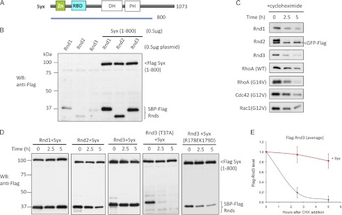
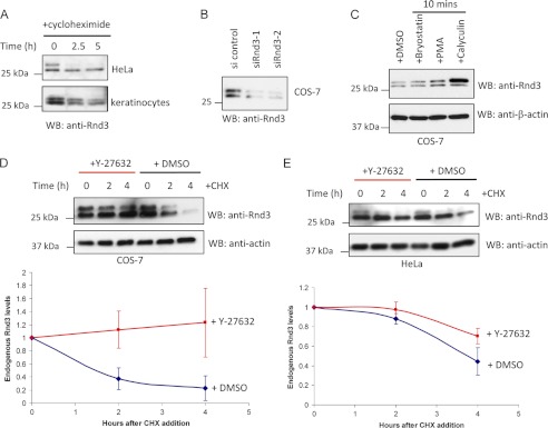
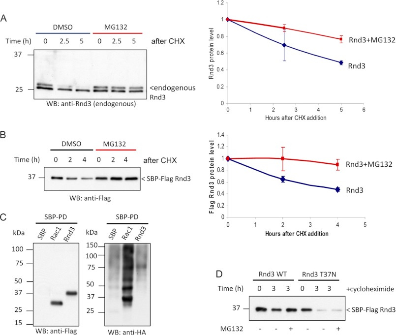
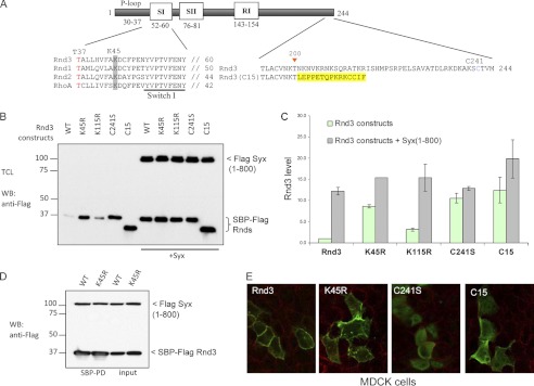
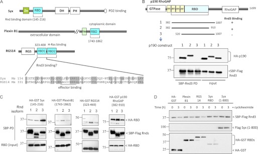
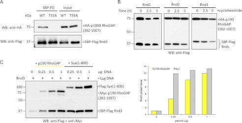
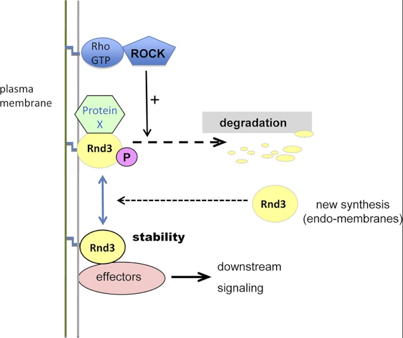
Rnd proteins are Rho family GTP-binding proteins with cellular functions that antagonize RhoA signaling. We recently described a new Rnd3 effector Syx, also named PLEKHG5, that interacts with Rnds via a Raf1-like "Ras-binding domain." Syx is a multidomain RhoGEF that participates in early zebrafish development. Here we demonstrated that Rnd1, Rnd2, and Rnd3 stability is acutely dependent on interaction with their effectors such as Syx or p190 RhoGAP. Although Rnd3 turnover is blocked by treatment of cells with MG132, we provide evidence that such turnover is mediated indirectly by effects on the Rnd3 effectors, rather than on Rnd3 itself, which is not significantly ubiquitinated. The minimal regions of Syx and p190 RhoGAP that bind Rnd3 are not sequence-related but have similar effects. We have identified features that allow for Rnd3 turnover including a conserved Lys-45 close to the switch I region and the C-terminal membrane-binding domain of Rnd3, which cannot be substituted by the equivalent Cdc42 CAAX sequence. By contrast, an effector binding-defective mutant of Rnd3 when overexpressed undergoes turnover at normal rates. Interestingly the activity of the RhoA-regulated kinase ROCK stimulates Rnd3 turnover. This study suggests that Rnd proteins are regulated through feedback mechanisms in cells where the level of effectors and RhoA activity influence the stability of Rnd proteins. This effector feedback behavior is analogous to the ability of ACK1 and PAK1 to prolong the lifetime of the active GTP-bound state of Cdc42 and Rac1.


Figures

FIGURE 1.
FIGURE 1.
Syx contributes to protein stability of Rnd3. A, schematic represents Syx construct used in 293T cell expression studies. B, 293T cells were transfected with Rnd plasmids in the absence or presence of Syx(1–800), lysed, and immunoblottted (WB). C, 293T cells were co-transfected with 0.5 μg of GFP-FLAG and 0.5 μg of the indicated plasmids. After 24 h, cells were incubated with cycloheximide (20 μg/ml) and then harvested at the indicated times with equal amounts of total protein loaded (40 μg/lane). Expression of GFP is marked by an asterisk (*). D, 293T cells were co-transfected with 0.5 μg of Rnd3 and 0.5 μg of Syx(1–800) plasmids and treated for the indicated times with cycloheximide, lysed, and immunoblottted. E, in the graphical representation, the y-axis corresponds to the ratio of Rnd3 relative to time 0. The average of three independent experiments is shown. Error bars, S.D.
FIGURE 2.
FIGURE 2.
ROCK activity stimulates Rnd3 turnover. A, HeLa cells and human keratinocytes treated for the indicated times with cycloheximide and then lysed and immunoblotted (WB) for endogenous Rnd3. B, Western blot analysis of Rnd3 levels in COS-7 cells transfected with control or two sets of Rnd3 siRNA duplexes for 24 h. C, COS cells treated with bryostatin (100 nm), phorbol 12-myristate 13-acetate (PMA) (100 nm), or calyculin (10 nm) for 10 min, lysed, and immunoblotted for endogenous Rnd3. D, Western blot analysis of endogenous Rnd3 protein in COS cells incubated in the presence or absence of 10 μm Y-27632 for 30 min before being treated with cycloheximide (CHX) for the indicated times. In the graphical representation, the y-axis corresponds to the ratio of Rnd3 relative to time 0. The average values derived from three independent experiments are shown with the level of the two bands normalized to time = 0 (using Image J). E, Western blot analysis of endogenous Rnd3 protein in HeLa cells treated and analyzed as in D. DMSO, dimethyl sulfoxide.
FIGURE 3.
FIGURE 3.
Rnd3 can be stabilized by proteasomal inhibition. A, HeLa cells were treated with cycloheximide (CHX) in the presence of 10 μm MG132 or dimethyl sulfoxide (DMSO), then lysed and immunoblotted (WB). In the graphical representation, the y-axis corresponds to the ratio of Rnd3 relative to time 0. The average of three independent experiments is shown. B, 293T cells overexpressing Rnd3 were treated for the indicated times with cycloheximide in the presence of 10 μm MG132 or dimethyl sulfoxide and analyzed as in A. C, HeLa cells were co-transfected with the indicated SBP-tagged plasmids and HA-ubiquitin. Cells were treated with 20 μm MG132 for 30 min prior to lysis. SBP pulldown was performed, and ubiquitinated proteins were visualized by immunoblotting using anti-HA. Conjugation of ubiquitin to Rnd3 was not detected. D, 293T cells overexpressing Rnd3(T37N) were treated for the indicated times with cycloheximide in the presence of 10 μm MG132 to assess whether the effect of proteasome inhibition is direct.
FIGURE 4.
FIGURE 4.
Lys-45 and membrane localization are important for Rnd3 turnover. A, schematic shows Rnd3 construct and amino acid substitutions made in this study. The highly conserved Lys-45 is highlighted in gray. The Rnd3(C15) chimeric construct in which the last 44 C-terminal residues of Rnd3 is replaced by the last 15 C-terminal residues of Cdc42 is in yellow. B, 293T cells overexpressing the indicated plasmids were treated for 3 h with cycloheximide, lysed, and immunoblottted. C, graph represents the average of two independent experiments. D, 293T cells were co-transfected with SBP-FLAG Rnd3 or Rnd3(K45R) with Syx(1–800) constructs. The purified protein complex was immunoblotted to visualize bound Syx. E, localizations of the indicated constructs in MDCK cells were immunolabeled with anti-FLAG antibodies and examined by confocal microscopy.
FIGURE 5.
FIGURE 5.
Rnd3 is not efficiently stabilized by all Rnd-interacting domains. A, schematic represents constructs used in 293T cell expression and Rnd interaction studies. Sequence alignment shows the highly conserved Raf1-like RBDs in mouse Syx (AAU04953) and RGS14 (NP_006471). B, p190B RhoGAP amino acid 382–910 is the minimum Rnd3-binding region. 293T cells were co-transfected with SBP-FLAG Rnd3 and HA or HA-GST p190B RhoGAP constructs. The purified protein complex was immunoblotted to visualize bound p190B RhoGAP. C, binding of Rnd isoforms to Plexin B1 was much weaker compared with Syx and p190 RhoGAP. 293T cells were transfected with expression vectors encoding SBP-FLAG Rnd1, Rnd2, or Rnd3 together with the indicated constructs and assessed for interaction. D, 293T cells overexpressing Rnd3 and the indicated plasmids were treated for 3 h with cycloheximide, lysed, and immunoblottted.
FIGURE 6.
FIGURE 6.
p190 RhoGAP is a genuine Rnd3 effector that stabilizes Rnd3. A, 293T cells were transfected as indicated, and SBP pulldown was performed. Associated proteins were visualized by immunoblotting. No interaction between Rnd3 effector mutant (T55A) and p190 RhoGAP was observed. B, 293T cells were co-transfected with Rnd1, Rnd2, or Rnd3 together with p190 RhoGAP(382–1007) and treated for the indicated times with cycloheximide, lysed, and immunoblottted (WB). C, the stabilizing effect of Rnd3 by Syx and p190 RhoGAP is dose-dependent. 293T cells were transfected with 0.5 μg of Rnd3 and increasing amounts of Syx or p190 RhoGAP plasmids as indicated. The levels of Rnd3 were visualized by immunoblotting with anti-FLAG antibody. Graphical representation of the experiment is shown.
FIGURE 7.
FIGURE 7.
Model showing Rnd3 regulation by the level of effectors and ROCK activity. Rnd3 is targeted for degradation by binding of unknown protein (X) which is sensitive to both the Rnd3(K45R) mutation and the membrane localization of Rnd3. This turnover of Rnd3 is sensitive to Y-27632, indicating that ROCK stimulates such turnover. Effector interaction can prevent the turnover of Rnd3 probably by competing for binding of protein X.

References

    1. Foster R., Hu K. Q., Lu Y., Nolan K. M., Thissen J., Settleman J. (1996) Identification of a novel human Rho protein with unusual properties: GTPase deficiency and in vivo farnesylation. Mol. Cell. Biol. 16, 2689–2699 - PMC - PubMed
    1. Guasch R. M., Scambler P., Jones G. E., Ridley A. J. (1998) RhoE regulates actin cytoskeleton organization and cell migration. Mol. Cell. Biol. 18, 4761–4771 - PMC - PubMed
    1. Chardin P. (2006) Function and regulation of Rnd proteins. Nat. Rev. Mol. Cell Biol. 7, 54–62 - PubMed
    1. Riou P., Villalonga P., Ridley A. J. (2010) Rnd proteins: multifunctional regulators of the cytoskeleton and cell cycle progression. Bioessays 32, 986–992 - PubMed
    1. Hansen S. H., Zegers M. M., Woodrow M., Rodriguez-Viciana P., Chardin P., Mostov K. E., McMahon M. (2000) Induced expression of Rnd3 is associated with transformation of polarized epithelial cells by the Raf-MEK-extracellular signal-regulated kinase pathway. Mol. Cell. Biol. 20, 9364–9375 - PMC - PubMed

MeSH terms