

Mild exercise increases dihydrotestosterone in hippocampus providing evidence for androgenic mediation of neurogenesis
- PMID: 22807478
- PMCID: PMC3420174
- DOI: 10.1073/pnas.1210023109
Mild exercise increases dihydrotestosterone in hippocampus providing evidence for androgenic mediation of neurogenesis
Abstract
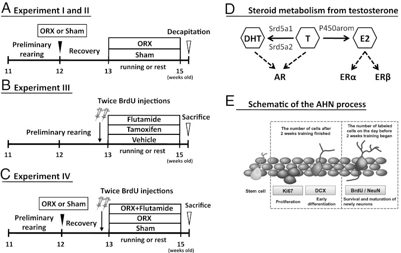
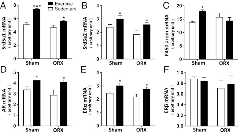
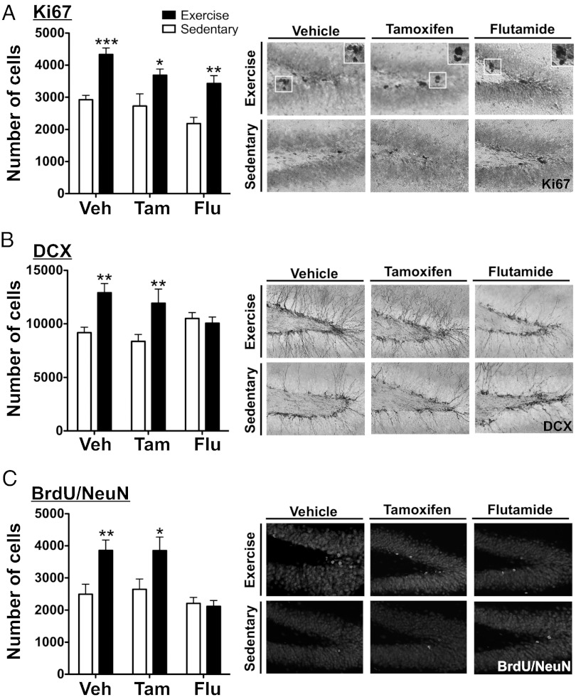
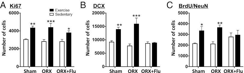
Mild exercise activates hippocampal neurons through the glutamatergic pathway and also promotes adult hippocampal neurogenesis (AHN). We hypothesized that such exercise could enhance local androgen synthesis and cause AHN because hippocampal steroid synthesis is facilitated by activated neurons via N-methyl-D-aspartate receptors. Here we addressed this question using a mild-intense treadmill running model that has been shown to be a potent AHN stimulator. A mass-spectrometric analysis demonstrated that hippocampal dihydrotestosterone increased significantly, whereas testosterone levels did not increase significantly after 2 wk of treadmill running in both orchidectomized (ORX) and sham castrated (Sham) male rats. Furthermore, analysis of mRNA expression for the two isoforms of 5α-reductases (srd5a1, srd5a2) and for androgen receptor (AR) revealed that both increased in the hippocampus after exercise, even in ORX rats. All rats were injected twice with 5'-bromo-2'deoxyuridine (50 mg/kg body weight, i.p.) on the day before training. Mild exercise significantly increased AHN in both ORX and Sham rats. Moreover, the increase of doublecortin or 5'-bromo-2'deoxyuridine/NeuN-positive cells in ORX rats was blocked by s.c. flutamide, an AR antagonist. It was also found that application of an estrogen receptor antagonist, tamoxifen, did not suppress exercise-induced AHN. These results support the hypothesis that, in male animals, mild exercise enhances hippocampal synthesis of dihydrotestosterone and increases AHN via androgenenic mediation.
Conflict of interest statement
The authors declare no conflict of interest.
Figures
Similar articles
-
Physical exercise increases adult hippocampal neurogenesis in male rats provided it is aerobic and sustained.J Physiol. 2016 Apr 1;594(7):1855-73. doi: 10.1113/JP271552. Epub 2016 Feb 24. J Physiol. 2016. PMID: 26844666 Free PMC article.
-
Testosterone and dihydrotestosterone, but not estradiol, enhance survival of new hippocampal neurons in adult male rats.Dev Neurobiol. 2007 Sep 1;67(10):1321-33. doi: 10.1002/dneu.20457. Dev Neurobiol. 2007. PMID: 17638384
-
5α-reduction of epitestosterone is catalysed by human SRD5A1 and SRD5A2 and increases androgen receptor transactivation.J Steroid Biochem Mol Biol. 2024 Jul;241:106516. doi: 10.1016/j.jsbmb.2024.106516. Epub 2024 Apr 4. J Steroid Biochem Mol Biol. 2024. PMID: 38582131
-
[Mild Exercise Results in Robust Brain Activation and Increased Memory Function].Brain Nerve. 2018 Jul;70(7):745-752. doi: 10.11477/mf.1416201075. Brain Nerve. 2018. PMID: 29997270 Review. Japanese.
-
Neurosteroids in Adult Hippocampus of Male and Female Rodents: Biosynthesis and Actions of Sex Steroids.Front Endocrinol (Lausanne). 2018 Apr 23;9:183. doi: 10.3389/fendo.2018.00183. eCollection 2018. Front Endocrinol (Lausanne). 2018. PMID: 29740398 Free PMC article. Review.
Cited by
-
The effects of peripheral hormone responses to exercise on adult hippocampal neurogenesis.Front Endocrinol (Lausanne). 2023 Nov 24;14:1202349. doi: 10.3389/fendo.2023.1202349. eCollection 2023. Front Endocrinol (Lausanne). 2023. PMID: 38084331 Free PMC article. Review.
-
Co-activator candidate interactions for orphan nuclear receptor NR2E1.BMC Genomics. 2016 Oct 26;17(1):832. doi: 10.1186/s12864-016-3173-5. BMC Genomics. 2016. PMID: 27782803 Free PMC article.
-
Light-exercise-induced dopaminergic and noradrenergic stimulation in the dorsal hippocampus: Using a rat physiological exercise model.FASEB J. 2024 Dec 13;38(24):e70215. doi: 10.1096/fj.202400418RRR. FASEB J. 2024. PMID: 39668509 Free PMC article.
-
Unraveling the complexity of human brain: Structure, function in healthy and disease states.Ageing Res Rev. 2024 Sep;100:102414. doi: 10.1016/j.arr.2024.102414. Epub 2024 Jul 15. Ageing Res Rev. 2024. PMID: 39002647 Free PMC article. Review.
-
Pubertally born neurons and glia are functionally integrated into limbic and hypothalamic circuits of the male Syrian hamster.Proc Natl Acad Sci U S A. 2013 Mar 19;110(12):4792-7. doi: 10.1073/pnas.1219443110. Epub 2013 Mar 4. Proc Natl Acad Sci U S A. 2013. PMID: 23460698 Free PMC article.
References
-
- Cameron HA, Gould E. The control of neuronal birth and survival. In: Shaw C, editor. Receptor Dynamics in Neural Development. Boca Raton, FL: CRC Press; 1996. pp. 141–157.
-
- Gage FH. Mammalian neural stem cells. Science. 2000;287:1433–1438. - PubMed
-
- Galea LA, Spritzer MD, Barker JM, Pawluski JL. Gonadal hormone modulation of hippocampal neurogenesis in the adult. Hippocampus. 2006;16:225–232. - PubMed
-
- MacLusky NJ, Hajszan T, Prange-Kiel J, Leranth C. Androgen modulation of hippocampal synaptic plasticity. Neuroscience. 2006;138:957–965. - PubMed
Publication types
MeSH terms
Substances
LinkOut - more resources
Full Text Sources
Research Materials
