. 2012;8(7):e1002797.
doi: 10.1371/journal.ppat.1002797. Epub 2012 Jul 12.

Structural mechanism of trimeric HIV-1 envelope glycoprotein activation

Affiliations

Structural mechanism of trimeric HIV-1 envelope glycoprotein activation

Erin E H Tran et al. PLoS Pathog. 2012.

Abstract

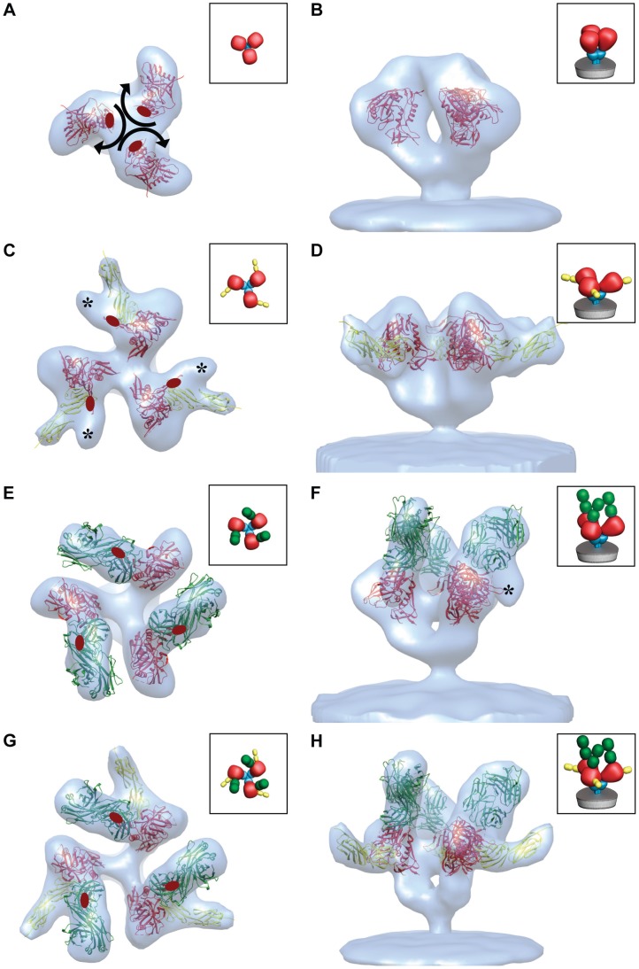
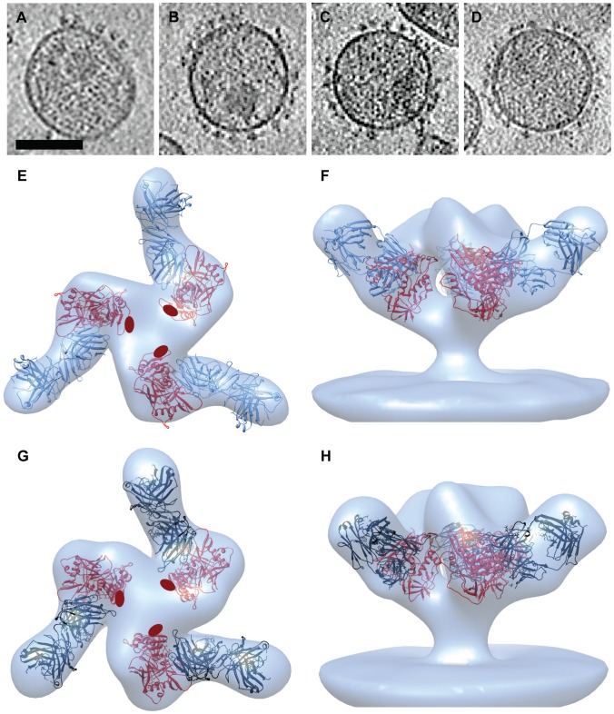
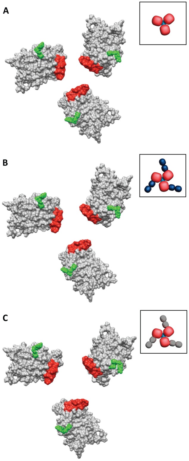
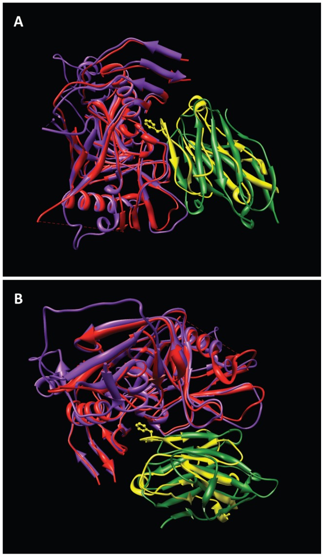
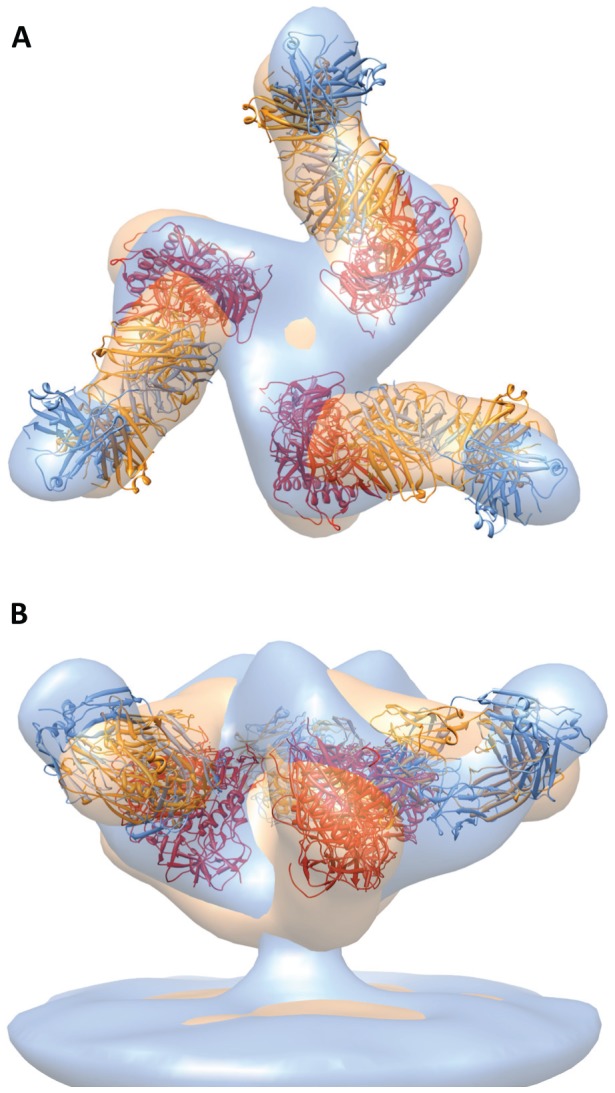
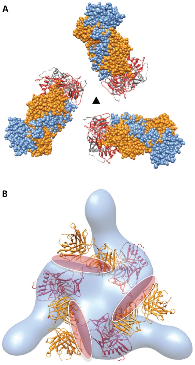
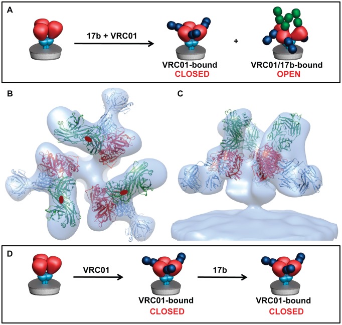
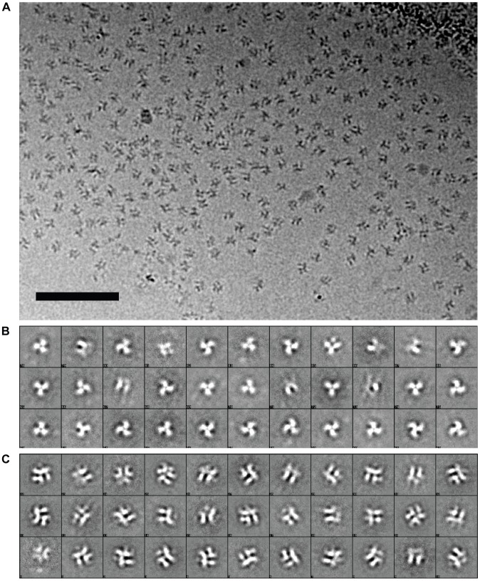
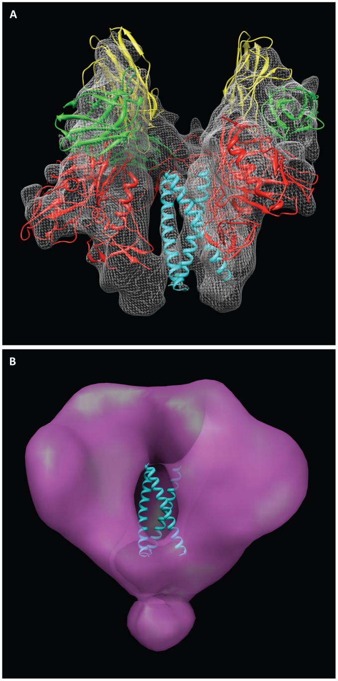
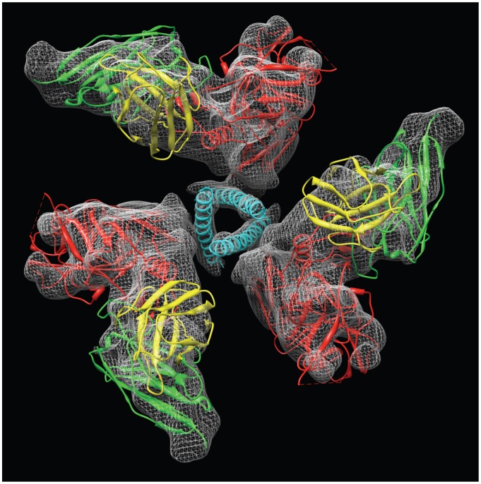
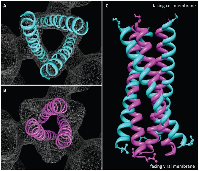
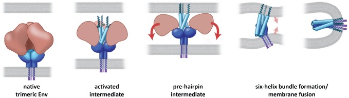
HIV-1 infection begins with the binding of trimeric viral envelope glycoproteins (Env) to CD4 and a co-receptor on target T-cells. Understanding how these ligands influence the structure of Env is of fundamental interest for HIV vaccine development. Using cryo-electron microscopy, we describe the contrasting structural outcomes of trimeric Env binding to soluble CD4, to the broadly neutralizing, CD4-binding site antibodies VRC01, VRC03 and b12, or to the monoclonal antibody 17b, a co-receptor mimic. Binding of trimeric HIV-1 BaL Env to either soluble CD4 or 17b alone, is sufficient to trigger formation of the open quaternary conformation of Env. In contrast, VRC01 locks Env in the closed state, while b12 binding requires a partial opening in the quaternary structure of trimeric Env. Our results show that, despite general similarities in regions of the HIV-1 gp120 polypeptide that contact CD4, VRC01, VRC03 and b12, there are important differences in quaternary structures of the complexes these ligands form on native trimeric Env, and potentially explain differences in the neutralizing breadth and potency of antibodies with similar specificities. From cryo-electron microscopic analysis at ∼9 Å resolution of a cleaved, soluble version of trimeric Env, we show that a structural signature of the open Env conformation is a three-helix motif composed of α-helical segments derived from highly conserved, non-glycosylated N-terminal regions of the gp41 trimer. The three N-terminal gp41 helices in this novel, activated Env conformation are held apart by their interactions with the rest of Env, and are less compactly packed than in the post-fusion, six-helix bundle state. These findings suggest a new structural template for designing immunogens that can elicit antibodies targeting HIV at a vulnerable, pre-entry stage.


Conflict of interest statement

The authors have declared that no competing interests exist.

Figures

Figure 1
Figure 1. Binding to sCD4 or 17b is sufficient for formation of the open quaternary Env conformation.
(a, b) Density map and molecular architecture for native trimeric Env from native HIV-1 BaL shown in top (a, from the apex) and side (b) views . The locations of the stumps of the V1V2 loop are shown by the red ovals in (a) and subsequent panels with top views, and the black arrows indicate the rotation of each gp120 monomer that must occur when the trimer transitions from closed to open conformations. (c–h) Top and side views of the sCD4-bound (c, d) 17b-bound (e, f) and sCD4/17b bound (g, h) Env density maps. Maps in all panels were fitted with subsets of the X-ray coordinates from the gp120-sCD4/17b complex (PDB ID:1GC1). The black asterisks (c, f) denote the location of the V1V2 loop. The insets schematically depict trimer conformation and bound ligands highlighting gp120 (red), gp41 (cyan), sCD4 (yellow) and 17b (green).
Figure 2
Figure 2. Structure of trimeric HIV-1 BaL Env bound to VRC01 and VRC03 antibodies.
(a–d) Slices through reconstructed cryo-electron tomograms of HIV-1 BaL in the unliganded state (a) or complexed with VRC01 (b), VRC02 (c) or VRC03 antibodies (d). Peripheral Env glycoprotein spikes are visible on the viral membrane. Scale bar is 100 nm. (e, f) Top and side views, respectively, of the VRC01-bound HIV-1 BaL Env density map fitted with X-ray coordinates of the gp120-VRC01 Fab complex (PDB ID:3NGB). (g, h) Top and side views, respectively, of the VRC03-bound Env density map fitted with X-ray coordinates for gp120-VRC03 Fab (PDB ID:3SE8). Coordinates show gp120 (red), VRC01 (blue) and VRC03 (black). In panels (e) and (g), the locations of the stumps of the V1V2 loop are shown by the red ovals.
Figure 3
Figure 3. Similar quaternary conformations of native unliganded HIV-1 Env and Env bound to VRC01 or VRC03.
(a–c) The gp120 portion of the gp120-VRC01 X-ray coordinates (PBD ID:3NGB) is shown after alignment to gp120 fitted to either the unliganded (a), VRC01-bound (b) or VRC03-bound (c) Env density maps to show the similarity in gp120 conformation in each of these states. The orientation of the stumps of the V1V2 (red) and V3 (green) loops on the gp120 surface provides a visual marker for gp120 conformation. Cartoon representations of the gp120 coordinates (unliganded or ligand-bound) are shown next to each set of coordinates, with gp120 (red), gp41 (cyan), VRC01 (blue) and VRC03 (dark grey).
Figure 4
Figure 4. Similarity in structure of regions of sCD4 and VRC01 that contact gp120.
(a, b) Two orthogonal views of the superposition of gp120 polypeptides derived from the complex with sCD4 and 17b (PDB ID: 1GC1) and the complex with VRC01 (PDB ID: 3NGB). For clarity of visualization, only those regions of VRC01 and sCD4 that are in close proximity to gp120 are shown. The gp120 chain and sCD4 chains from the 1GC1 structure are shown in red, and yellow, respectively, while the gp120 and VRC01 chains from the 3NGB structure are shown in magenta and green, respectively.
Figure 5
Figure 5. Comparison of structures of trimeric Env complexed to either VRC01 or b12.
(a, b) Top and side views, respectively, showing the superposition of VRC01-bound (blue) or b12-bound (orange) Env density maps fitted with the corresponding gp120-VRC01 (PDB ID:3NGB) or gp120-b12 (PDB ID:2NY7) coordinates. The gp120 components of the coordinates are shown in red for both complexes, while the VRC01 and b12 Fab fragments are shown in blue and orange, respectively.
Figure 6
Figure 6. Binding of VRC01 retains Env in the closed state, but b12 binding requires an in-plane rotation of gp120.
(a) Top view of the molecular coordinates for gp120 bound to VRC01 (gp120 in red, VRC01 in blue, PDB ID: 3NGB) or bound to b12 (gp120 in grey, b12 in orange, PDB ID: 2NY7). Env binding to b12 results in a small in-plane rotation of gp120, while VRC01 binding retains the unliganded gp120 conformation. (b) The gp120 portion of the b12-bound complex was aligned to the gp120 portion of the VRC01-bound complex to model b12 binding in the VRC01-bound, closed, conformation. Coordinates of the gp120-b12 complex are shown with the VRC01-bound Env density map. The projected steric overlap between b12 Fab and neighboring gp120 protomers in the closed state is highlighted for visualization purposes with the red elliptical patches. Coordinates for gp120, b12 and VRC01 are in red, orange and blue, respectively.
Figure 7
Figure 7. Conformational states of trimeric Env in the presence of VRC01 and 17b.
(a) Schematic representation of the Env maps obtained after incubation of HIV-1 BaL with 17b and VRC01. (b, c) Top and side views, respectively, of the VRC01/17b-bound Env density map. X-ray coordinates for the gp120-VRC01 complex (PDB ID:3NGB) were fitted into the map and subsequently aligned to the gp120-CD4/17b (PDB ID:1GC1) complex. (d) Schematic illustration of the finding that when VRC01 is pre-bound to HIV-1 BaL, all detectable Env is in a closed VRC01-bound state, and this is not altered upon subsequent addition of 17b.
Figure 8
Figure 8. Cryo-electron microscopy of the complex formed between soluble KNH1144 trimeric Env and 17b Fab fragments.
(a) Projection image recorded on a Titan Krios electron microscope operated at 80 kV using a 4K×4K CCD camera. Scale bar is 50 nm. (b, c) Representative examples of initial 2D class averages separating unbound or partially occupied complexes (b) from those that appear to have full occupancy of 17b (c).
Figure 9
Figure 9. Structure of the open conformation of trimeric Env at sub-nanometer resolution.
(a) Side view of the structure of trimeric Env bound to 17b Fab. The map was fitted with three copies of the X-ray structures for the gp120-17b portion of the 1GC1 coordinates with gp120 (red) and 17b Fv fragments (light chain: yellow, heavy chain: green). One copy of the gp41 N-terminal helix (cyan) of 1AIK coordinates (N34) was fitted individually into each of the three densities, which occupy the central region of the spike that is essentially a cavity in the unliganded state. (b) Side view of the density map from unliganded native trimeric Env , , , , with the three gp41 N-terminal helices (cyan) superposed to show that in the open conformation, they occupy the solvent filled cavity in the density map of the unliganded state.
Figure 10
Figure 10. View of the central gp41 helices in the open conformation of trimeric Env bound to 17b Fab.
The map was fitted with three copies of the X-ray structures for the gp120-17b portion of the 1GC1 coordinates with color scheme as in Figure 9.
Figure 11
Figure 11. Mechanism of activation of trimeric Env.
(a) Top view of the locations of the three N-terminal helices in the open conformation (as in Figure 10). (b) Top view of the locations of the same helices in the crystal structure (1AIK) of the six-helix bundle formed by the three N- and three C-terminal helices, illustrating the more compact packing in the post-fusion state. (c) Superposition of the arrangement of the three N-terminal helices in the open quaternary Env conformation (cyan) derived from cryo-electron microscopy of soluble trimeric Env with that in the post-fusion conformation (magenta) derived by X-ray crystallography of the six-helix bundle.
Figure 12
Figure 12. Model for the mechanism of Env activation.
The CD4- or co-receptor-triggered activation of the Env spike forms an activated intermediate in which the N-terminal gp41 helices are vulnerable to neutralizing ligands. The pre-hairpin intermediate is formed upon insertion of the fusion peptide into the target cell membrane and dissociation of gp120, leading subsequently to formation of the six-helix bundle state and subsequent fusion between viral and target cell membranes.

Comment in

References

    1. Dalgleish AG, Beverley PC, Clapham PR, Crawford DH, Greaves MF, et al. The CD4 (T4) antigen is an essential component of the receptor for the AIDS retrovirus. Nature. 1984;312:763–767. - PubMed
    1. Chan DC, Kim PS. HIV entry and its inhibition. Cell. 1998;93:681–684. - PubMed
    1. Sattentau QJ, Moore JP, Vignaux F, Traincard F, Poignard P. Conformational changes induced in the envelope glycoproteins of the human and simian immunodeficiency viruses by soluble receptor binding. J Virol. 1993;67:7383–7393. - PMC - PubMed
    1. Stamatatos L, Cheng-Mayer C. Evidence that the structural conformation of envelope gp120 affects human immunodeficiency virus type 1 infectivity, host range, and syncytium-forming ability. J Virol. 1993;67:5635–5639. - PMC - PubMed
    1. Trkola A, Dragic T, Arthos J, Binley JM, Olson WC, et al. CD4-dependent, antibody-sensitive interactions between HIV-1 and its co-receptor CCR-5. Nature. 1996;384:184–187. - PubMed

Publication types

MeSH terms