. 2012;8(7):e1002807.
doi: 10.1371/journal.ppat.1002807. Epub 2012 Jul 12.

HPV 5 and 8 E6 abrogate ATR activity resulting in increased persistence of UVB induced DNA damage

Affiliations

HPV 5 and 8 E6 abrogate ATR activity resulting in increased persistence of UVB induced DNA damage

Nicholas A Wallace et al. PLoS Pathog. 2012.

Abstract

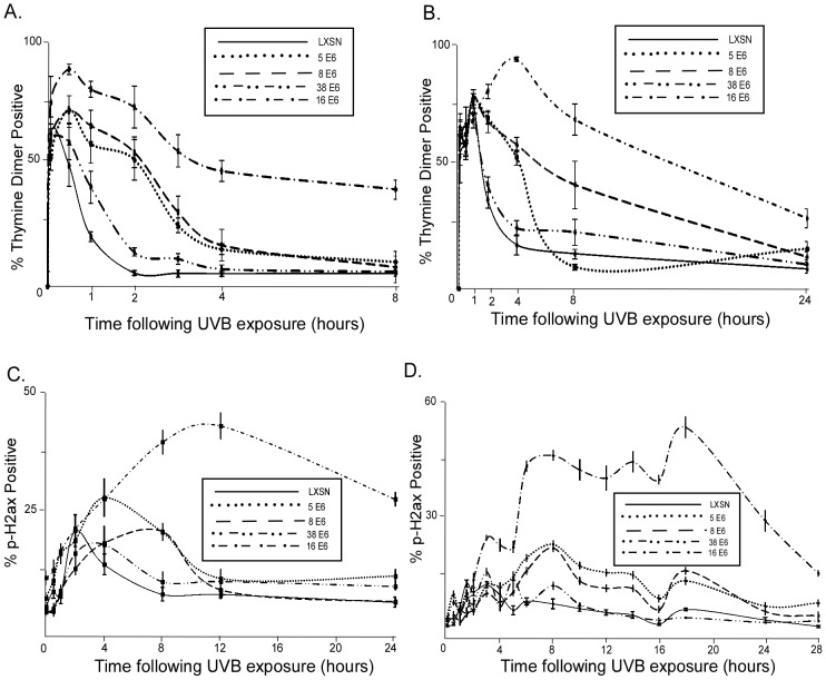
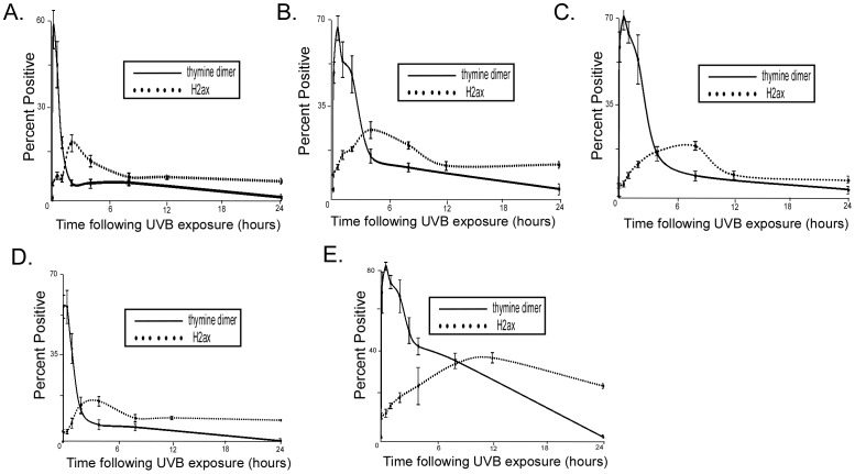
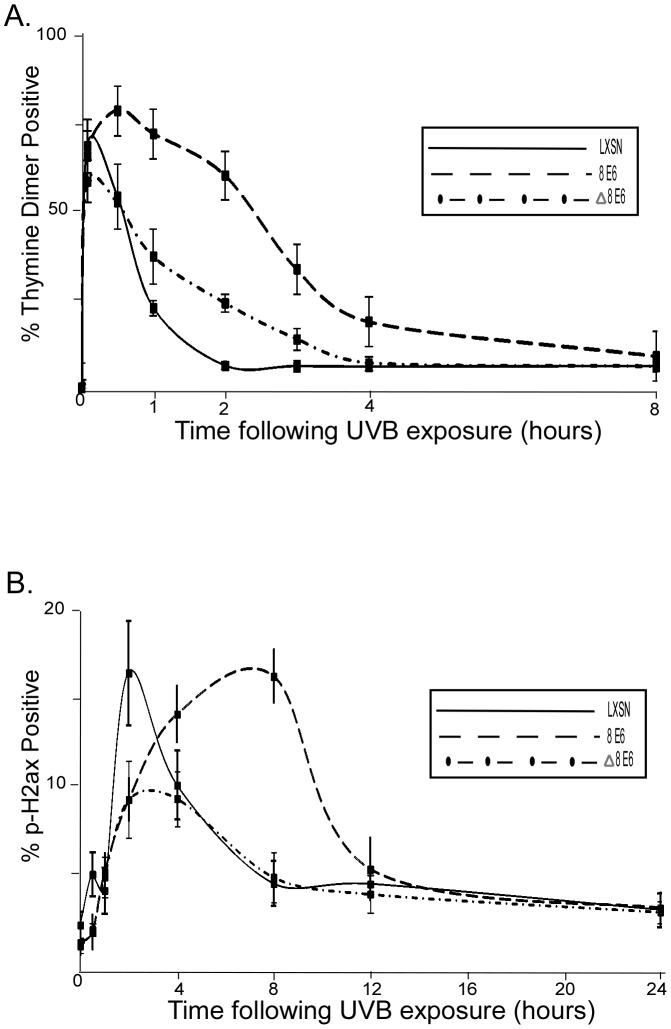
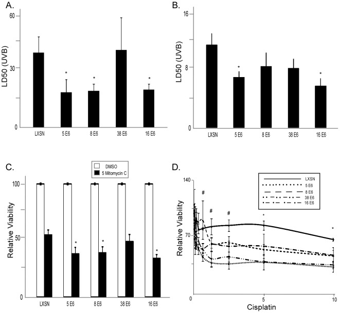
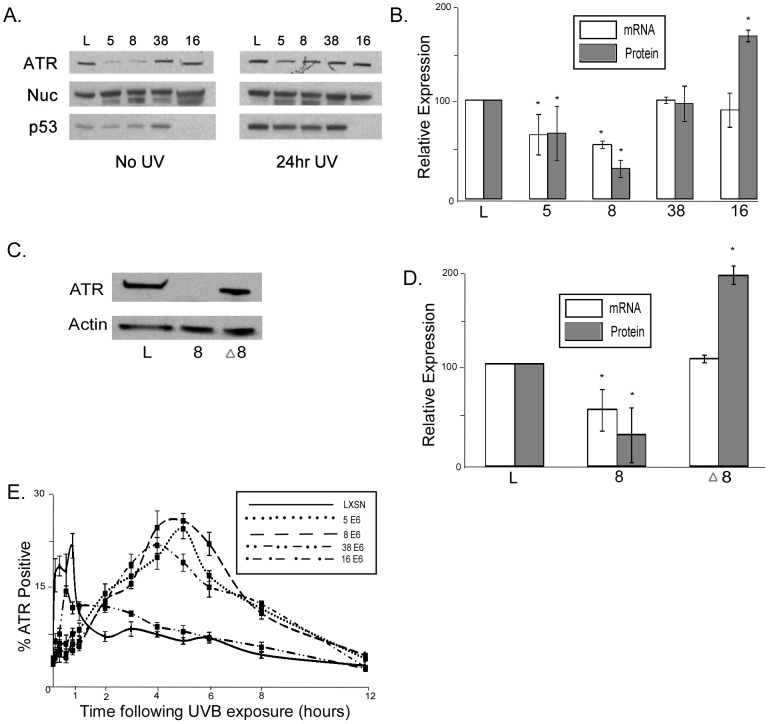
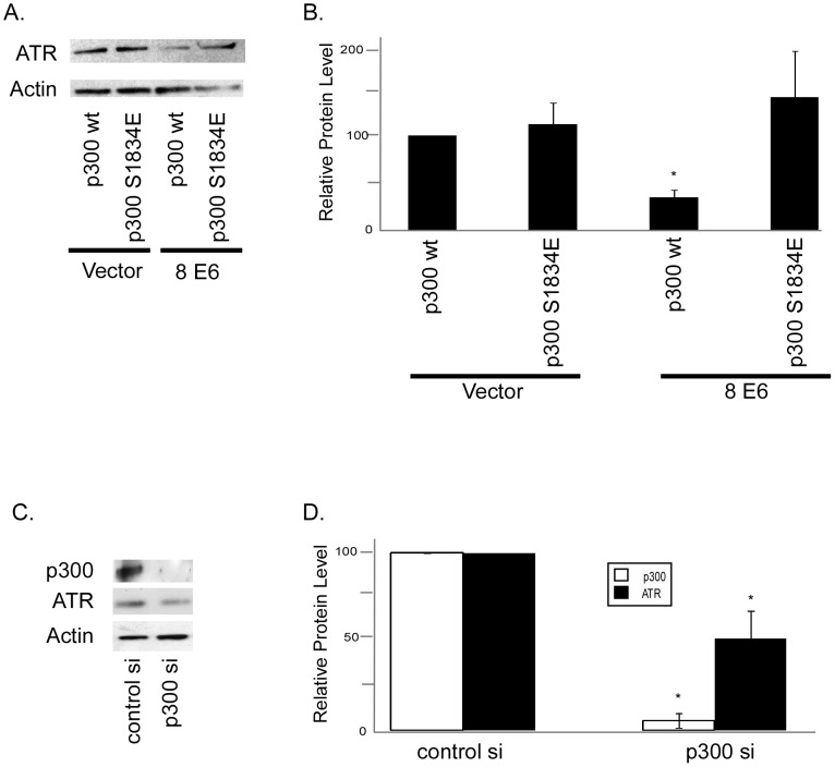
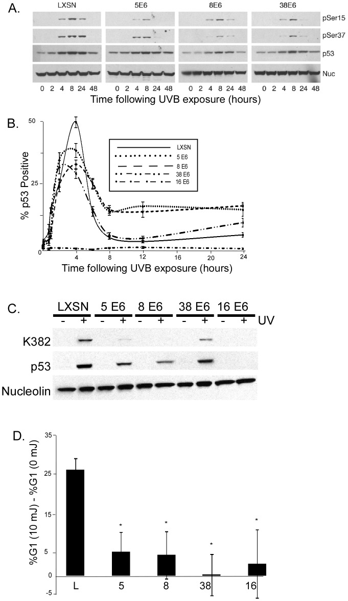
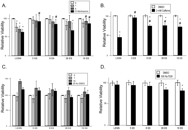
The role of the E6 oncoprotein from high-risk members of the α human papillomavirus genus in anogenital cancer has been well established. However, far less is known about the E6 protein from the β human papillomavirus genus (β-HPVs). Some β-HPVs potentially play a role in non-melanoma skin cancer development, although they are not required for tumor maintenance. Instead, they may act as a co-factor that enhances the carcinogenic potential of UV damage. Indeed, the E6 protein from certain β-HPVs (HPV 5 and 8) promotes the degradation of p300, a histone acetyl transferase involved in UV damage repair. Here, we show that the expression of HPV 5 and 8 E6 increases thymine dimer persistence as well as the likelihood of a UVB induced double strand break (DSB). Importantly, we provide a mechanism for the increased DNA damage by showing that both extended thymine dimer persistence as well as elevated DSB levels are dependent on the ability of HPV 8 E6 to promote p300 degradation. We further demonstrate that HPV 5 and 8 E6 expression reduces the mRNA and protein levels of ATR, a PI3 kinase family member that plays a key role in UV damage signaling, but that these levels remain unperturbed in cells expressing a mutated HPV 8 E6 incapable of promoting p300 degradation. We confirm that the degradation of p300 leads to a reduction in ATR protein levels, by showing that ATR levels rebound when a p300 mutant resistant to HPV 8 mediated degradation and HPV 8 E6 are co-transfected. Conversely, we show that ATR protein levels are reduced when p300 is targeted for degradation by siRNA. Moreover, we show the reduced ATR levels in HPV 5 and 8 E6 expressing cells results in delayed ATR activation and an attenuated ability of cells to phosphorylate, and as a result accumulate, p53 in response to UVB exposure, leading to significantly reduced cell cycle arrest. In conclusion, these data demonstrate that β-HPV E6 expression can enhance the carcinogenic potential of UVB exposure by promoting p300 degradation, resulting in a reduction in ATR levels, which leads to increased thymine dimer persistence and increased UVB induced DSBs.


Conflict of interest statement

The authors have declared that no competing interests exist.

Figures

Figure 1
Figure 1. HPV E6 increases thymine dimer persistence and DSB prevalence following UVB exposure.
A. HFK cells were exposed to 10 mJ/cm2 UVB and immunofluorescence was used to measure the percentage of cells positive for thymine dimers above background. B. HT1080 cells were exposed to 10 mJ/cm2 UVB and immunofluorescence was used to measure the percentage of cells positive for thymine dimers above background. C. HFK cells were exposed to 10 mJ/cm2 UVB and immunofluorescence was used to measure the percentage of cells positive for phospho H2AX above background. D. HT1080 cells were exposed to 10 mJ/cm2 UVB and immunofluorescence was used to measure the percentage of cells positive for phospho H2AX above background. For all portions of this figure, n≥5, error bars represent standard errors of the mean.
Figure 2
Figure 2. Temporal relationship between thymine dimer and phospho H2AX detection.
HFK cells were exposed to 10 mJ/cm2 UVB and immunofluorescence was used to measure the percentage of cells positive for thymine dimers and phospho H2AX above background. Graph shows percentage of HFK cells staining positive for either thymine dimers (solid line) or phospho H2AX (dotted line) after indicated period of time following exposure to 10 mJ/cm2 UVB. A. LXSN HFK cells B. 5 E6 HFK cells C. HPV 8 E6 HFK cells D. HPV 38 E6 HFK cells E. HPV 16 E6 HFK cells. n≥5. Error bars represent standard errors of the mean.
Figure 3
Figure 3. Increased persistence of UVB induced DNA damage in HPV 8 E6 expressing cells is dependent on p300 degradation.
A. HFK cells were exposed to 10 mJ/cm2 UVB and immunofluorescence was used to measure the percentage of cells positive for thymine dimers above background. B. HFK cells were exposed to 10 mJ/cm2 UVB and immunofluorescence was used to measure the percentage of cells positive for phospho H2AX above background. For all portions of this figure, n≥5, error bars represent standard errors of the mean, and Δ8 E6 denotes cells expressing a mutant form of 8 E6 incapable of promoting p300 degradation.
Figure 4
Figure 4. HPV E6 expression increases sensitivity to crosslinking agents.
A. Median Lethal dose of UVB for HPV E6 expressing primary human foreskin keratinocytes (HFKs) was measured following exposure to a range of UVB doses (0–20 mJ/cm2). n≥5. Error bars represent standard errors of the mean. B. Median Lethal dose of UVB for HT1080 cells expressing the same HPV E6s was measured following a range of UVB doses (0–10 mJ/cm2). n≥5. Error bars represent standard errors of the mean. C. Relative viability of HPV E6 expressing HT1080 cells was measured after exposure to mitomycin C (µM). n≥5. Error bars represent standard errors of the mean. D. Relative viability was measured in HPV E6 expressing HT1080 cells following exposure to a range of cisplatin doses (µM). n≥5. Error bars represent standard errors of the mean. Linear regression lines were calculated using GraphPad Prism software. # denotes a statistical significant difference from LXSN for HPV 5, 38, and 16 E6 expressing cells (p≤0.05). * denotes a statistical significant difference from LXSN for all HPV E6 expressing cells (p≤0.05).
Figure 5
Figure 5. HPV E6 expression decreases ATR expression and activity.
A. Representative immunoblot showing ATR protein levels in HFK cells either untreated or exposed to 10 mJ/cm2 UVB. p53 is shown as a control for HPV 16 E6 expression, which results in p53 degradation. Nuc denotes Nucleolin. B. Densitometry of immunoblots (n = 3) and measurements by quantitative RT-PCR of ATR mRNA levels (n = 3) in HPV E6 expressing HFK cells. Levels were normalized to GAPdh (mRNA) and Nucleolin (protein) for each experiment. Error bars represent standard errors of the mean. * denotes a statistically significant difference from LXSN cells. C. Representative immunoblot showing ATR protein levels in HFK cells exposed to 10 mJ/cm2 UVB. D. Densitometry of immunoblots (n = 3) and measurements by quantitative RT-PCR of ATR mRNA levels (n = 3) in HPV E6 expressing HFK cells. Levels were normalized to GAPdh (mRNA) or β Actin (protein) for each experiment. Error bars represent standard errors of the mean. * denotes a statistically significant difference from LXSN cells. E. ATR foci formation was measured using immunofluorescence following 10 mJ/cm2 UVB exposure. Graph shows percentage of cells positive for ATR foci, above background levels.
Figure 6
Figure 6. Reduced ATR protein levels in HPV 5 and 8 E6 expressing cells is dependent on p300 degradation.
A. Representative immunoblot showing ATR levels in HFK cells cotransfected with either p300wt or p300 S1834E and vector control or HPV 8 E6. β Actin was used as a loading control. B. Densitometry of immunoblots (n = 3) of ATR protein levels in HPV E6 expressing HFK cells cotransfected with either p300wt or p300 S1834E and vector control or HPV 8 E6. Levels were normalized to β actin for each experiment. Error bars represent standard errors of the mean. * denotes a statistically significant difference from HFK cells cotransfected with vector control and p300wt. C. p300 was knocked down in HFK cells by transfection with a pool of 4 siRNAs targeting p300 or a pool of 4 non-targeting siRNAs. Representative immunoblot showing ATR and p300 levels in these cells HFK cells 72 hours post transfection. β Actin was used as a loading control. D. Densitometry of immunoblots (n = 3) of ATR and p300 protein levels in HFK cells transfected with a pool of 4 p300 targeting siRNAs or a pool of 4 non-targeting siRNAs. Levels were normalized to β actin for each experiment. Error bars represent standard errors of the mean. * denotes a statistically significant difference from HFK cells transfected with control siRNAs.
Figure 7
Figure 7. UVB induced p53 response is attenuated in HPV E6 expressing cells.
A. Representative immunoblot showing total p53 and phosphorylated p53 (Ser15 and Ser37) levels in β-HPV E6 expressing HFKs following 10 mJ/cm2 UVB exposure. Nucleolin levels were used as a loading control. B. p53 levels were measured by immunofluorescence following 10 mJ/cm2 UVB exposure in HPV E6 expressing HFKs following 10 mJ/cm2 UVB exposure. Graph shows percentage of cells with p53 levels above background. n≥5, and error bars represent standard errors of the mean. In this experiment, HPV 16 E6 served as a negative control because of its ability to degrade p53. C. Representative immunoblot showing total p53 and acetylated p53 (Lys382) levels in HPV E6 expressing HFK either 24 hours after exposure to 10 mJ/cm2 UVB or mock exposure. D. Following density arrest in G1, HT1080 cells were either exposed to 10 mJ/cm2 UVB or left untreated. This graph shows the difference between the percentage of cells in G1 in UVB exposed cells compared to untreated cells 24 hours after UVB exposure. n≥3. Error bars represent standard errors of the mean. * denotes a statistically significant difference from LXSN cells.
Figure 8
Figure 8. HPV E6 expression reduces sensitivity to combined ATR inhibition and UVB exposure.
A. Relative viability of HFK cells expressing HPV 5, 8, 38, and 16 E6 was measured following exposure to 10 mJ/cm2 UVB in the presence of a gradient of Wortmannin concentrations (µM). Data was normalized to cells exposed to UVB in the absence of Wortmannin for each cell line. n≥3. Error bars represent standard errors of the mean. B. Relative viability of HFK cells expressing HPV 5, 8, 38, and 16 E6 was measured following exposure to 10 mJ/cm2 UVB in the presence or absence of Caffeine (2 mM). Data was normalized to cells exposed to UVB in the absence of Wortmannin for each cell line. n≥3. Error bars represent standard errors of the mean. C. Relative viability of HFK cells expressing HPV 5, 8, 38, and 16 E6 was measured following exposure to 10 mJ/cm2 UVB in the presence of a gradient of Ku 55933 concentrations (nM). Data was normalized to cells exposed to UVB in the absence of Ku 55933 for each cell line. n≥3. Error bars represent standard errors of the mean. D. Relative viability of HFK cells expressing HPV 5, 8, 38, and 16 E6 was measured following exposure to 10 mJ/cm2 UVB with or without Nu 7026 (nM). Data was normalized to cells exposed to UVB in the absence of Nu 7026 for each cell line. n≥3. Error bars represent standard errors of the mean. * denotes a statistically significant difference between cells exposed with and without inhibitor. # denotes a statistically significant difference from LXSN cells for the same concentration of inhibitor.

Similar articles

Cited by

References

    1. Bernard HU, Burk RD, Chen Z, van Doorslaer K, Hausen H, et al. Classification of papillomaviruses (PVs) based on 189 PV types and proposal of taxonomic amendments. Virology. 2010;401:70–79. - PMC - PubMed
    1. zur Hausen H. Papillomaviruses in human cancers. Proc Assoc Am Physicians. 1999;111:581–587. - PubMed
    1. Cogliano V, Baan R, Straif K, Grosse Y, Secretan B, et al. Carcinogenicity of human papillomaviruses. Lancet Oncol. 2005;6:204. - PubMed
    1. Lorincz AT, Reid R, Jenson AB, Greenberg MD, Lancaster W, et al. Human papillomavirus infection of the cervix: relative risk associations of 15 common anogenital types. Obstet Gynecol. 1992;79:328–337. - PubMed
    1. Bouwes Bavinck JN, Feltkamp M, Struijk L, ter Schegget J. Human papillomavirus infection and skin cancer risk in organ transplant recipients. J Investig Dermatol Symp Proc. 2001;6:207–211. - PubMed

Publication types

MeSH terms

Substances