. 2012;8(7):e1002768.
doi: 10.1371/journal.pgen.1002768. Epub 2012 Jul 12.

Three dopamine pathways induce aversive odor memories with different stability

Affiliations

Three dopamine pathways induce aversive odor memories with different stability

Yoshinori Aso et al. PLoS Genet. 2012.

Abstract

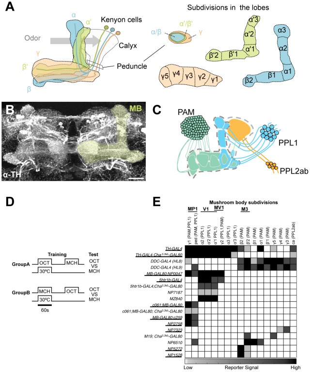
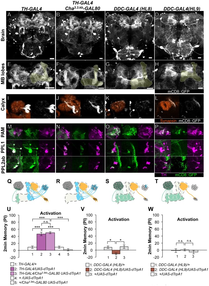
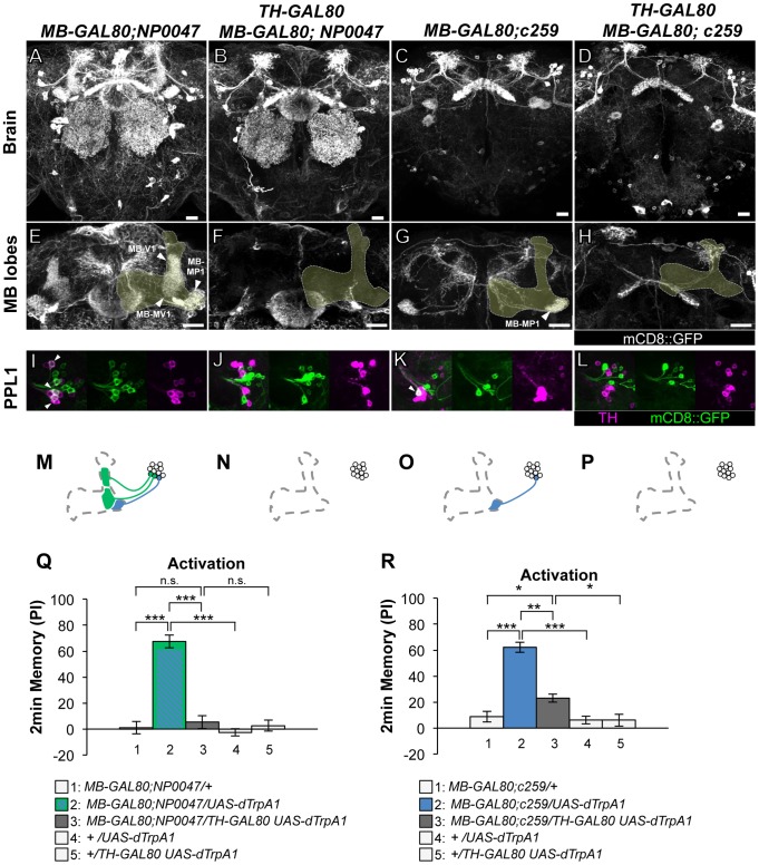
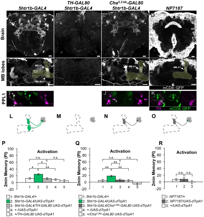
Animals acquire predictive values of sensory stimuli through reinforcement. In the brain of Drosophila melanogaster, activation of two types of dopamine neurons in the PAM and PPL1 clusters has been shown to induce aversive odor memory. Here, we identified the third cell type and characterized aversive memories induced by these dopamine neurons. These three dopamine pathways all project to the mushroom body but terminate in the spatially segregated subdomains. To understand the functional difference of these dopamine pathways in electric shock reinforcement, we blocked each one of them during memory acquisition. We found that all three pathways partially contribute to electric shock memory. Notably, the memories mediated by these neurons differed in temporal stability. Furthermore, combinatorial activation of two of these pathways revealed significant interaction of individual memory components rather than their simple summation. These results cast light on a cellular mechanism by which a noxious event induces different dopamine signals to a single brain structure to synthesize an aversive memory.


Conflict of interest statement

The authors have declared that no competing interests exist.

Figures

Figure 1
Figure 1. GAL4 drivers for dopaminergic neurons that project to the mushroom body.
(A) Schematic diagrams of the mushroom body (left) and subdivisions in the lobe system of the MB (right). Kenyon cells are the major intrinsic neurons of the MB. They have dendritic terminals forming the structure called calyx, where odor signals are conveyed from the antennal lobes. From the calyx, Kenyon cell axons project anteriorly through the peduncle to the lobes. α, α', β, β', and γ indicate the lobes of the MB. In the peduncle, the parallel axon fibers of Kenyon cell subtypes are organized in concentric layers. The spur is contributed exclusively by the neurons projecting to the γ lobe (γ1). The terminals of the extrinsic neurons define the subdivision in the lobes along the longitudinal axis of Kenyon cell axon bundles . (B) Confocal projection of the MB region (rectangle in A; light green overlay) labeled with the antibody against tyrosine hydroxylase (TH; frontal view; dorsal up). Many types of dopaminergic neurons project to the entire MB. Scale bar represents 20 µm. (C) The MB is supplied by three distinct clusters of TH-immunoreactive cells (PAM, PPL1, and PPL2ab) , . (D) The conditioning protocol for dTRPA1-induced memory (see Material and Methods for detail). (E) Terminal distribution of dopamine neurons in the MB in GAL4 drivers used in this study. The gray scale represents subjectively determined intensity of terminals in the MB. Drivers that induced significant aversive memory are underlined. See Figure S2 for the fraction of GAL4 expressing cells per cluster. See , for the description of the clusters.
Figure 2
Figure 2. Memories induced by thermo-activation with drivers that label many types of dopamine neurons.
(A–D) Expression of mCD8::GFP in the central brain in drivers described above (frontal view; dorsal up). (E–H) Magnification of the anterior brain region including the MB-lobes (shaded). Scale bars represent 20 µm. (I–L) Magnification of the posterior regions centered on the MB calyx. (M–P) TH-immunoreactivity in three clusters of dopamine neurons that project to the MB. (Q–T) Diagrams illustrate the dopamine neurons projecting to the MB labeled in each driver. (U–W) Immediate memories induced by transient thermo-activation of dTRPA1-expressing cells. Memory of experimental group GAL4/UAS-dTrpA1 is compared with that of control groups (i.e. GAL4/+, +/UAS-dTrpA1). For experiments with GAL80, the performance of GAL4/GAL80 UAS-dTrpA1 is compared with those of GAL4/UAS-dTrpA1, GAL4/+ and +/GAL80 UAS-dTrpA1. n = 9–20. Bars and error bars represent the mean and s.e.m., respectively. * P<0.05; *** P<0.001; n.s. not significant.
Figure 3
Figure 3. Memories induced by thermo-activation with MB-GAL80;NP0047 and MB-GAL80;c259.
(A–D) Expression of mCD8::GFP in the central brain in drivers described above (frontal view; dorsal up). (E–H) Magnification of the anterior brain region including the MB-lobes (shaded). Scale bars represent 20 µm. (I–L) TH-immunoreactivity in the PPL1 cluster of dopamine neurons. Arrowheads indicate colocalization of TH and GFP. (M–P) Diagrams illustrate the dopamine neurons projecting to the MB labeled in each driver. (Q–R) Immediate memories induced by transient thermo-activation of dTrpA1 expressing cells. (Q) n = 12–16. (R) n = 16–17. Bars and error bars represent the mean and s.e.m., respectively. * P<0.05; *** P<0.001; n.s. not significant.
Figure 4
Figure 4. Memories induced by thermo-activation with 5htr1b-GAL4 and NP7187.
(A–D) Expression of mCD8::GFP in the central brain in drivers described above (frontal view; dorsal up). (E–H) Magnification of the anterior brain regions including the MB-lobes (shaded). Scale bars represent 20 µm. (I–K) TH-immunoreactivity in the PPL1 cluster of dopamine neurons. Arrowheads indicate colocalization of TH and GFP. (L–O) Diagrams illustrate the dopamine neurons projecting to the MB labeled in each driver. (P–R) Immediate memories induced by transient thermo-activation of dTrpA1 expressing cells. (P) n = 20–22. (Q) n = 16. (R) n = 12. Bars and error bars represent the mean and s.e.m., respectively. * P<0.05; ** P<0.01; n.s. not significant.
Figure 5
Figure 5. Memories induced by thermo-activation with NP7323 and MZ19;Cha3.3kb-GAL80.
(A–B) Expression of mCD8::GFP in the central brain in drivers described above (frontal view; dorsal up). (C–D) Magnification of the anterior brain region including the MB-lobes (shaded). Scale bars represent 20 µm. (E–F) TH-immunoreactivity in the PAM cluster of dopamine neurons. Arrowheads indicate colocalization of TH and GFP. (G–H) Diagrams illustrate the dopamine neurons projecting to the MB labeled in each driver. (I–J) Immediate memories induced by transient thermo-activation of dTrpA1 expressing cells. (I) n = 14–16. (J) n = 12. Bars and error bars represent the mean and s.e.m., respectively. * P<0.05; n.s. not significant.
Figure 6
Figure 6. Identity of labeled cells in the PPL1 cluster.
(A) TH-immunoreactivity in the PPL1 cluster of dopamine neurons in the combination of GAL4 drivers. Arrowheads indicate colocalization of TH and GFP. Scale bar represents 20 µm. (B) Counting of mCD8::GFP-labeled dopamine neurons in the PPL1 cluster. * P<0.05; n.s. not significant. (C) A diagram illustrating the identity and the number of dopamine neurons labeled in respective GAL4 drivers. See , for the data of c061, NP2758, and MZ840.
Figure 7
Figure 7. Projection of specific dopamine neurons that induce aversive odor memory.
Double labeling of dopamine neurons by membrane and presynaptic markers UAS-Syt::HA;UAS-mCD8::GFP driven by (A) NP2758, (B) 5htr1b-GAL4, (C) NP6510. Scale bars represent 20 µm. (D) Projection of registered brains labeling dopamine neurons that induce aversive memory. The anterior inferior medial protocerebrum (aimpr) is commonly innervated by these neurons (arrowhead). (E) Registration of control brains that do not induce aversive memory. The prominent processes project in the lateral protocerebrum (arrowhead). (F) The two functional groups of dopamine neurons are separately pooled and presented in different colors (green and magenta, respectively) for comparison. The MB lobes are shown in gray. Scale bars represent 20 µm.
Figure 8
Figure 8. Requirement of the identified dopamine neurons for shock reinforcement.
Output of the targeted neurons is blocked with Shits1 during conditioning by shifting up temperature to 33°C for 30 min prior to the training. Memory is tested immediately following the training or two or nine hours after keeping flies at permissive temperature (25°C). (A) NP5272/UAS-shits1 shows significantly impaired 2-hour memory, while the effect on immediate and 9-hour memory is marginal. n = 12–16. (B) Blocking with 5htr1b-GAL4 preferentially impaired 9-hour memory, whereas 2-min and 2-hour memory is not significantly affected. Because of memory impairment at permissive temperature with three copies of UAS-shits1, one copy of UAS-shits1 was used (see also Figure S5). n = 16–28. (C) Memories at all retention times are significantly impaired by blocking with c061;MB-GAL80. n = 16–24. Bars and error bars represent the mean and s.e.m., respectively. * P<0.05; ** P<0.01; n.s. not significant.
Figure 9
Figure 9. Temporal requirement of the MB-MP1 and MB-MV1/MB-V1 during training.
(A–D) Output of MB-MV1/V1 is selectively required for the formation of consolidated memory (9 h). At permissive temperature, 5htr1b-GAL4/UAS-shi ts1 show indistinguishable memory performance compared to the control genotypes (A). The transient block for 30 min including the training phase causes a slight but significant impairment of 9 hour memory (B), while the same blockade immediately after training (C) or during test (D) did not (n = 12–28). (E–H) c061;MB-GAL80/UAS-shi ts1 show impaired 2-h memory, only when they are trained at restrictive temperature (F). Permissive temperature (E), the same temperature shift after training (G), or during test (H) does not significantly affect the memory performance (n = 12–20).
Figure 10
Figure 10. Magnitude of immediate memory depends on the cell type and dTRPA1 activation.
Thermo-activation of individual dopamine pathways with various temperatures from 25°C to 30°C induces different degrees of immediate memories. c061;MB-GAL80/UAS-dTrpA1 formed significant memory at 27°C compared to flies with no activation (25°C). Memory performance steeply increased with the elevation of activation temperature. In contrast, 5htr1b-GAL4 and NP5272 cause more modest immediate memory with the lowest activation temperature for inducing significant memory at 28°C and 30°C, respectively. Bars and error bars represent the mean and s.e.m., respectively. * P<0.05; ** P<0.01; *** P<0.001 by Dunnett's multiple comparison test with 25°C control following one-way ANOVA. n = 12–16.
Figure 11
Figure 11. Individual dopamine pathways induce memories with distinct retention dynamics.
Flies expressing dTRPA1 in different dopamine neurons are trained at 30°C and tested at various retention times. c061;MB-GAL80/UAS-dTrpA1 are also trained at 27°C. For comparison, averaged performance of all these genotypes after electric shock conditioning is also plotted (gray; see Figure S7 for memory dynamics of individual genotype). Memories induced by different dopamine neurons decayed with significantly distinct temporal dynamics (P<0.05; significant interaction [genotype×retention time] in two-way ANOVA). Points and error bars represent the mean and s.e.m., respectively. n = 16–26.
Figure 12
Figure 12. Combinatorial activation of different dopamine neurons shapes the stability of odor memory.
Retention of memories by activating different combinations of dopamine neurons. Diagram above each graph depicts dopamine neurons targeted by the driver combination. (A) Memory of MB-GAL80;NP0047/NP5272 UAS-dTrpA1 is significantly higher than that of MB-GAL80;NP0047/UAS-dTrpA1 at 2 hours, but not at the other tested time points. n = 16–22. (B) Memory of 5htr1b-GAL4 NP5272/UAS-dTrpA1 is significantly higher than that of 5htr1b-GAL4/UAS-dTrpA1 specifically at 2-hour retention. n = 16–26. (C) Flies do not form significant 2 h memory with MZ840 that label MB-V1, but not MB-MV1. The addition of NP5272 does not improve the memory. n = 16. (D) Memory of c061;MB-GAL80 NP5272/UAS-dTrpA1 is not significantly different from that of c061;MB-GAL80/UAS-dTrpA1 at all retention time, although it tended to be higher. n = 16–26. (E) Compared to c061;MB-GAL80/UAS-dTrpA1, combinatorial activation with c061;MB-GAL80 5htr1b-GAL4 does not significantly improve the performance at all retention times, if cells are activated at 30°C. n = 16. (F) Milder dTRPA1-activation at 28°C reveals that the performance of c061;MB-GAL80/UAS-dTrpA1 and 5htr1b-GAL4/UAS-dTrpA1 is redundant and interdependent for immediate and 9-hour memories, respectively. This resembles the increasing memory impairments upon blocking with 5htr1b-GAL4 (Figure 8B). n = 16–22. Bars and error bars represent the mean and s.e.m., respectively. * P<0.05; n.s. not significant.
Figure 13
Figure 13. ARM after combinatorial activation of different dopamine neurons.
(A–D) Flies are trained by transient activation of dTrpA1 expressing cells using respective GAL4 drivers and tested 2 hours later. (A) n = 12–26. (B) n = 26. (C) n = 16–26. (D) 16–24. (E–H) At permissive temperature (25°C), 2-h memory with electric shock is not significantly different in flies expressing dTrpA1 with single and specific combinations of drivers that are used for thermo-activation. Bars and error bars represent the mean and s.e.m., respectively (n = 12–18). * P<0.05; n.s. not significant. For (E), bars and error bars represent median and interquartile range, because the data points were not normally distributed.

Similar articles

Cited by

References

    1. Frost WN, Castellucci VF, Hawkins RD, Kandel ER. Monosynaptic connections made by the sensory neurons of the gill- and siphon-withdrawal reflex in Aplysia participate in the storage of long-term memory for sensitization. Proc Natl Acad Sci U S A. 1985;82:8266–8269. - PMC - PubMed
    1. Medina JF, Repa JC, Mauk MD, LeDoux JE. Parallels between cerebellum- and amygdala-dependent conditioning. Nat Rev Neurosci. 2002;3:122–131. - PubMed
    1. McGuire SE, Deshazer M, Davis RL. Thirty years of olfactory learning and memory research in Drosophila melanogaster. Progress in neurobiology Prog Neurobiol. 2005;76:328–347. - PubMed
    1. Heisenberg M. Mushroom body memoir: from maps to models. Nat Rev Neurosci. 2003;4:266–275. - PubMed
    1. Gerber B, Tanimoto H, Heisenberg M. An engram found? Evaluating the evidence from fruit flies. Curr Opin Neurobiol. 2004;14:737–744. - PubMed

Publication types

MeSH terms

LinkOut - more resources