

Management of a small paracentral corneal perforation using iatrogenic iris incarceration and tissue adhesive
- PMID: 22807912
- PMCID: PMC3398097
- DOI: 10.1159/000341094
Management of a small paracentral corneal perforation using iatrogenic iris incarceration and tissue adhesive
Abstract
Background: Surgical intervention for corneal perforation is indicated when the anterior chamber does not reform within a short period of time. Herein, we report the successful management of a small paracentral corneal perforation using autologous iris incarceration and tissue adhesive.
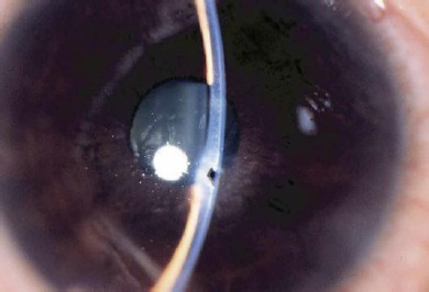
Case: A 41-year-old man developed a small paracentral corneal perforation (0.5 mm in size) in the right eye, while the treating physician attempted to remove the residual rust ring after removal of a piece of metallic foreign body.
Observations: The eye was initially managed with a bandage soft contact lens to ameliorate the aqueous leakage; however, without success. Iatrogenic iris incarceration of the wound was first induced, followed by application of cyanoacrylate tissue adhesive to the perforated site. As a result, the anterior chamber was immediately reformed and maintained. Complete corneal epithelialization of the perforation was achieved in 2 months without visual compromises.
Conclusions: Cyanoacrylate tissue adhesive with iatrogenic incarceration of the autologous iris was effective in treating this type of small corneal perforation. This technique is simple and potentially useful for small paracentral corneal perforations outside the visual axis and without good apposition.
Keywords: Corneal perforation; Cyanoacrylate; Iris incarceration; Tissue adhesive.
Figures

References
-
- Leibowitz HM. Hydrophilic contact lenses in corneal disease. IV. Penetrating corneal wounds. Arch Ophthalmol. 1972;88:602. - PubMed
-
- Parrish CM, Chandler JW. Corneal trauma. In: Kaufman HE, Barron BA, McDonald MB, editors. Cornea. Second edition. Washington: Butterworth-Heinemann; 1997. pp. 633–672.
-
- Webster RG, Slansky HH, Refojo MF, et al. The use of adhesive for closure of corneal perforations. Arch Ophthalmol. 1968;80:705–709. - PubMed
-
- Taravella MJ, Chang CD. 2-Octyl cyanoacrylate medical adhesive in treatment of a corneal perforation. Cornea. 2001;20:220–221. - PubMed
-
- Duchesne B, Tahi H, Galand A. Use of human fibrin glue and amniotic membrane transplant in corneal perforation. Cornea. 2001;20:230–232. - PubMed
Publication types
LinkOut - more resources
Full Text Sources
Research Materials
