. 2012 Jul 12:3:200.
doi: 10.3389/fimmu.2012.00200. eCollection 2012.

Marginal reticular cells: a stromal subset directly descended from the lymphoid tissue organizer

Affiliations

Marginal reticular cells: a stromal subset directly descended from the lymphoid tissue organizer

Tomoya Katakai. Front Immunol. .

Abstract

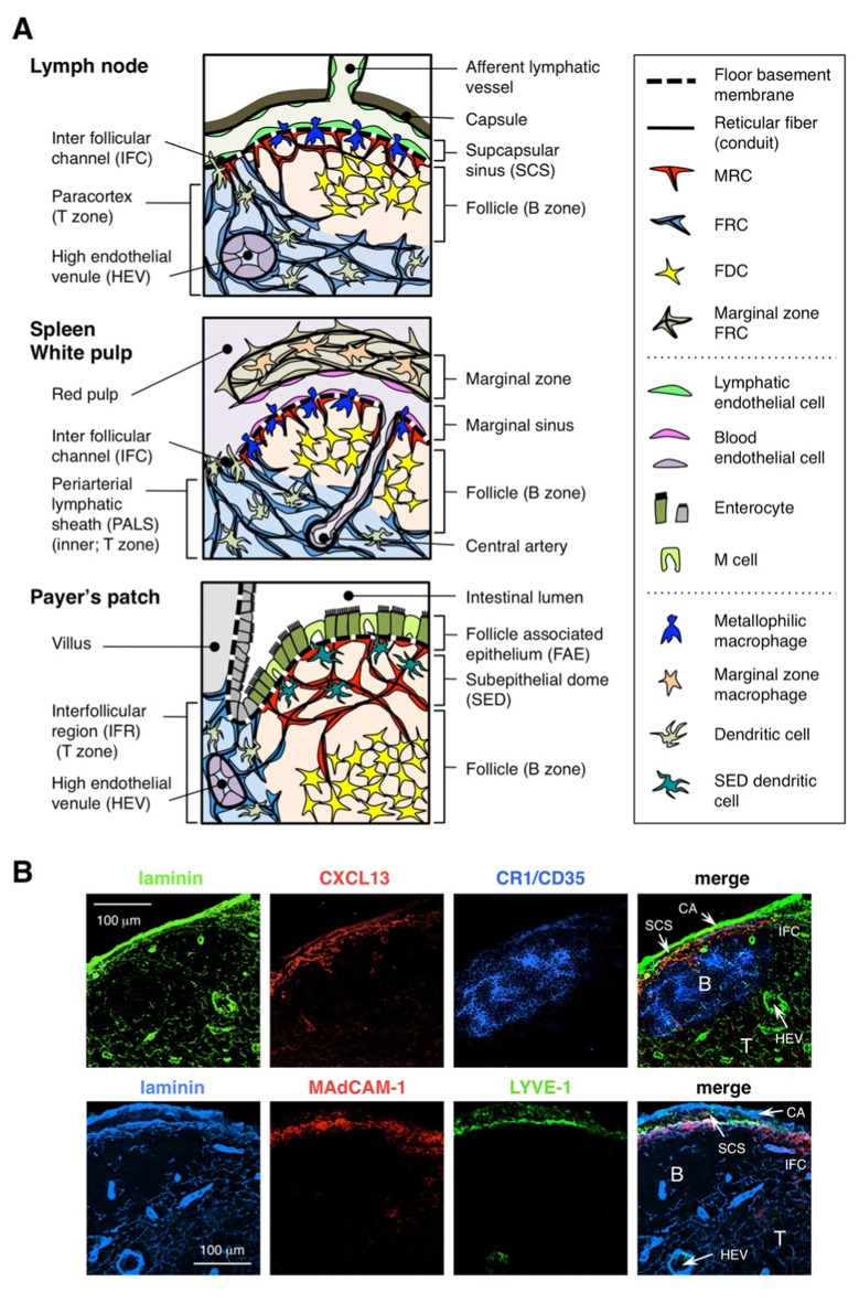
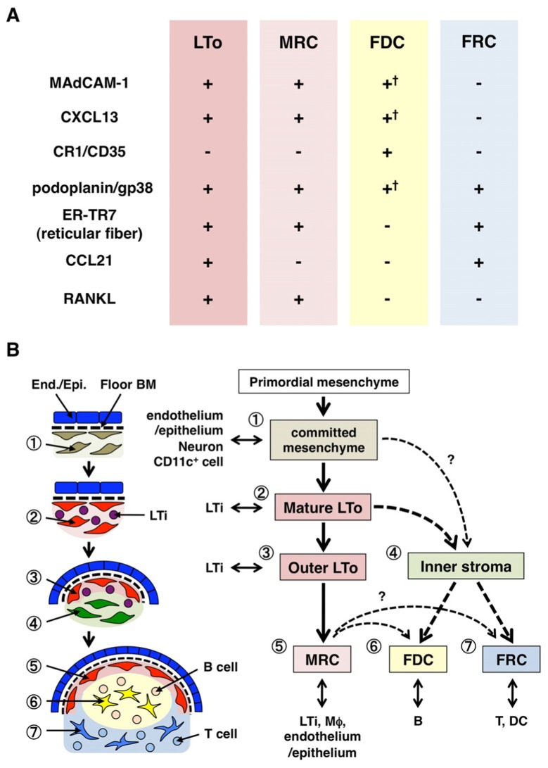
The architecture of secondary lymphoid organs (SLOs) is supported by several non-hematopoietic stromal cells. Currently it is established that two distinct stromal subsets, follicular dendritic cells and fibroblastic reticular cells, play crucial roles in the formation of tissue compartments within SLOs, i.e., the follicle and T zone, respectively. Although stromal cells in the anlagen are essential for SLO development, the relationship between these primordial cells and the subsets in adulthood remains poorly understood. In addition, the roles of stromal cells in the entry of antigens into the compartments through some tissue structures peculiar to SLOs remain unclear. A recently identified stromal subset, marginal reticular cells (MRCs), covers the margin of SLOs that are primarily located in the outer edge of follicles and construct a unique reticulum. MRCs are closely associated with specialized endothelial or epithelial structures for antigen transport. The similarities in marker expression profiles and successive localization during development suggest that MRCs directly descend from organizer stromal cells in the anlagen. Therefore, MRCs are thought to be a crucial stromal component for the organization and function of SLOs.

Keywords: CXCL13; fibroblastic reticular cell; follicular dendritic cell; lymph node; marginal reticular cell; organizer; secondary lymphoid organ; stromal cell.


Figures

FIGURE 1
FIGURE 1
Stromal cell subsets and tissue structures of SLOs. (A) Schematic representation of tissue structures in mouse SLOs, with an emphasis on stromal cells and the antigen-transporting apparatus. Stromal elements, including mesenchymal, endothelial, and epithelial cells as well as myeloid cells such as macrophages and dendritic cells (listed in the right panel), are drawn for emphasis. (B) Upper: The tissue framework constructed by three different types of stromal cells in the outer cortical region of LN. Fluorescent immunostaining of a mouse LN section. In the upper micrograph, there is a small resting follicle (B) without a germinal center, which is supported by FDCs expressing CR1/CD35 but undetectable levels of CXCL13. Instead, MRCs form a laminin+CXCL13+ reticular network in the outer edge of the follicle underneath the capsule (CA) and SCS. The network constructed by FRCs in the paracortex (T) is laminin+CXCL13-CR1/CD35-. Lower: MAdCAM-1+ MRC layer is closely associated with LYVE-1+ lymphatic endothelial layer in the SCS. Note that MRC layer is extended to the IFC area.
FIGURE 2
FIGURE 2
Relationship between MRCs and other stromal cell subsets in SLOs. (A) Pattern of marker expression in stromal cell subsets. Expression levels of MAdCAM-1, CXCL13, and podoplanin/gp38 in FDCs are often weak or undetectable by immunohistochemistry (†). (B) Schematic of a generalized model of SLO development with the locations of stromal cell subsets (left) and a putative genealogy of stromal cell subsets (right). Numbers indicating cell elements in the left drawing represent stromal cell subsets shown in the right scheme. BM, basement membrane; End./Epi., endothelial or epithelial layer.

References

    1. Bajénoff M., Egen J. G., Koo L. Y., Laugier J. P., Brau F., Glaichenhaus N., Germain R. N. (2006). Stromal cell networks regulate lymphocyte entry, migration, and territoriality in lymph nodes. Immunity 25 989–1001 - PMC - PubMed
    1. Bajénoff M., Germain R. N. (2009). B-cell follicle development remodels the conduit system and allows soluble antigen delivery to follicular dendritic cells. Blood 114 4989–4997 - PMC - PubMed
    1. Balogh P., Balázs M., Czömpöly T., Weih D. S., Arnold H. H., Weih F. (2007). Distinct roles of lymphotoxin-beta signaling and the homeodomain transcription factor Nkx2.3 in the ontogeny of endothelial compartments in spleen. Cell Tissue Res. 328 473–486 - PubMed
    1. Bénézech C., White A., Mader E., Serre K., Parnell S., Pfeffer K., Ware C. F., Anderson G., Caamaño, J. H. (2010). Ontogeny of stromal organizer cells during lymph node development. J. Immunol. 184 4521–4530 - PMC - PubMed
    1. Braun A., Worbs T., Moschovakis G. L., Halle S., Hoffmann K., Bölter J., Münk A, Förster R. (2011). Afferent lymph-derived T cells and DCs use different chemokine receptor CCR7-dependent routes for entry into the lymph node and intranodal migration. Nat. Immunol. 12 879–887 - PubMed

LinkOut - more resources