. 2012;7(7):e40439.
doi: 10.1371/journal.pone.0040439. Epub 2012 Jul 10.

Impact of combined mTOR and MEK inhibition in uveal melanoma is driven by tumor genotype

Affiliations

Impact of combined mTOR and MEK inhibition in uveal melanoma is driven by tumor genotype

Alan L Ho et al. PLoS One. 2012.

Abstract

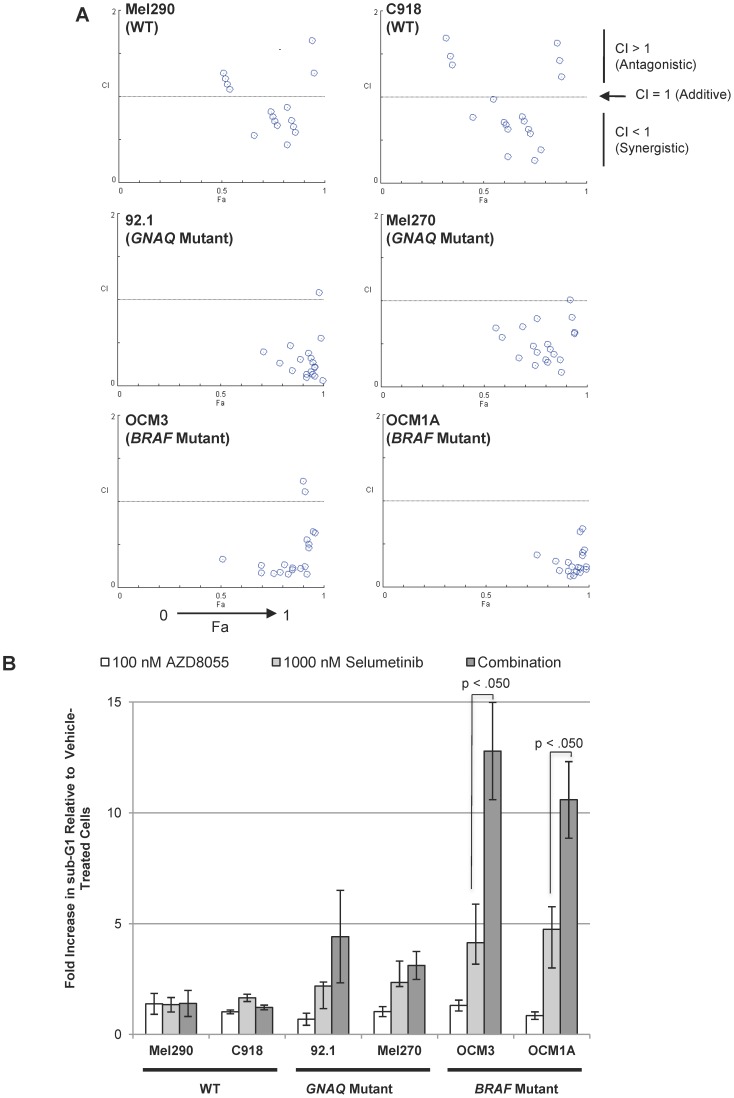
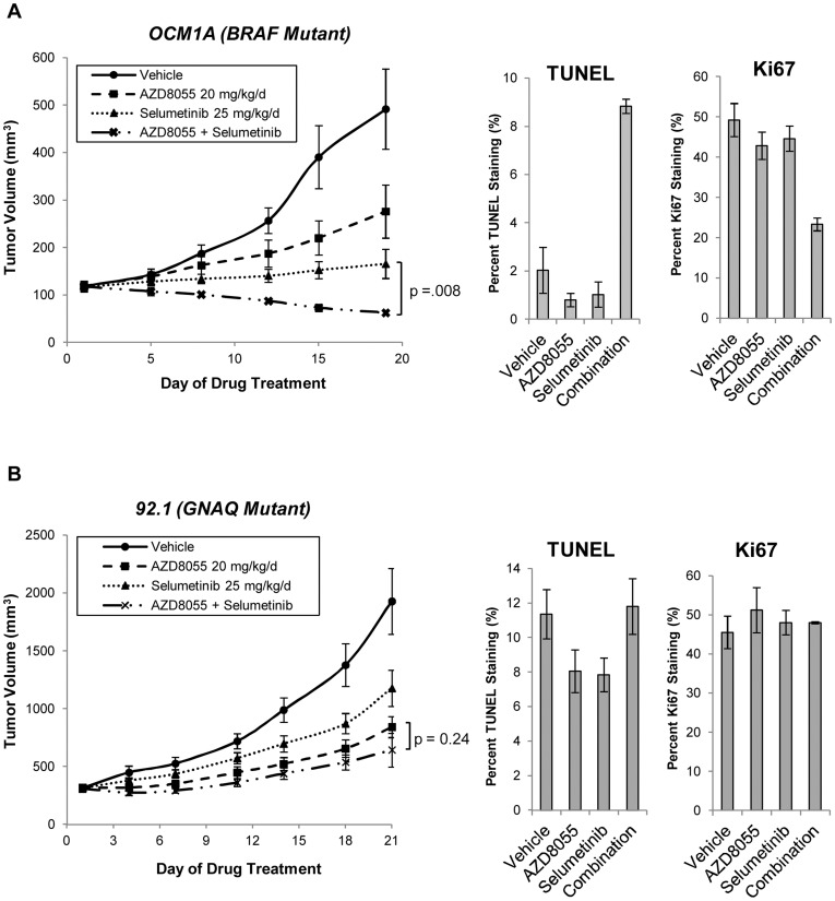
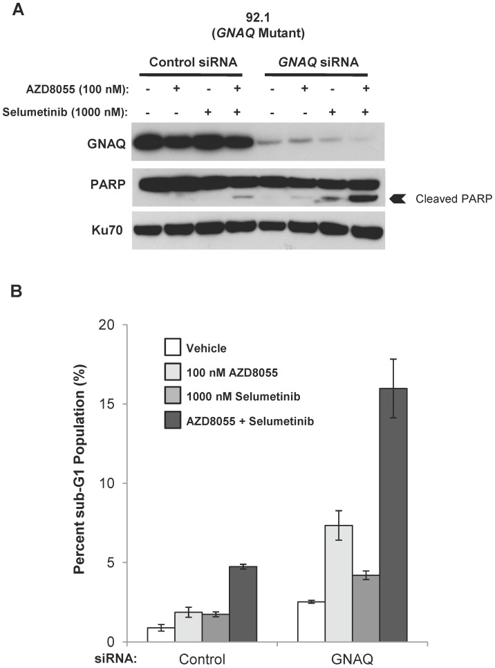
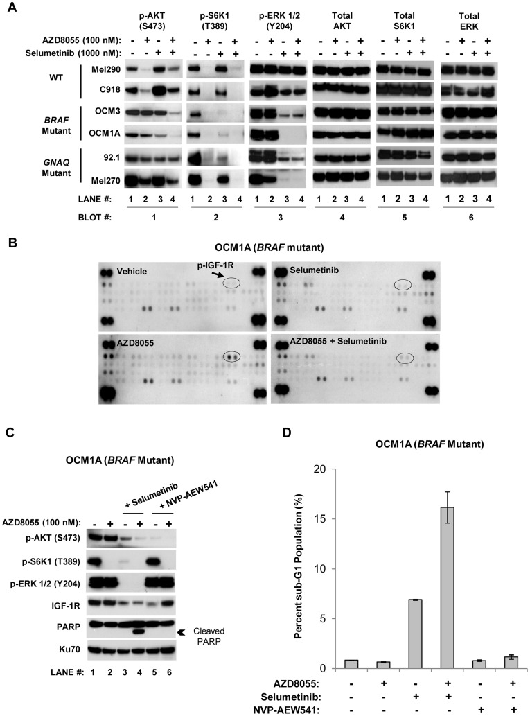
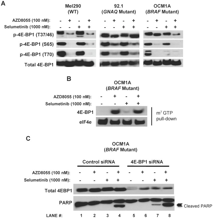
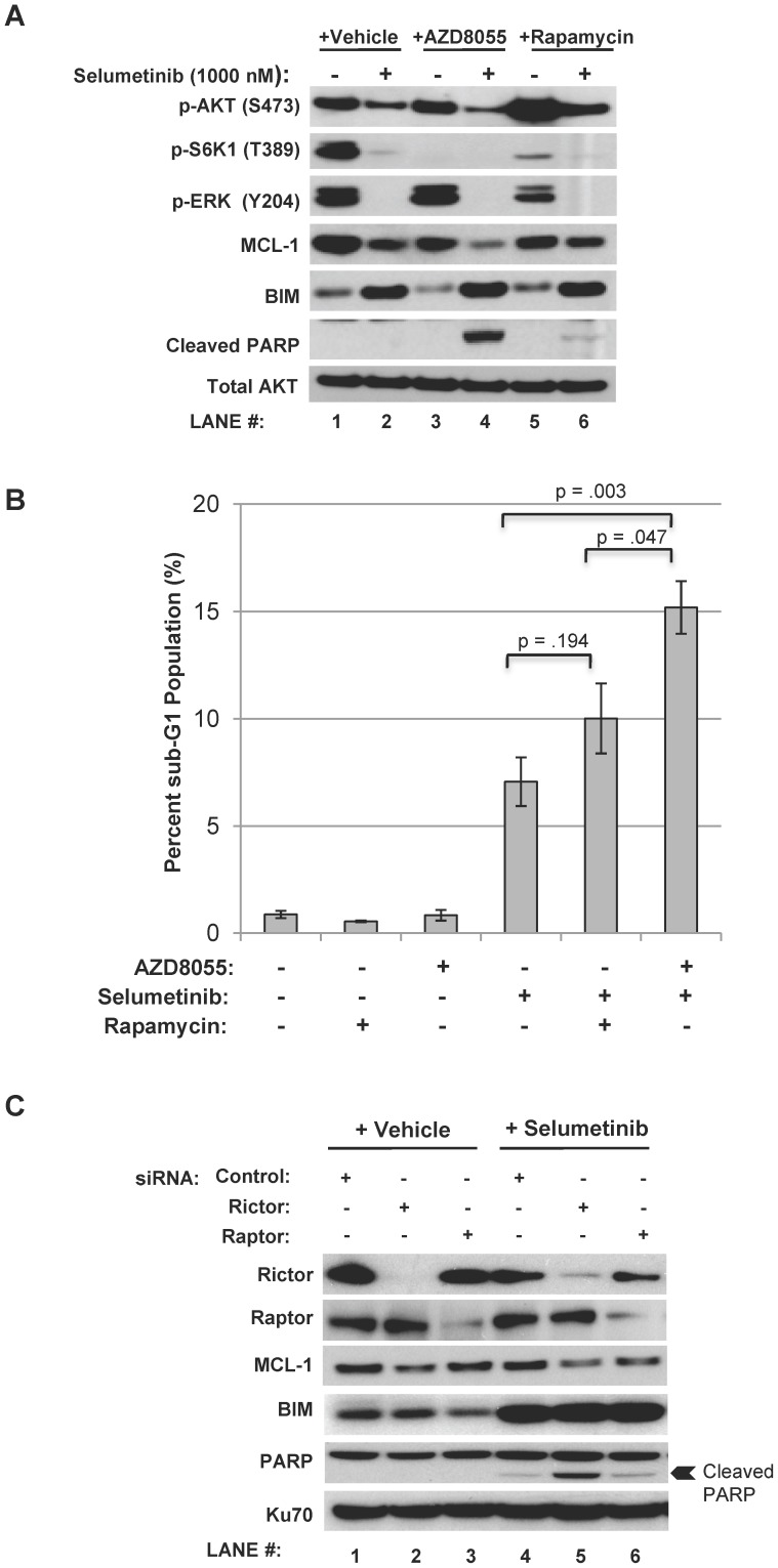
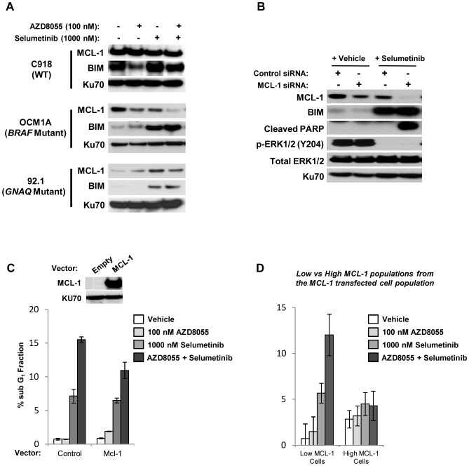
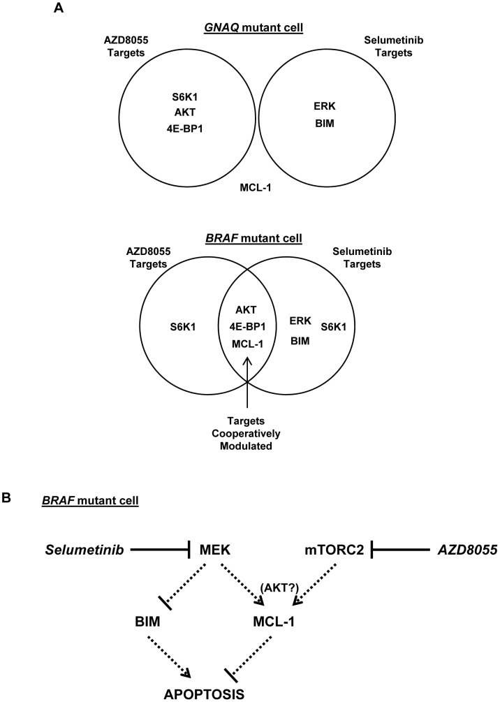
Uveal melanomas possess activation of the mitogen-activated protein kinase (MAPK) and phosphoinositide 3-kinase (PI3K)/AKT/mammalian Target of Rapamycin (mTOR) pathways. MAPK activation occurs via somatic mutations in the heterotrimeric G protein subunits GNAQ and GNA11 for over 70% of tumors and less frequently via V600E BRAF mutations. In this report, we describe the impact of dual pathway inhibition upon uveal melanoma cell lines with the MEK inhibitor selumetinib (AZD6244/ARRY-142886) and the ATP-competitive mTOR kinase inhibitor AZD8055. While synergistic reductions in cell viability were observed with AZD8055/selumetinib in both BRAF and GNAQ mutant cell lines, apoptosis was preferentially induced in BRAF mutant cells only. In vitro apoptosis assay results were predictive of in vivo drug efficacy as tumor regressions were observed only in a BRAF mutant xenograft model, but not GNAQ mutant model. We went on to discover that GNAQ promotes relative resistance to AZD8055/selumetinib-induced apoptosis in GNAQ mutant cells. For BRAF mutant cells, both AKT and 4E-BP1 phosphorylation were modulated by the combination; however, decreasing AKT phosphorylation alone was not sufficient and decreasing 4E-BP1 phosphorylation was not required for apoptosis. Instead, cooperative mTOR complex 2 (mTORC2) and MEK inhibition resulting in downregulation of the pro-survival protein MCL-1 was found to be critical for combination-induced apoptosis. These results suggest that the clinical efficacy of combined MEK and mTOR kinase inhibition will be determined by tumor genotype, and that BRAF mutant malignancies will be particularly susceptible to this strategy.


Conflict of interest statement

Competing Interests: The authors have read the journal’s policy and have the following conflicts: one-time paid consultancy (AstraZeneca), research funding (AstraZeneca) was provided to Alan Ho. To clarify, funding in both instances were not used or applicable to the study described in this submission, but instead to other, unrelated clinical research. Selumetinib and AZD8055 were supplied by AstraZeneca for this study. There are no other patents, products in development or other marketed products to declare. This does not alter the authors' adherence to all the PLoS ONE policies on sharing data and materials, as detailed online in the guide for authors.

Figures

Figure 1
Figure 1. The impact of AZD8055/selumetinib upon uveal tumor cell viability and apoptosis.
A, AZD8055/selumetinib synergistically inhibited BRAF and GNAQ mutant tumor cell viability after 96 hours of drug exposure. The graphs are Chou-Talalay plots (X-axis: Fa, or fractional activity, reflects the fraction of cellular viability relative to vehicle controls affected by the drug treatment; Y-axis, combination index (CI) with <1, >1, and  = 1 indicating synergistic, antagonistic, and additive effects, respectively). Each point represents a different combination of drug concentrations tested. Concentrations tested: AZD8055 (0, 20, 50, 100, 1000 nM) and selumetinib (0, 20, 50, 100, 1000, 5000 nM). B, sub-G1 fractions were quantified in the uveal melanoma cell line panel following the indicated 48 hour-drug treatments. AZD8055/selumetinib induced apoptosis over either drug alone only in BRAF mutant uveal tumor cells, and not in GNAQ mutant cells. Results represent the mean of three independent experiments.
Figure 2
Figure 2. AZD8055/selumetinib induces tumor regression in a BRAF mutant, but not GNAQ mutant, xenograft model.
A, AZD8055/selumetinib cooperatively induced tumor regression relative to baseline in a xenograft model with the BRAF mutant OCM1A cell line. For detailed methods, please see the Materials and Methods section. Briefly, athymic mice were subcutaneously injected with OCM1A cells. Drug treatments began after the tumors were about 100 mm3. Animals with established tumors were treated once daily with AZD8055 (20 mg/kg/d) or selumetinib (25 mg/kg/d) alone or in combination for 5 days each week for a total of 3 weeks. Tumors were measured with calipers every 2 to 3 days. Tumor volume was compared between groups of mice at various points in time. Each value represents the mean measurement of 3 to 5 animals. p-value = .008 (Wilcoxon Rank Sum test) for the comparison of selumetinib alone versus the combination or AZD8055 alone versus the combination at Day 19. Also, after the fifth drug(s) or vehicle treatment, two animals from each cohort were sacrificed and the tumors were assessed for TUNEL and Ki67 staining. Results in the graphs represent the mean percentages from 4 randomly selected fields; at least 100 cells were counted from each field. B, AZD8055/selumetinib failed to induce tumor regressions in the GNAQ mutant 92.1 cell line xenograft model. The experiment was conducted as described in A. p-value = 0.24 (Wilcoxon Rank Sum test) for the comparison of AZD8055 alone versus the combination. Error bars, SE.
Figure 3
Figure 3. Suppression of GNAQ expression in GNAQ mutant cells augments AZD8055/selumetinib-induced apoptosis.
A, suppression of GNAQ expression in the GNAQ mutant cell line 92.1 with pooled siRNA constructs resulted in increased PARP cleavage with the AZD8055/selumetinib combination. 92.1 cells were transfected with pooled GNAQ targeting siRNA constructs or unrelated control constructs for 24 hours and then treated with drugs for 24 hours (vehicle (denoted by “-”), 100 nM AZD8055, 1000 nM selumetinib, or the combination). Cell lysates were created and Western blots were then performed. The nuclear protein Ku70 was used as a loading control. B, suppression of GNAQ expression increased the sub-G1 population induced by the AZD8055/selumetinib combination in 92.1 cells. Cells were treated as detailed in A and then analyzed by flow cytometry for DNA content. The percentage of sub-G1 cells was quantified. Results are the mean of two independent experiments. Error bars, SE.
Figure 4
Figure 4. Selumetinib suppresses AKT phosphorylation in AZD8055-treated BRAF mutant cells.
A, uveal melanoma cell lines produced distinct biochemical responses to AZD8055 and selumetinib exposure. Cells were treated with the indicated drugs or vehicle (denoted by “-”) for 24 hours and Western blots were then performed. Total AKT was used as a loading control. Of note, selumetinib alone inhibited S6K1 phosphorylation in BRAF cells and to a lesser extent in GNAQ cells (lane 1 versus 3 in Blot #2). Given how effectively AZD8055 inhibited S6K1 phosphorylation, though, it is unlikely that this selumetinib effect significantly contributed to the impact of the combination in BRAF cells. B, IGF-1R phosphorylation increased with AZD8055 treatment and was suppressed by selumetinib in the BRAF mutant cell line OCM1A. OCM1A cells were treated as described in A, and cellular lysates were created and analyzed with phosphorylated receptor tyrosine kinase (RTK) antibody array blots. The blots reflect the phosphorylation status of 42 RTKs. Duplicate spots in the corners of each blot are positive controls. IGF-1R duplicate spots are circled. C, inhibition of IGF-1R blocked AKT phosphorylation in OCM1A cells, but did not induce PARP cleavage in combination with AZD8055. Cells were treated with the same drugs and concentrations as detailed in A in addition to the IGF-1R small molecule inhibitor NVP-AEW541 at 1000 nM. Cells were treated for 24 hours before Western blots were performed. Ku70 was used as a loading control. D, IGF-1R inhibition with NVP-AEW541 failed to induce apoptosis in combination with selumetinib in OCM1A cells. Cells were treated with drugs for 48 hours and analyzed by flow cytometry for DNA content. The percentages of sub-G1 cells were quantified. Results are the mean of two independent experiments. Error bars, SE.
Figure 5
Figure 5. 4E-BP1 phosphorylation is modulated by AZD8055/selumetinib, but does not regulate BRAF mutant cell survival.
A, the AZD8055/selumetinib combination cooperatively suppressed 4E-BP1 phosphorylation at T37/46 in the BRAF mutant cell line OCM1A. Cells were treated with the indicated drugs for 24 hours. Western blots were then performed. Total 4E-BP1 was used as a loading control. B, the capability of 4E-BP1 binding to the mRNA cap complex was not impacted by the AZD8055/selumetinib combination in OCM1A cells. A cap binding assay was performed (referenced in the Materials and Methods section). Briefly, cells were treated with the indicated drugs for 24 hours. Cell lysates were created and then incubated with m7 GTP sepharose beads to capture all 4E-BP1 and eIF4E proteins that are capable of binding to an mRNA cap complex. The bead-associated proteins were analyzed by Western blot. C, suppression of 4E-BP1 expression did not diminish apoptosis as assessed by PARP cleavage in OCM1A cells. Cells were transfected with siRNA constructs targeting 4E-BP1 or unrelated control constructs for 48 hours. Cells were then treated with the indicated drugs for 24 hours. Western blots were performed.
Figure 6
Figure 6. mTORC2 inhibition in combination with selumetinib induces apoptosis in BRAF mutant cells.
A, mTORC1 inhibitor rapamycin in combination with selumetinib failed to induce apoptosis as evidenced by a lack of PARP cleavage in the BRAF mutant cell line OCM1A. Cells were treated with the indicated drugs (vehicle, 100 nM AZD8055, 1000 nM selumetinib, and 10 nM rapamycin) for 24 hours. Western blots were then performed. Total AKT was used as a loading control. B, rapamycin in combination with selumetinib failed to substantially increase the sub-G1 fraction in the OCM1A cell line. Cells were treated as detailed in A and then analyzed by flow cytometry for DNA content; the sub-G1 fraction was quantified. Results are the mean of four independent experiments. Error bars, SE. C, suppression of Rictor and mTORC2 activity led to MCL-1 downregulation and PARP cleavage in combination with selumetinib in the OCM1A cell line. Cells were transfected with pooled siRNA constructs targeting Rictor or Raptor or an unrelated control construct for 48 hours. Cells were then treated with either vehicle or 1000 nM selumetinib for 24 hours and a Western blot was then performed. Ku70 was used as a loading control.
Figure 7
Figure 7. Modulation of MCL-1 by AZD8055/selumetinib contributes to apoptosis in BRAF mutant cells.
A, the AZD8055/selumetinib combination cooperatively suppressed MCL-1 and induced BIM in the BRAF mutant OCM1A cell line. Cells were treated with the indicated drugs (vehicle denoted by “-”) for 24 hours and Western blots were then performed. Ku70 was used as a loading control. B, suppression of MCL-1 levels via targeting siRNA constructs in combination with selumetinib treatment was sufficient to induce apoptosis as evidenced by increased PARP cleavage in the OCM1A cell line. Cells were transfected with pooled siRNA constructs targeting MCL-1 or unrelated control constructs for 48 hours and then treated with 1000 nM selumetinib for 24 hours. Western blots were then performed. Ku-70 was used as a loading control. C, MCL-1 overexpression in the OCM1A cell line reduced AZD8055/selumetinib induced apoptosis. Cells were transiently transfected with an MCL-1 cDNA expression plasmid under the control of a constitutively active viral promoter or an empty vector for 48 hours. Western blot was performed to confirm overexpression of MCL-1. Cells were then treated with the indicated drugs for 24 hours. Cells were analyzed by flow cytometry for DNA content; sub-G1 populations were quantified. D bi-parametric flow cytometry demonstrated that apoptosis was preferentially induced by AZD8055/selumetinib in low MCL-1 expressing cell populations. Cells were transiently transfected with MCL-1 as detailed in C. The low- and high- MCL-1 expressing cell populations from these MCL-1 transfected cells were then detected and analyzed by bi-parametric flow cytometry for DNA content in addition to MCL-1 expression level (please see Figure S6 for the flow cytometry plots). Depicted results are representative of two independent experiments. Error bars, SE.
Figure 8
Figure 8. Diagrammatic representation of signaling proteins and pathways modulated by AZD8055 and selumetinib.
A, This study revealed that AZD8055 and selumetinib impacted the regulation of several signaling proteins differently in GNAQ mutant versus BRAF mutant cells. The proteins AKT, 4E-BP1, and MCL-1 were cooperatively regulated by the AZD8055/selumetinib combination in BRAF mutant cells, while none of the candidate targets examined were found to be cooperatively regulated in GNAQ mutant cells. B, Diagram of the signaling pathways impacted by selumetinib and AZD8055 to induce apoptosis in BRAF mutant cells. Our data suggests that BIM upregulation and MCL-1 downregulation are necessary for apoptosis achieved with dual pathway inhibition. While BIM was induced by selumetinib, MCL-1 was decreased by combined mTORC2 inhibition and MEK inhibition by AZD8055 and selumetinib, respectively, possibly through cooperative AKT inhibition. The dotted arrows indicate that drug mediated inhibition of MEK and mTORC2 likely influence BIM and MCL-1 through molecular intermediates.

References

    1. Patel M, Smyth EC, Chapman PB, Wolchok JD, Schwartz GK, et al. Therapeutic Implications of the Emerging Molecular Biology of Uveal Melanoma. Clin Cancer Res. 2011. - PubMed
    1. Weber A, Hengge UR, Urbanik D, Markwart A, Mirmohammadsaegh A, et al. Absence of mutations of the BRAF gene and constitutive activation of extracellular-regulated kinase in malignant melanomas of the uvea. Lab Invest. 2003;83:1771–1776. - PubMed
    1. Zuidervaart W, van Nieuwpoort F, Stark M, Dijkman R, Packer L, et al. Activation of the MAPK pathway is a common event in uveal melanomas although it rarely occurs through mutation of BRAF or RAS. Br J Cancer. 2005;92:2032–2038. - PMC - PubMed
    1. Edmunds SC, Cree IA, Di Nicolantonio F, Hungerford JL, Hurren JS, et al. Absence of BRAF gene mutations in uveal melanomas in contrast to cutaneous melanomas. Br J Cancer. 2003;88:1403–1405. - PMC - PubMed
    1. Malaponte G, Libra M, Gangemi P, Bevelacqua V, Mangano K, et al. Detection of BRAF gene mutation in primary choroidal melanoma tissue. Cancer Biol Ther. 2006;5:225–227. - PubMed

Publication types

MeSH terms

Substances